Introduction
Lymphatic filariasis (LF) is caused by three species of thread-like filarial nematodes (Wuchereria bancrofti, Brugia malayi and B. timori) and, of these three, 90% of all infections result from W. bancrofti (Ravindran, Reference Ravindran2003). The World Health Organization (WHO) estimates that in 81 countries over 120 million people are infected, and approximately 1.34 billion are at risk in those tropical and subtropical regions endemic to filariasis. About 25 million men have contracted urogenital swelling, mainly scrotal hydrocoele, and almost 15 million (mostly women) display lymphoedema/elephantiasis of the leg (WHO, 2010).
The disease is transmitted through the bite of infected mosquitoes, including species of Culex, Aedes, Anopheles, Mansonia and Coquilletidia spp., and infective third-stage larvae (L3) that subsequently migrate to the lymphatic system. After reaching sexual maturity in 6–12 months, the adult female worms release millions of microfilariae (mf) into the bloodstream. The life cycle is completed when the intermediate host, a mosquito vector, ingests the microfilariae in a blood meal (Garcia, Reference Garcia2007).
Zoonosis is a disease usually found in animals that is transmissible to humans. Zoonotic filariasis is a term reserved specifically for humans infected by filariae of parasites normally originating in mammals (Orihel & Eberhard, 1998). In the majority of cases, human infection is subclinical. The identification of the species is often problematic insofar as the mf may take up residence in a variety of tissues and elicit no overt immunological response and rarely any discernible symptoms, with the possible exception of ocular infection involving eyelids, conjunctiva, anterior chamber or uvea. Rao et al. (Reference Rao, Mahapatra, Pattnayak and Pattnaik2008) report the case of a live intravitreal filarial worm successfully removed from the area between the optic disc and macula of the left eye of a patient and, by elimination, identified as B. malayi ‘as this is the only other filarial worm found in the Indian subcontinent’ (Rao et al., Reference Rao, Mahapatra, Pattnayak and Pattnaik2008). Yen & Mak (Reference Yen and Mak1978) have proposed using acid phosphatase activity to reliably differentiate between mf of closely related species of Filariae. However, identification of mf by subtle morphological measurements is often difficult, tedious and time consuming, requiring expertise to microscopically differentiate between the two Brugia species.
Brugia pahangi is a common parasite of dogs and cats, which often resides in the lymphatic system (and in this respect is similar to B. malayi in humans) but is usually restricted to these animals. However, an early study involving the experimental transmission of B. pahangi concluded that this particular parasite can be ‘potentially infective to man’ (Edeson et al., Reference Edeson, Wilson, Wharton and Laing1960).
The present study describes a case of Brugian filariasis outside the endemic areas in Surat Thani Province, southern Thailand, and possible sources of this cryptic infection are discussed.
Case report
In November 2009, a 2-year-old boy from Natrear Village, Ban Nasan District in Surat Thani Province developed a high fever (39°C) and was taken to a private hospital in the neighbouring Mueang District. On physical examination, there was no neck stiffness, the heart and lungs sounded normal and the abdominal findings were normal. There was no evidence of lymphoedema. A peripheral blood examination showed haemoglobin of 12.7 g/dl, a total leucocyte count of 8470 cells/ml, differential leucocyte percentages of 41% neutrophils, 43% lymphocytes, 6% eosinophils, 6% monocytes and 4% basophils, and a platelet count of 588,000 cells/ml. Urine was examined and found to be normal. Thick blood smear staining was performed to exclude malaria and lymphatic filarial infection. Two mf were detected from the blood and identified as B. malayi (sheathed mf length 250 μm, with an elongated cephalic space and two terminal nuclei at the tip of the tail), whereas no malaria was found.
The boy was immediately referred to a government-run general hospital in the Mueang District where a repeat screening using nocturnal blood was carried out and examined. Microfilariae of B. malayi were detected. After treatment with diethylcarbamazine citrate and albendazole, his symptoms resolved completely, and he was discharged the next day. As regulations require, the incident of Brugian filariasis was reported to the Vector Borne Disease Control Center (VBDCC) of the Ministry of Public Health located in Surat Thani Province.
Since this case of Brugian filariasis was found outside the endemic areas of LF in this province, health officials from the VBDCC were contacted regarding the possible source of this infection. An investigation was carried out from January to March 2010, during which time a complete family history was elicited. The boy resided with his parents and three siblings in the Natrear village of the Nasan District. His father's occupation was that of a furniture maker which necessitated frequent trips by the entire family away from home without staying overnight. Consequently, the family's travel history within the past year, including that of the boy, was thoroughly reviewed. They had close relatives living nearby in the Huai Mut community (Nasan District, Surat Thani Province), who they visited a few times in the first 2 months of 2009. In addition, once in April 2009 for about 1 h at sunset, they visited the Hart Charoen village, Trikeung subdistrict of the Prasang District to have dinner with friends.
Additionally, night blood was collected by finger prick from 128 residents, including the patient's family, living within a 1 km radius from the family home, and examined for the presence of mf. The peripheral thick smear was negative in all cases.
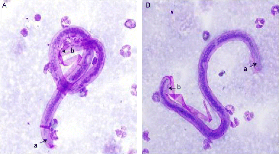
There is still no study on the periodicity of Brugia spp. infection in cats. Thus, to investigate the possibility that they were reservoir hosts, ear blood was collected from domestic cats residing or straying within a 1 km radius at night time, according to the recommendations of a WHO committee (WHO, 1962). Thick blood smear staining was performed for mf detection in 17 cats. Of the 17 cats, two were found to be positive for mf of the Brugia spp. (fig. 1).
(colour online) Sheathed microfilariae of Brugia spp. from a thick blood smear of two cats (A, B). a, Elongated cephalic space; b, nuclei near the tip of the tail.

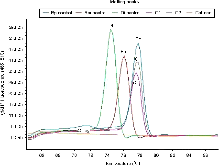
To accurately identify the specific species, a high-resolution melting real-time polymerase chain reaction (PCR) assay targeting the mitochondrial 12S rRNA gene, which is highly conserved and contains genus- and species-specific sequence variation useful for identifying B. malayi, B. pahangi and Dirofilaria immitis, was performed at the Department of Parasitology, Faculty of Medicine, Siriraj Hospital, Mahidol University, according to the method of Wongkamchai et al. (Reference Wongkamchai, Munkong, Mahannob, Taweethavonsawat, Loymak and Fungladda2012). The mf from the two positive cats could be recognized by the same melting temperature as the B. pahangi reference control. Thus, the mf were identified as B. pahangi (fig. 2).
(colour online) High-resolution melting peaks of microfilariae from positive cat samples (C1 and C2), and control species. Bm, Brugia malayi; Bp, B. pahangi; Di, Dirofilaria immitis.
In an attempt to identify the possible vector(s) of this parasite, the human-landing-catch method was employed (Ndiath et al., Reference Ndiath, Mazenot, Gaye, Konate, Bouganali, Ousmane, Sokhna and Trape2011) by staff from the VBDCC, no. 11.3 Surat Thani Province. Four male adult volunteers served as bait or lures for attracting mosquitoes and were supervised by nursing personnel during the collection period. Collection took place on two consecutive evenings and in two different outdoor locales in close proximity to the patient's house (within 15 m). Since Mansonia spp. and Anopheles spp. were reported by WHO (1962) as vectors of B. malayi, mosquitoes were collected for 3 h, i.e. from 18.00 to 21.00 hours. Thus, sampling of the mosquito density resulted in a total of 24 man-hours spent in the collection process. These data were converted to the number of mosquitoes collected for every 10 man-hours spent collecting, according to the following formula:
 $$\begin{eqnarray} D = N /( t /10), \end{eqnarray}$$
where D is density, expressed as the number of mosquitoes per 10 man-hours of collecting; N is the total number of mosquitoes collected, summed over the number of volunteers, the hours spent serving as bait and number of nights in the field; t is the total time in hours across all volunteers and nights spent collecting; and 10 is the conversion factor, 10 man-hours.
This formula essentially equalizes any variability in the number of volunteers or time spent with volunteers serving as collection agents, and is used extensively in different studies in southern Thailand for cross-study comparisons.
The collected mosquitoes were sent to the VBDCC, no. 11.3 Surat Thani Province for species identification under stereomicroscopy. A total of 10 different mosquito species were collected and, of these, Armigeres spp. and Culex gelictus were the most prevalent, with the two Mansonia species ranking third and fourth in frequency. However, the number of all the individual species collected was relatively low, based on a general rule of thumb used in the field, where (a) if the mosquito density is < 50, there is a low probability of filarial transmission; (b) if the mosquito density is between 51 and 100, it is intermediate; and (c) if the density is >100, it is high and filariasis transmission is more probable.
Additionally, the general environment surrounding the patient's house was surveyed. Geographically, the centre of the province is the coastal plain of the Tapi River, which is mostly grassland, interspersed with rubber and coconut trees. The delta of the two main rivers (Tapi and the Phum Duang) flowing through the centre of the province consists of several channels with small islands covered predominantly with mangrove and orchards. There is an open swamp forest located close to the boy's home.
Discussion
The present case history deals with a young boy who was found to be infected with mf identified as B. malayi using Giemsa staining of thick blood smears. Subsequently the local VBDCC, alerted by this finding beyond the known endemic areas for this parasite, launched an intensive investigation. The possibility arose that the initially identified parasite was not B. malayi but B. pahangi, since differentiating between these two mf using blood-staining procedures is often difficult, and could lead to a misdiagnosis. Unfortunately, at the time of the initial blood collection, PCR was not performed.
Tan et al. (Reference Tan, Fong, Mahmud, Muslim, Lau and Kamarulzaman2011) described five Malaysian patients diagnosed with LF on the basis of clinical symptoms and positive serological findings. The major presenting symptoms were recurring descending lymphangitis and cellulitis of the lower limbs which were resolved after treatment with diethylcarbamazine (2 weeks), albendazole (single dose) and, in one patient, after an 8-week course of deoxycycline. Only two patients were found to be PCR positive for B. pahangi cytochrome c oxidase subunit 1 (COXI) gene sequence when the test was performed (a) on fresh nocturnal blood of one patient (P5), and (b) on the retrieved blood of patient P4. Of the three mosquito vectors surveyed within a 2 km radius of the patients' residential area, only Armigeres subalbatus carried filariasis larvae whose COXI DNA sequence was identical to the B. pahangi larvae and that of patient P4. The other three patients were classified as probable cases of human B. pahangi infection. The reservoir host was determined to be one of five cats testing positive for B. pahangi mf based on morphology and length, and verified by exhibiting identical COXI to that from patient P4, and of the larvae found in A. subalbatus (Tan et al., Reference Tan, Fong, Mahmud, Muslim, Lau and Kamarulzaman2011).
In the present study, from the family's travel history there were two probable locales in Surat Thani Province where the patient may have contracted the infection: the Hart Charoen village, in the Trikeung subdistrict of the Prasang District, and the Huai Mut village of the Nasan District. The Prasang District has long been known as an endemic area of Malayan filariasis (Nuchprayoon et al., Reference Nuchprayoon, Junpee, Nithiuthai, Chungpivat, Suvannadabba and Poovorawan2006). There are palm oil and rubber plantations in close proximity to a large swamp forest, serving as convenient ecosystems for the breeding of Mansonia mosquitoes. In this district, Nuchprayoon et al. (Reference Nuchprayoon, Junpee, Nithiuthai, Chungpivat, Suvannadabba and Poovorawan2006) examined 52 cats and identified, by restriction fragment length polymorphism-PCR, five cats of which four exhibited B. pahangi and one D. immitis. Additionally, two infected cats were detected by restriction fragment length polymorphism-PCR but were not identified by microscopy (Nuchprayoon et al., Reference Nuchprayoon, Junpee, Nithiuthai, Chungpivat, Suvannadabba and Poovorawan2006). This low percentage (7.7%) makes it unlikely that the boy became infected in this area, which was extensively treated by mass drug administration between 2000 and 2005 through Thailand's National Program for the Elimination of LF. Currently, the programme is in the post-mass drug treatment-evaluation phase. No children between the ages of 2 and 6 years have been found to be infected with LF. Moreover, the boy did not stay overnight in this area. His length of stay was about 1 h. In general terms, the intensity of transmission (infection prevalence) is determined by (a) the cumulative past exposure to infection bites by filarial vectors, (b) the rate of parous biting mosquitoes, and (c) the proportion of the L3 larvae invading the host from one bite (Srividya et al., Reference Srividya, Das, Subramanian, Ramaiah, Grenfell, Michael and Bundy1996), which is negligible in the now endemic-free Prasang District due to repeated mass treatments (Michael & Gambhir, Reference Michael and Gambhir2010).
The Huai Mut community of the Nasan District where the family visited relatives is approximately 1 km from the patient's home. While there were no instances of filariasis in this area, it is a known entomological site for Mansonia spp. Close to the Huai Mut community is the Huai Yai village, which has had a history of filariasis but in 2009 was considered endemic free. Data obtained by the VBDCC officers surveying residents within a 1-km radius of the patient's home also showed no evidence of B. malayi mf in analysed thick blood smears.
Only two cats out of the 17 cats surveyed in the Natrear village were positive for B. pahangi. Further, the density of the mosquitoes trapped in the Natrear village was low, indicating a low transmission potential.
Thus the conundrum surrounding the aetiology of the boy's infection remains unsolved at present. It can only be concluded that a further re-investigation coupled with human and animal surveillance by the PCR procedure be instituted in both the old and new endemic areas of Brugian filariasis in the Natrear village, and the Huai Mut and Hart Chareon communities, for the re-emergence of this disease.