Introduction
Meningiomas are slow-growing, benign tumours that originate from the arachnoid villi of the meninges. They account for 13 to 26 per cent of all intracranial neoplasms.Reference Whittle, Smith, Navoo and Collie1 Less than 1 per cent of all meningiomas consist of primary extracranial tumours of the ear and temporal bone.Reference Ayache, Trabalzini, Bordure, Gratacap, Darrouzet and Schmerber2
Intracranial meningiomas extending to the middle-ear cleft are uncommon, with fewer than 100 cases reported to date.Reference Chang, Cheung and Jackler3 These patients usually present with hearing loss, tinnitus, dizziness, vertigo or facial nerve weakness.Reference Chang, Cheung and Jackler3, Reference Civantos, Ferguson, Hemmati and Gruber4 Secretory otitis media secondary to eustachian tube obstruction has also been reported.Reference Civantos, Ferguson, Hemmati and Gruber4
We have previously reported a case of a meningioma extending into the middle ear and mimicking chronic otitis media.Reference Khalaila, Braun, Lurie, Zaaroor and Luntz5 Since then, we have seen two further patients presenting with similar clinical and radiological findings. In this paper, we present these three unique patients in whom chronic otitis media was the presenting clinical sign of skull base meningioma. We also discuss the imaging characteristics and management considerations.
Case reports
Case one
A 49-year-old woman was referred to our department with right chronic otitis media and hearing loss, both of more than three years' duration.
Otoscopy revealed an opaque tympanic membrane with a solid mass visible behind it.
Audiometry showed mixed conductive and sensorineural hearing loss.
There were no neurological symptoms or deficits.
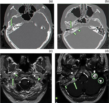
High-resolution computed tomography (CT) demonstrated an opacified middle ear, antrum and mastoid air-cell system, without significant bony destruction (Figure 1a and 1b).
(a) and (b): Axial high resolution computed tomography scans showing opacified right middle ear and mastoid (long arrows), and displaying a pathological appearance of the posterior aspect of the petrous bone (short arrows, part b). (c) and (d): Axial, T1-weighted magnetic resonance imaging scans with gadolinium enhancement, showing an enhancing lesion (long arrows) surrounding the right internal carotid artery (small arrow, part d) and compressing the right internal jugular vein, which is seen only on the normal left side (arrowhead, part c). The lesion borders the clivus medially (arrowhead, part d) and the sigmoid sinus laterally (short arrow, part d). There is no enhancement of the right jugular vein or sigmoid sinus, contrary to the normal appearance of the left side (circles, part d).

In view of her history and atypical otoscopic findings, the patient underwent middle-ear exploration and mastoidectomy. At surgery, the mastoid was found to contain only fluid. The attic and the middle ear were occupied by a non-bleeding mass suspected to be a soft tissue neoplasm. Frozen section analysis pointed to the possibility of a benign non-epithelial tumour.
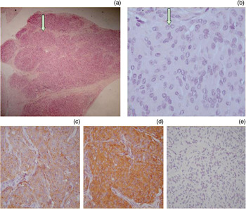
Histological studies of permanent sections demonstrated epithelioid-like cellular aggregates with a whorling pattern, comprising cells with indistinct borders and round-to-oval nuclei. There was no evidence of nuclear pleomorphism or mitotic activity (Figure 2a).
Haematoxylin and eosin staining showing (a) cellular aggregates with a whorling pattern (arrow) (× 100) and (b) indistinct, interdigitating cell borders, round to oval nuclei, delicate chromatin and nuclear clearing nuclei (arrow) (× 400). Immunohistochemical studies show positivity for (c) epithelial membrane antigen (× 200) and (d) vimentin (× 200) (note brown staining for both), but (e) negativity for S100 (× 200).

Immunohistochemical analysis showed positive staining for epithelial membrane antigen and vimentin, but negative staining for S100 protein and cytokeratin (Figure 2b).
The morphological features and immunohistochemical profile of the tumour were typical of meningioma.
Magnetic resonance imaging (MRI) showed an enhancing lesion surrounding the right internal carotid artery and compressing the right internal jugular vein. The lesion bordered the clivus medially and the sigmoid sinus laterally. In contrast to the normal left side, there was no enhancement of the right jugular vein or sigmoid sinus (Figure 1c and 1d). These imaging findings were consistent with the diagnosis of meningioma.
In view of the absence of clinical signs of neurological involvement or brain compression, it was decided to adopt a conservative ‘wait and scan’ management policy.
Case two
A 41-year-old woman was referred to our department with right chronic otitis media and hearing loss, both of seven years' duration.
Otoscopy revealed a solid mass filling the right external ear canal, with otorrhoea.
Audiometry disclosed severe conductive hearing loss.
There were no neurological symptoms or deficits.
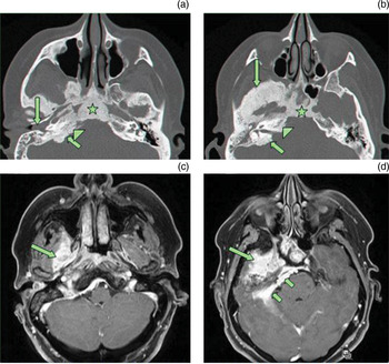
High resolution CT demonstrated an opacified right middle ear and mastoid with a mass filling the external ear canal, without bony destruction. There was marked hyperostosis involving the temporal bone, the greater wing of the sphenoid, the petrous pyramid and the clivus, with a hairy aspect of the margins of the affected bone (Figure 3a and 3b).
(a) and (b): Axial, high resolution computed tomography scans showing opacified right middle ear and mastoid with a mass filling the external ear canal (long arrow, part a). There is marked hyperostosis involving the temporal bone (short arrows, parts a and b), the greater wing of the sphenoid (long arrow, part b), the petrous pyramid (arrowhead, parts a and b) and the clivus (star, parts a and b), with a hairy aspect of the margins of the affected bone. (c) and (d): Axial, T1-weighted magnetic resonance imaging scans with gadolinium contrast, showing an enhancing lesion in the right infratemporal fossa (long arrows) with linear enhancement on the posterior aspect of the petrous pyramid (short arrows, part d), typical of en plaque meningioma.

Magnetic resonance imaging showed an enhanced lesion in the right infratemporal fossa, with linear enhancement on the posterior aspect of the petrous pyramid (Figure 3c and 3d).
Imaging characteristics were typical of an en plaque meningioma.
In view of the lack of neurological symptoms, it was decided that the patient should undergo repeated imaging without any surgical intervention.
Case three
A 59-year-old man was referred to our department with left chronic otitis media and hearing loss, both of three years' duration.
Otoscopy revealed a middle-ear effusion.
Endoscopy showed a normal nasopharynx.
Audiometry disclosed moderate conductive hearing loss.
There were no neurological symptoms or deficits.
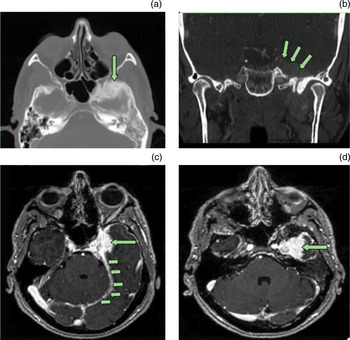
High resolution CT demonstrated an opacified left middle ear and mastoid without bony destruction. There was hyperostosis involving the greater wing of the sphenoid, with hairy margins of the affected bone, and an enhancing left parasellar mass (Figure 4a and 4b).
(a) Axial and (b) coronal high resolution computed tomography scans showing opacified left middle ear and mastoid. There is hyperostosis involving the greater wing of the sphenoid (arrow, part a), with hairy margins of the affected bone. Part (b) shows an enhancing, left, para-sellar mass (arrows). (c) and (d): Axial, T1-weighted magnetic resonance imaging scans with gadolinium contrast, showing an enhancing, left, parasellar mass extending to the left cavernous sinus (long arrows, parts c and d) and surrounding the internal carotid artery. Meningeal thickening and the hyper-intense signal of the tentorium represent the ‘dural tail’ (short arrows, part c).

Magnetic resonance imaging showed that this mass extended to the left cavernous sinus and surrounded the internal carotid artery. Meningeal thickening and a hyperintense signal of the tentorium represented the ‘dural tail’ typical of meningioma (Figure 4c and 4d).
Despite the extent of the lesion, and in view of the absence of brainstem compression and cranial nerve involvement, it was decided that the patient should undergo repeated imaging without surgical intervention.
Discussion
Meningiomas comprise the second largest group of primary brain tumours after gliomas. Middle-ear or temporal bone involvement by meningioma is extremely rare. Extension of meningiomas into the middle ear or mastoid can occur via several pathways: tegmen tympani, posterior fossa plate, internal auditory canal or jugular foramen.Reference Chang, Cheung and Jackler3
Computed tomography in conjunction with MRI can reliably determine the diagnosis and extent of a temporal bone meningioma. Magnetic resonance imaging with gadolinium contrast typically shows an enhancing, dural-based, soft tissue mass, while CT shows a hyperostotic bone reaction and a hairy aspect of the margins of the affected bone.Reference Ayache, Trabalzini, Bordure, Gratacap, Darrouzet and Schmerber2
Histologically, meningioma is characterised by cellular aggregates in a whorling pattern and psammoma bodies (Figure 2a). A lack of nuclear pleomorphism or mitotic activity, positive immunohistochemical staining with vimentin and epithelial membrane antigen, and negative staining with cytokeratin confirm the diagnosis (Figure 2b).
Nager'sReference Nager6 1966 paper delineated two types of temporal bone meningiomas: type one, extradural extension of an intracranial meningioma; and type two, primary extradural meningioma. There are also two types of intracranial meningiomas: globular and en plaque. The most common form is the globular type (80 per cent), which presents as a bulky mass compressing adjacent neural structures. En plaque meningiomas infiltrate the dura without bulging into the cerebellopontine angle, and spread along the surface of the temporal bone, most often the posterior surface of the petrous pyramid.
Approximately 90 per cent of meningiomas are benign, 6 per cent are atypical and 2 per cent are malignant.Reference Rowland and Rowland7
The classical approach to meningioma treatment is tumour excision, as this offers the highest chance of permanent tumour-free survival.Reference Marosi, Hassler, Roessler, Reni, Sant and Mazza8 Gross total resection reportedly results in five-, 10- and 15-year overall survival rates of approximately 85, 75 and 70 per cent, respectively, with five-, 10- and 15-year progression-free survival rates of 90, 80 and 67 per cent, respectively.Reference Johnson, Loredo and Slater9
Meningiomas often infiltrate the bone along the haversian canals, and clear margins are rarely apparent. Hence, complete resection is considered impossible in many cases, because of the likelihood of significant functional impairment of hearing and balance and the high risk of severing important neural and vascular structures.
Radiotherapy as a treatment modality for meningioma has long been controversial. It is generally regarded as a standard treatment for atypical, malignant or recurrent meningioma, based on level three type evidence (i.e. evidence obtained from well-designed, quasi-experimental studies such as nonrandomised, controlled, single group, pre-post, cohort, and time or matched case–control series).Reference Marosi, Hassler, Roessler, Reni, Sant and Mazza8, Reference Johnson, Loredo and Slater9 Radiotherapy may be administered either as conventional external-beam radiation or stereotactically by linear accelerator, Leksell gamma knife, or Cyberknife(TM) radiosurgery. Advocates of stereotactic radiotherapy prefer it to surgery, especially in patients with meningiomas in surgically inaccessible locations, elderly patients and other high-risk patients. Radiotherapy is known to cause an early somnolence syndrome but may also cause late sequelae, in particular a delayed leuko-encephalopathy with cognitive dysfunction and radiation necrosis.Reference Marosi, Hassler, Roessler, Reni, Sant and Mazza8
In cases in which a meningioma is unresectable or where surgery and/or radiotherapy have failed, hormonal therapy or chemotherapy may be considered.Reference Marosi, Hassler, Roessler, Reni, Sant and Mazza8
• Meningiomas involving the middle ear are extremely rare
• Diagnosis is with computed tomography plus magnetic resonance imaging
• Biopsy enables the histological features, characteristic immunophenotype and ultrastructural analysis to aid definitive diagnosis
• If neurological symptoms are absent, ‘wait and scan’ is often the best management
Ayache et al. Reference Ayache, Trabalzini, Bordure, Gratacap, Darrouzet and Schmerber2 described 10 patients with temporal en plaque meningiomas involving the middle ear or mastoid, whose main symptoms suggested serous otitis media. These authors concluded that, in cases of asymptomatic meningioma or when treatment modalities carry a significant risk of morbidity, conservative follow-up management is a safe decision. Given that the long-term prognosis for most meningiomas is good, with an overall five-year survival exceeding 80 per cent,Reference Whittle, Smith, Navoo and Collie1, Reference Marosi, Hassler, Roessler, Reni, Sant and Mazza8 and since asymptomatic meningiomas show even slower growth rates, with approximately 60 per cent exhibiting no tumour growth,Reference Yano and Kuratsu10 conservative management with serial imaging follow up seems to be the best management strategy in such cases.Reference Whittle, Smith, Navoo and Collie1, Reference Marosi, Hassler, Roessler, Reni, Sant and Mazza8, Reference Yano and Kuratsu10
In all of our three presented cases, complete resection of the tumour was impossible without serious injury to surrounding neural or vascular structures. Therefore, we chose to closely follow these patients with serial imaging, especially given their relative lack of neurological symptoms. Follow up after two years revealed no significant changes.
Conclusion
Meningioma involving the middle ear is an extremely rare condition. Computed tomography in conjunction with MRI determines the diagnosis. Histological features, characteristic immunophenotype and ultrastructural analysis are valuable tools yielding a definitive diagnosis for middle-ear tumours subjected to biopsy. In cases of aggressive meningioma causing brainstem compression or multiple cranial nerve deficits, surgical treatment is required before complications ensue. A ‘wait and scan’ policy can be adopted in selected cases of neurologically asymptomatic patients, and when treatment modalities carry a significant risk of morbidity.