Regular physical exercise is recognised for its numerous health benefits, including improving cardiovascular fitness, increasing muscle mass, regulating body weight and promoting general well-being(Reference Sallam and Laher1). However, when performed exhaustively it can trigger a series of adverse effects, ranging from extreme fatigue to damage to cellular and tissue structures. Among the affected systems, intestinal health emerges as an area of interest, as exhaustive exercise (EE) can compromise the integrity of this biological barrier(Reference Steinbacher and Eckl2,Reference Suzuki3) .
Exhaustive physical exercise leads to changes in the immune system, causing oxidative stress and inflammation, resulting in a series of negative changes in intestinal health, including dysfunction of tight junctions, inflammation of intestinal tissue, reduction of SCFA, changes in intestinal morphology and increased oxidative stress(Reference Engel, Ackermann and Chtourou4). These changes can compromise normal intestinal function, leading to symptoms such as abdominal discomfort, diarrhoea and impaired nutrient absorption(Reference Soares5).
Given this, these changes can be influenced through adjustments in the diet, including the incorporation of food components with modulating potential, such as whey protein (WP). WP has been studied as a dietary supplement capable of modulating several physiological responses to physical exercise, including those related to intestinal health, as its composition rich in branched-chain amino acids and bioactive peptides has been associated with preserving the integrity of the intestinal barrier, reduced inflammation and increased SCFA concentration(Reference Boscaini, Skuse and Nilaweera6).
In addition to WP, another component that can influence intestinal health is curcumin (CCM), a bioactive compound found in turmeric, which has demonstrated anti-inflammatory, antioxidant and immune system modulating properties. CCM can protect intestinal health by reducing inflammation, preserving the integrity of the intestinal mucosa and reducing oxidative stress induced by exhaustive physical exercise, acting on intestinal permeability(Reference Gupta, Patchva and Aggarwal7,Reference Scazzocchio, Minghetti and D’archivio8) .
Considering the relevance of the relationship between exhaustive physical exercise and intestinal health, as well as the potential modulating role of WP and CCM, this study seeks to fill an important gap in the scientific literature. Thus, our main objective is to evaluate the combined effects of WP and CCM on intestinal health variables in Wistar rats submitted to EE. The results of this study are expected to provide a more comprehensive understanding of the mechanisms by which these supplements may influence gut health during periods of intense physical stress.
The use of Wistar rats as an experimental model is widely recognised in biomedical research due to their physiological and metabolic relevance for studies on nutrition and intestinal health. This model allows for the controlled evaluation of the effects of consuming whey protein concentrate (WPC) with added CCM in a context of intense physical stress, mimicking inflammatory and oxidative responses similar to those observed in humans undergoing EE. In addition, the intestinal physiology of rats, including the composition of the microbiota and the production of SCFA, presents similarities to that of humans, making them a suitable system for investigating nutritional interventions aimed at maintaining the integrity of the intestinal barrier and redox balance(Reference Domínguez-Oliva, Hernández-Ávalos and Martínez-Burnes9,Reference Mukherjee, Roy and Ghosh10) .
Material and methods
Feedstock
Curcumin
The CCM used was Theracurmin® (Theravalues), which has high absorption and is dispersed in colloidal nanoparticles. A detailed compositional analysis of the CCM is provided in the Supplementary Material. Theracurmin® is composed of 30 w/w% CCM, showing higher bioavailability than conventional CCM.
Whey protein concentrate
The WPC used in this study was prepared and characterised as described in detail by Gomes et al. (2021), including its compositional and functional properties(Reference Gomes, de Oliveira and Pereira11). Briefly, the milk used to manufacture WPC was purchased from the stable of Universidade Federal de Viçosa (UFV). To obtain the whey, the milk was submitted to microfiltration, and then dried in a single-level, single-model spray dryer (model MSD 1·0), according to the methodology of Perrone et al. (2013)(Reference Perrone, Simeão and Rodrigues Júnior12).
Curcumin-added whey protein concentrate
To formulate WP admixture of CCM (WPC + CCM), Theracurcumin® was incorporated into WPC to achieve a concentration of 2·7 g of Theracurcumin® per 100 g of WPC, which represents a concentration of 0·8 g of CCM per 100 g of WPC. Thus, protein intake, through WPC, corresponded to 30 % of protein needs for rats in the maintenance phase(Reference Reeves, Nielsen and Fahey13). This formulation was developed considering a mean of daily food intake for Wistar rats of 20 g, and final body weight of 400 g(Reference Novelli, Diniz and Galhardi14), which represents a consumption of 18 mg of CCM/kg/day for Wistar rats, and a consumption of 2·90 mg of CCM/kg/day for humans(Reference Nair and Jacob15), value near to the maximum value established by Acceptable Daily Intake (3·0 mg/kg/day)(Reference Stankovic16). This weight-based scale was used to align with previous WPC + CCM studies in rodents and ensure comparability.
Biological assay
Animals
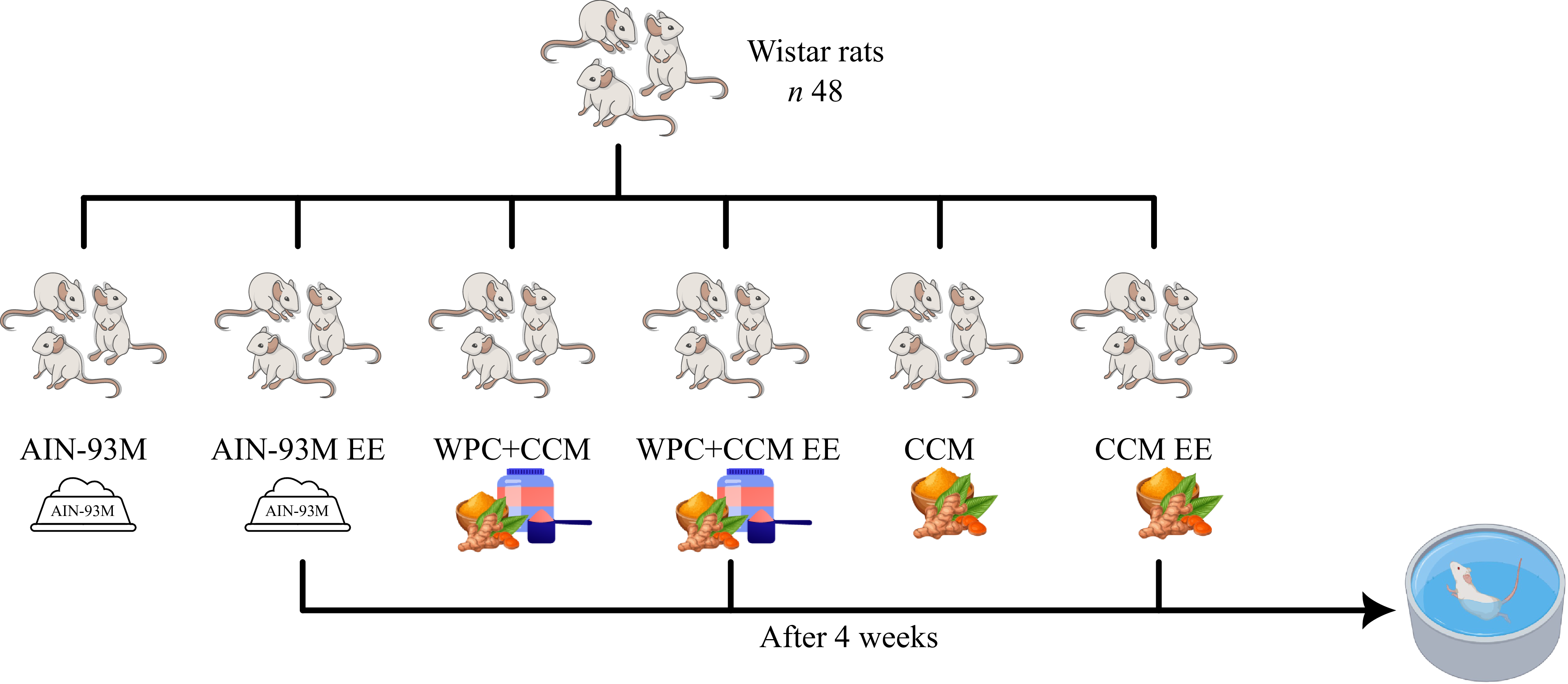
Forty-eight male Wistar rats (young adults) (Rattus novergicus, variety albinus, Rodentia) were used, coming from the Central Animal Facility of the Center of Biological and Health Sciences, at UFV. The animals were housed in individual stainless-steel cages, in an environment with controlled temperature (22 ± 2℃) and photoperiod (12 h), according to CONCEA(17), and, after 12 weeks, they were randomly divided, according to weight, into six groups (n 8): G1: the control group that received a standard diet (AIN-93M); G2: the control group that received a standard diet submitted to the EE (AIN-93M EE); G3: the CCM-added WPC group (WPC + CCM); G4: the CCM-added WPC group submitted to the EE (WPC + CCM EE); G5: the CCM group (CCM) and G6: the CCM group submitted to the EE (CCM EE) (Figure 1). These animals received distilled water and their respective experimental diets ad libitum, during 4 weeks.
Experimental design. AIN-93M: group that received a standard diet; AIN-93M EE: group submitted to the exhaustive exercise that received a standard diet; WPC + CCM: group not submitted to EE that received curcumin-added whey protein concentrate; WPC + CCM EE: group submitted to the exhaustive exercise that received curcumin-added whey protein concentrate; CCM: group not submitted to EE that received curcumin; CCM EE: group submitted to the exhaustive exercise that received curcumin; EE: exhaustive exercise. CCM, curcumin; EE, exhaustive exercise; WPC, whey protein concentrate.

The animals were allocated according to the method proposed by Conagin(Reference Conagin18). For the sample calculation, the following equation was used: r = 2 × s2 × (tα + tβ) × (tα + tβ)/d2, with the reference data for the sample calculation taken from Maithilikarpagaselvi et al. (2016)(Reference Maithilikarpagaselvi, Sridhar and Swaminathan19), obtaining eight animals per experimental group. During the study, confounding factors were not controlled and caregivers were not blinded.
Experimental diets
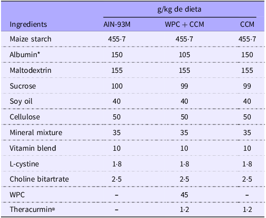
The experimental diets were formulated in accordance with the AIN-93M recommendations, proposed by the American Institute of Nutrition (1993)(Reference Reeves, Nielsen and Fahey13) for the maintenance of adult animals, namely: AIN-93M diet (control), WPC + CCM diet (30 % of the protein requirement coming from the WPC) and CCM diet (AIN-93M + CCM) (Table 1), and were offered in powder form.
Composition of experimental diets

CCM, curcumin; WPC, whey protein concentrate.
Source: Reeves et al., 1993; WPC: whey protein concentrate. *Albumin was considered to be 80 % protein.
Exhaustive exercise protocol
After 4 weeks, animals in groups G2, G4 and G6 underwent an exhaustive swimming. The animals were forced to swim until exhaustion, supporting a weight of 5 % of their body weight attached to the tail(Reference Venditti and Di Meo20). Exhaustion was defined when the rat remained submerged for 10 s or when they were no longer able to maintain their body movements(Reference Dawson and Horvath21). The exercise was carried out in a tiled masonry tank, with a water height of 45 cm maintained at around 28°C ± 1°C.
Euthanasia
Twenty-four hours after carrying out the EE, the animals from all experimental groups were anesthetised with 5 % inhaled isoflurane (Isoforine, Cristália®) and euthanised by cardiac puncture, with a 12-h fasting period. The entire procedure was carried out with the aim of promoting a humane death of the animals(22). Colon and feces from the cecum were collected for further analysis.
Analysis
SCFA
The fecal contents of propionic, acetic and butyric acids were determined following Siegfried et al. (1984)(Reference Siegfried, Rückemann and Stumpf23) with modifications. Cecal feces were suspended in water, centrifuged and treated sequentially with calcium hydroxide, cupric sulphate and sulphuric acid before analysis by liquid chromatography (Shimadzu) using a Bio-Rad HPX-87H column. Identification and quantification were performed by comparison with external standards and standard curves.
Oxidative stress
For oxidative balance analyses, a homogenate of intestinal tissue (colon) of all animals was prepared, and the tissues were homogenised in a phosphate buffer and subsequently centrifuged. The resulting supernatants were then stored to conduct analyses of malondialdehyde (MDA), nitric oxide (NO), superoxide dismutase (SOD), glutathione-S-transferase (GST) and catalase (CAT), while the pellet was reserved for analysis of carbonyl protein (PCN).
MDA levels, indicating lipid peroxidation, were measured using a 1,1,3,3-tetramethoxypropane standard curve, by Wallin et al. (1993)(Reference Wallin, Rosengren and Shertzer24). NO production was estimated by NO2/NO₃ quantification via the Griess method and sodium nitrite standards, by Tsikas et al. (2007)(Reference Tsikas25). Protein carbonyl content was determined with 2,4-dinitrophenylhydrazine, performed by Levine et al. (1990)(Reference Levine, Garland and Oliver26). Antioxidant enzyme activities were assessed as follows: SOD by free radical elimination, by the method of Dieterich et al. (2000)(Reference Dieterich, Bieligk and Beulich27); GST via conjugation of 1-chloro-2,4-dinitrobenzene with glutathione, by Habig and Jakoby (1981)(Reference Habig and Jakoby28) and CAT by the decomposition rate of hydrogen peroxide, by Hadwan and Abed (2016)(Reference Hadwan and Abed29).
Real-time PCR
For intestinal gene expression analysis, RNA was extracted with TRIzol Reagent (Invitrogen), and then used for cDNA synthesis, which is performed by Livak and Schmittgen (2001) Reference Livak and Schmittgen30).
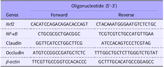
The mRNA expression levels of the genes were analysed by RT-qPCR, using the SYBR Green PCR master mix (Applied Biosystems), and the analyses were performed on the QuantStudio 1 Real-Time PCR System (Thermo Fisher Scientific). The primer sequences, sense and antisense were ordered for amplification of Nrf2, NF-κB, claudin and occludin. Relative mRNA expression levels were normalised relative to the endogenous β-actin control (Table 2).
Sequence of primers used in the RT-PCR analysis

Nrf2: nuclear factor erythroid 2-related factor 2; NF-κB: nuclear factor kappa B.
Intestinal histomorphometry
Intestinal fragments were fixed in formaldehyde, dehydrated in ethanol and embedded in Historesin® (Leica), by Sabarense et al. (2012)(Reference Sabarense, Rocha and Rosa31). Semi-serial 3 μm sections were cut with a Leica microtome, stained and examined under a light microscope (Olympus CX31) at 4× and 10× magnification. Crypt depth and thickness, muscular layers, mucosa, submucosa and goblet cell number and area were analysed using ImagePro-Plus® v4.5.
Statistical analysis
Statistical analyses and graph construction were performed using GraphPad Prism software, version 9.0.2 (GraphPad Prism Inc). Data normality was assessed using the Shapiro–Wilk test. The t test was used to assess differences within the same group (not submitted to EE × submitted to EE). Intragroup differences (AIN-93M, WPC + CCM and CCM) were submitted to ANOVA, followed by the Newman–Keuls mean test. A P value < 0·05 was adopted. The results are expressed as mean and standard deviation (mean ± sd).
Results
SCFA
Acetic acid remained stable in the exercised groups compared with controls. However, the AIN-93M group showed higher concentrations compared with the groups that received WPC + CCM and CCM (P < 0·05). For propionic and butyric acids, there were no significant differences among the groups (Table 3).
Quantification of acetic, propionic and butyric acids in cecal samples

CCM, curcumin; WPC, whey protein concentrate.
AIN-93M: group that received a standard diet; AIN-93M EE: group submitted to the exhaustive exercise that received a standard diet; WPC + CCM: group not submitted to EE that received curcumin-added whey protein concentrate; WPC + CCM EE: group submitted to the exhaustive exercise that received curcumin-added whey protein concentrate; CCM: group not submitted to EE that received curcumin; CCM EE: group submitted to the exhaustive exercise that received curcumin; EE: exhaustive exercise. *Indicates significant differences between the same group (sedentary × EE), according to the t test (P < 0·05). Different lowercase letters (a – b) indicate significant differences within groups, according to ANOVA, followed by the Newman–Keuls test, at 5 % probability. Data expressed as mean (standard deviation).
Oxidative balance
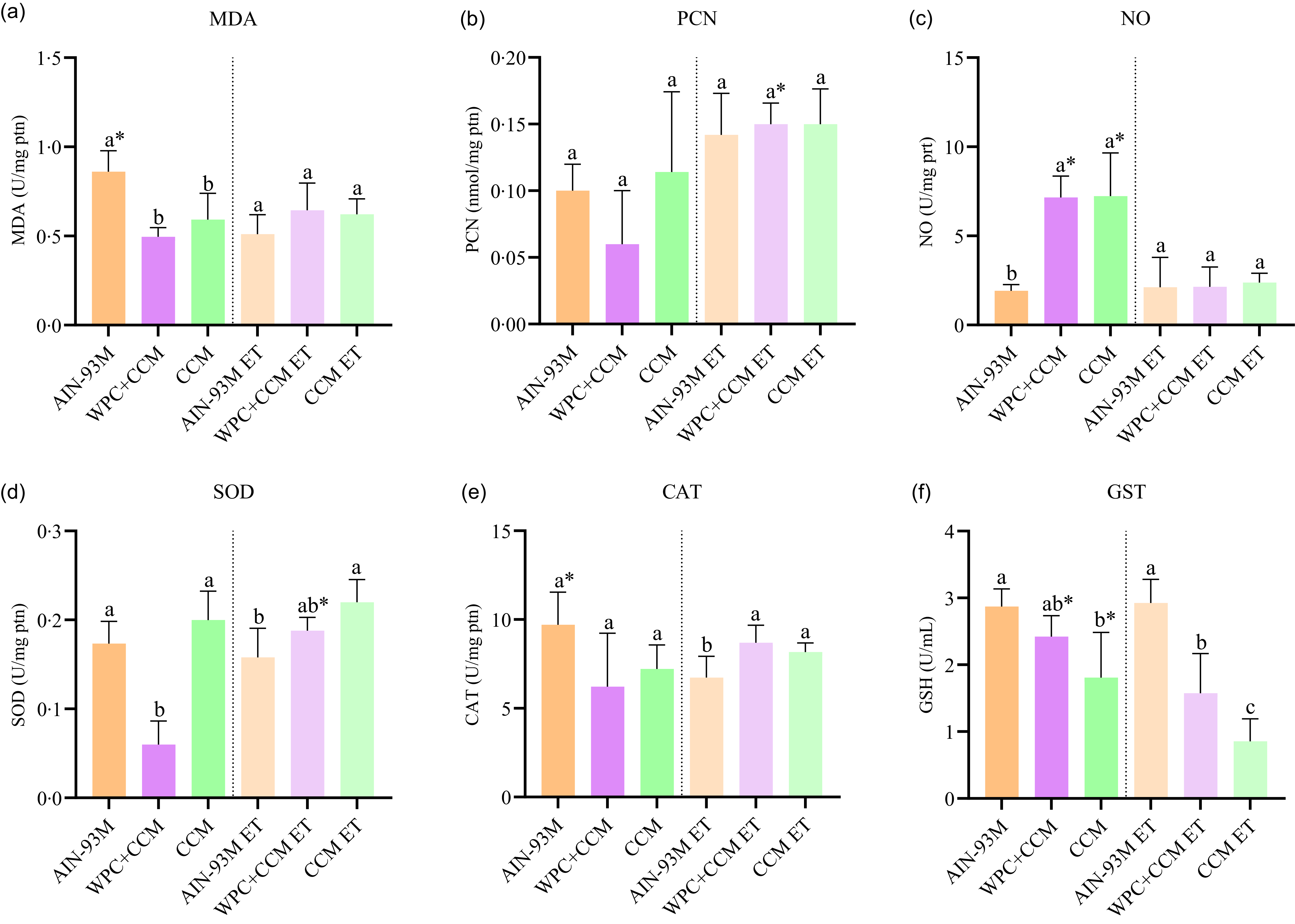
MDA concentrations were lower in the groups that received CCM, isolated or associated with WPC (P < 0·05). When evaluating the effect of exercise, MDA levels were maintained in the groups that received the WPC + CCM and CCM diets, while the non-exercised AIN-93M group showed higher MDA (P < 0·05) (Figure 2(a)).
Analysis of oxidative balance in the intestine (colon). AIN-93M: group that received a standard diet; AIN-93M EE: group submitted to the exhaustive exercise that received a standard diet; WPC + CCM: group not submitted to EE that received curcumin-added whey protein concentrate; WPC + CCM EE: group submitted to the exhaustive exercise that received curcumin-added whey protein concentrate; CCM: group not submitted to EE that received curcumin; CCM EE: group submitted to the exhaustive exercise that received curcumin; EE: exhaustive exercise. The graphs show (a) Malondialdehyde, (b) Carbonylated protein, (c) Nitric oxide, (d) Superoxide dismutase, (e) Catalase, (f) Glutathione S-transferase. *Indicates significant differences between the same group (sedentary × EE), according to the t test (P < 0·05). Different lowercase letters (a – c) indicate significant differences within groups, according to ANOVA, followed by the Newman–Keuls test, at 5 % probability. Data expressed as mean (standard deviation). CCM, curcumin; EE, exhaustive exercise; WPC, whey protein concentrate.

As for PCN analysis, the WPC + CCM EE group showed higher concentrations compared with the WPC + CCM group (P < 0·05). No differences were observed intragroup, both in the groups that performed EE and those that did not (Figure 2(b)). In the NO analysis, it was observed that among the non-exercised groups there was an increase in NO in the groups that received CCM, isolated or associated with WPC, while EE reduced nitric oxide concentrations in these groups (P < 0·05) (Figure 2(c)).
For the SOD analysis, lower concentrations were observed in the group that received WPC + CCM (P < 0·05) compared with the other groups without EE. However, there was a significant increase compared with the trained group (P < 0·05). Furthermore, among the exercised groups, the group that received CCM isolated had the highest SOD concentrations (P < 0·05) (Figure 2(d)).
The non-exercised groups did not show significant differences in the concentrations of CAT. However, among the exercise groups, those who received CCM, isolated or combined, showed higher concentrations of this enzyme (P < 0·05). Furthermore, EE reduced CAT concentrations in the control group (P < 0·05) (Figure 2(e)). For GST, the AIN-93M groups showed higher concentrations, while exercise reduced concentrations in the WPC + CCM and CCM groups (P < 0·05) (Figure 2(f)).
Real-time PCR
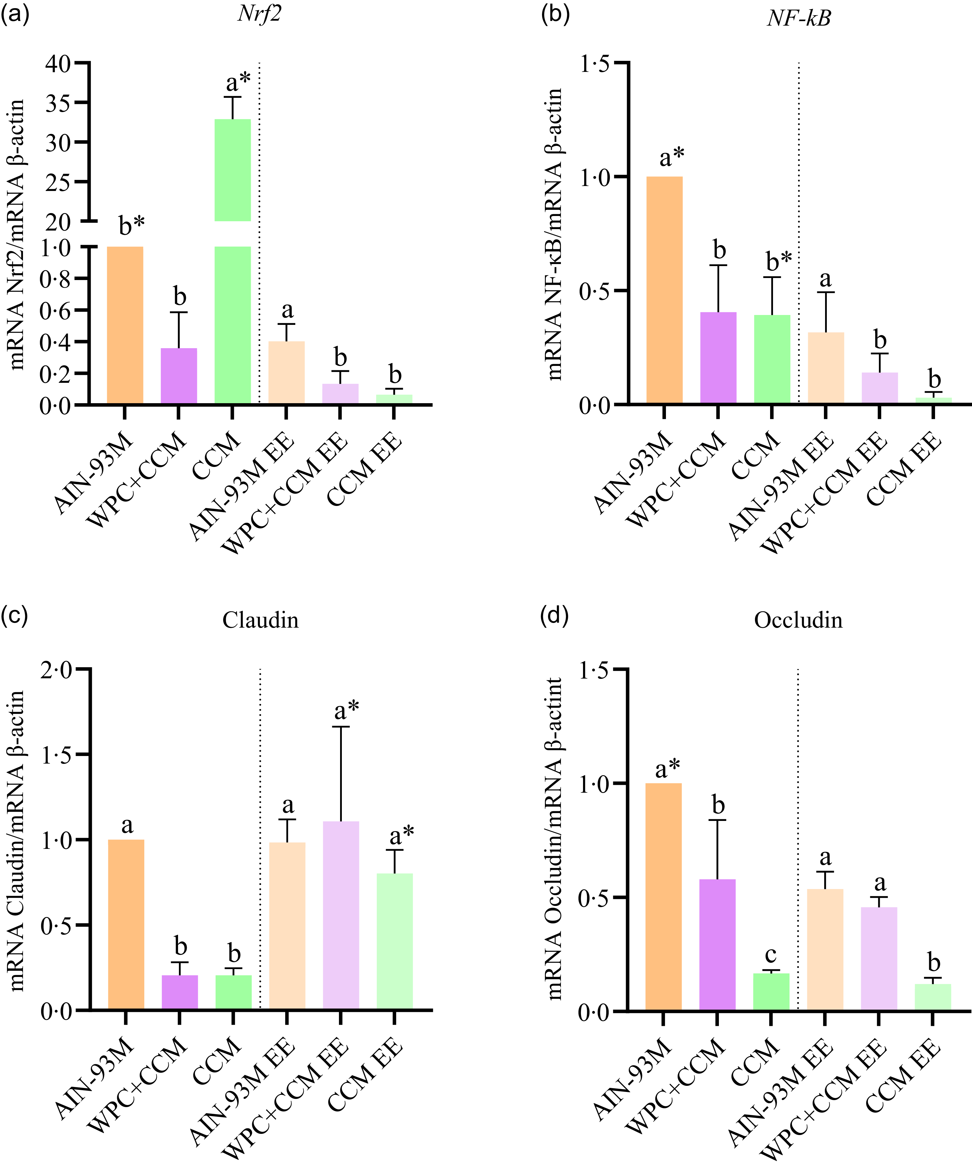
CCM group presented higher expression of Nrf2 and lower expression of occludin compared with the other groups that were not submitted to EE (Figures 3(a) and (d)). In addition, among the groups that performed EE, WPC + CCM EE and CCM EE presented lower expressions of antioxidant and inflammatory markers compared with AIN-93M EE (Figures 3(a) and (b)). WPC + CCM EE increased the expression of claudin, similarly to AIN-93M EE (Figure 3(c)).
Gene expression of Nrf2, NF-κB, claudin and occludin in intestine. AIN-93M: group that received a standard diet; AIN-93M EE: group submitted to the exhaustive exercise that received a standard diet; WPC + CCM: group not submitted to EE that received curcumin-added whey protein concentrate; WPC + CCM EE: group submitted to the exhaustive exercise that received curcumin-added whey protein concentrate; CCM: group not submitted to EE that received curcumin; CCM EE: group submitted to the exhaustive exercise that received curcumin; EE: exhaustive exercise. The graphs show (a) Nrf2, (b) NF-κB, (c) Claudin and (d) Occludin. *Indicates significant differences between the same group (sedentary × EE), according to the t test (P < 0·05). Different lowercase letters (a – c) indicate significant differences within groups, according to ANOVA, followed by the Newman–Keuls test, at 5 % probability. Data expressed as mean (standard deviation). CCM, curcumin; EE, exhaustive exercise; WPC, whey protein concentrate.

Regarding the effect of EE, the AIN-93M and CCM groups presented higher expressions of Nrf2 and NF-κB compared with their respective groups submitted to EE (Figures 3(a) and (b)). On the other hand, the groups that received WPC + CCM, whether or not submitted to EE, did not show any difference in the expression of these markers (Figures 3(a) and (b)). Furthermore, we observed that the WPC + CCM EE and CCM EE groups showed an increase in the expression of claudin compared with their respective groups that did not perform the exercise (Figure 3(c)).
Intestinal histomorphometry
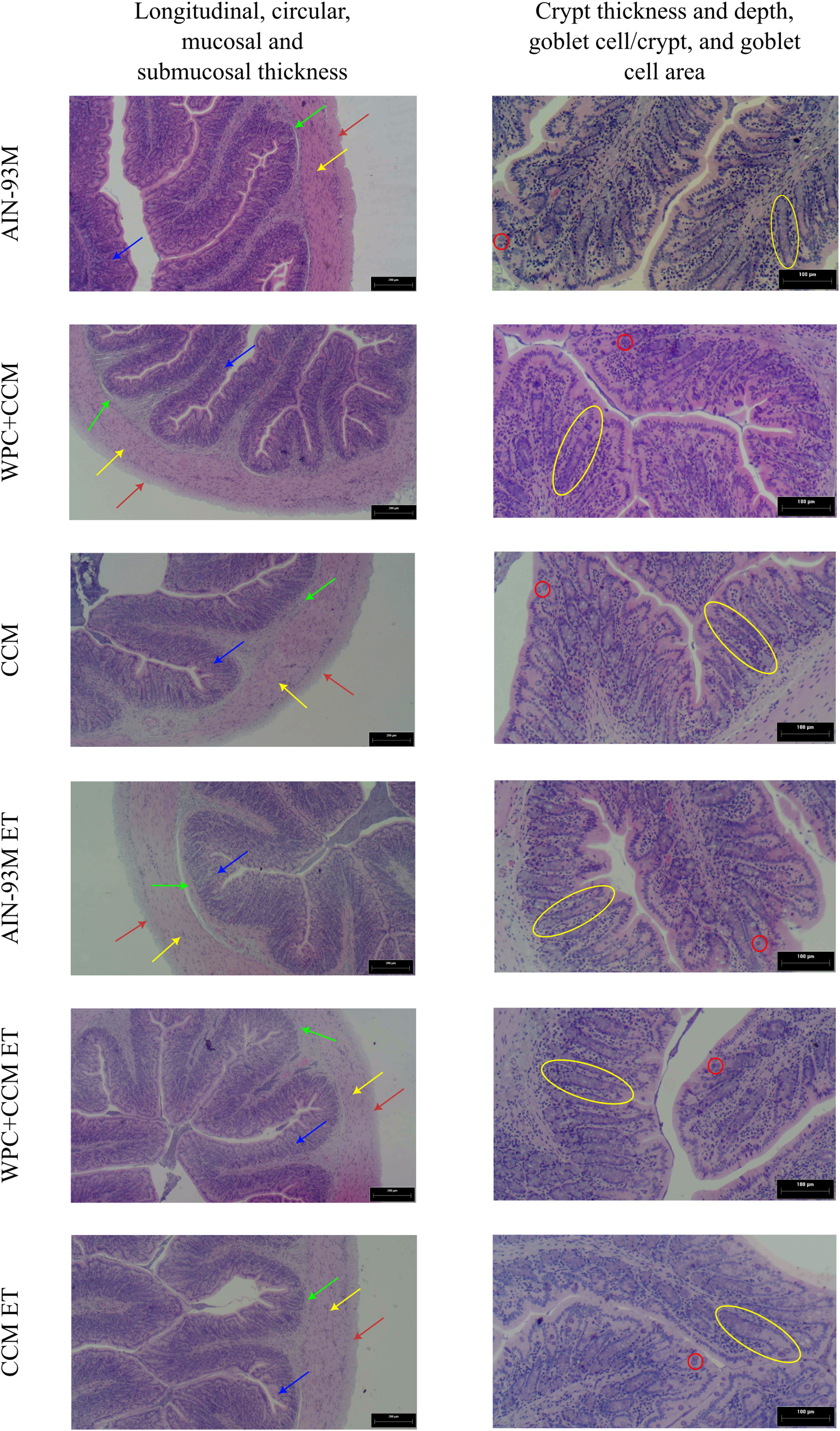
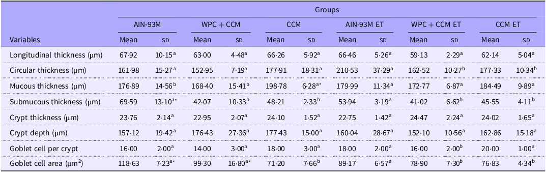
The longitudinal muscular layer thickness and the crypts depth and thickness did not differ among the groups. The circular muscle layer showed no difference in thickness among the non-exercised groups, but among the groups that were submitted to the EE, the WPC + CCM EE and CCM EE groups showed smaller thicknesses compared with the AIN-93M EE group (Figure 4, Table 4).
Representative photomicrographs of the colon stained with hematoxylin and eosin in rats from experimental groups. AIN-93M: group that received a standard diet; AIN-93M EE: group submitted to the exhaustive exercise that received a standard diet; WPC + CCM: group not submitted to EE that received curcumin-added whey protein concentrate; WPC + CCM EE: group submitted to the exhaustive exercise that received curcumin-added whey protein concentrate; CCM: group not submitted to EE that received curcumin; CCM EE: group submitted to the exhaustive exercise that received curcumin; EE: exhaustive exercise. Red arrows: longitudinal thickness; Yellow arrows: circular thickness; Green arrows: submucosa; Blue arrows: mucosa; Yellow circles: crypts; Red circles: coblet cell. CCM, curcumin; EE, exhaustive exercise; WPC, whey protein concentrate.

Histomorphometry of the colon of rats submitted or not to exhaustive exercise

CCM, curcumin; EE, exhaustive exercise; WPC, whey protein concentrate.
AIN-93M: group that received a standard diet; AIN-93M EE: group submitted to the exhaustive exercise that received a standard diet; WPC + CCM: group not submitted to EE that received curcumin-added whey protein concentrate; WPC + CCM EE: group submitted to the exhaustive exercise that received curcumin-added whey protein concentrate; CCM: group not submitted to EE that received curcumin; CCM EE: group submitted to the exhaustive exercise that received curcumin; EE: exhaustive exercise. *Indicates significant differences between the same group (sedentary × EE), according to the t test (P < 0·05). Different lowercase letters (a – b) indicate significant differences within groups, according to ANOVA, followed by the Newman–Keuls test, at 5 % probability. Data expressed as mean (standard deviation).
CCM consumption increased mucosal thickness (P < 0·05). However, under physical stress, consumption of CCM, isolated or combined with WPC, resulted in mucosal thickness similar to the control group. Additionally, the consumption of WPC + CCM and CCM maintained the thickness of the submucosa under physical stress, although this thickness was lower than that of the control group (P < 0·05). The control group, on the other hand, was unable to maintain submucosal thickness under the same stress (P < 0·05) (Figure 4, Table 4).
Administration of CCM, isolated or combined with WPC, was effective in maintaining goblet cell numbers during EE. Notably, the use of CCM in its isolated form demonstrated superior efficacy (P < 0·05) (Figure 4, Table 4). Regarding the area of goblet cells, the consumption of isolated CCM was able to maintain the area of these cells after EE, although they were still smaller compared with the control group (P < 0·05). Conversely, the combined use of CCM with WPC did not achieve similar results, showing a reduction in cell area after exercise (P < 0·05) (Figure 4, Table 4).
Discussion
The production and absorption of SCFA are essential for intestinal health, regulating cellular processes such as differentiation, proliferation, apoptosis, gene expression and anti-inflammatory mechanisms. These acids are produced by a variety of intestinal bacteria, each contributing in a specific way to their synthesis. For example, acetate is mainly produced by enteric bacteria such as Blautia hydrogenotrophica, while propionate can be generated by Firmicutes and Bacteroidetes, and butyrate is produced by bacteria in the phylum Firmicutes(Reference Liu, Li and Yang32).
Changes in the diet have the potential to influence the production and absorption of these SCFAs, as the composition of the diet can modify the composition of the intestinal microbiota. Therefore, it is crucial to understand how different dietary interventions or external factors can affect gut health, especially in situations of physiological stress such as EE. EE is known to induce oxidative stress and inflammation in the body, affecting intestinal health, and consequently increasing intestinal permeability(Reference Engel, Ackermann and Chtourou4,Reference Rosa33) .
Although a significantly higher concentration of acetic acid was observed in the AIN-93M group compared with the WPC + CCM and CCM groups, no differences were found in the levels of propionic and butyric acids after EE among groups after EE. Thus, the ability of the experimental groups to maintain the levels of these acids may indicate an effective regulation of intestinal permeability, ensuring SCFA production is adequate for metabolic requirements. This preservation of SCFA levels can be attributed to several potential mechanisms, such as the fact that exercise can improve the diversity and abundance of certain bacterial genera, such as those from the Firmicutes phylum(Reference Dalton, Mermier and Zuhl34), which is directly related to production of these two SCFA, and also the fact that WPC admixture with CCM may have a protective or preservation role for these acids. The fermentation of WP by intestinal bacteria leads to an increase in the concentration of SCFA, and CCM also has a regulatory effect on the composition of the intestinal microbiota, favouring the growth of beneficial strains and butyrate-producing bacteria(Reference Boscaini, Skuse and Nilaweera6,Reference Dalton, Mermier and Zuhl34) .
The interaction between CCM and WPC is particularly interesting in this context. CCM, known for its low bioavailability, has been used in a form with high absorption and bioavailability in this study. Its combination with WPC may enhance the overall antioxidant and anti-inflammatory effects due to complementary mechanisms of action. CCM has been shown to modulate the gut microbiota, promoting the growth of beneficial bacteria and improving gut barrier function, while WPC, a rich source of bioactive peptides and amino acids, supports muscle recovery and immune function. Together, they provide a synergistic effect, enhancing the production of SCFA and potentially improving the intestinal barrier integrity. This synergism justifies the inclusion of both components in a single formulation, as they may work together to offer enhanced protection against oxidative stress and inflammation induced by EE.
The possible decrease in acetic acid compared with the control group can be justified by the fact that most of this acid produced by the microbiota was readily transported to the liver and metabolised, or used by skeletal muscle as a source of energy, as highlighted by Yang et al.(2024)(Reference Yang, Zhang and Liu35). In this context, during EE, the body can direct more metabolic resources to meet immediate energy demands, resulting in an increase in hepatic metabolism and, consequently, in the accelerated metabolisation of acetic acid. Despite this difference in relation to the control group, acetic acid levels remained stable during exercise. This indicates that, although there is greater use and metabolisation of acetic acid due to increased energy demands, the production of this acid by the intestinal microbiota continues to adequately meet these needs(Reference Boscaini, Skuse and Nilaweera6).
Furthermore, SCFA, particularly butyrate, play a role in intestinal homeostasis, directly influencing the redox balance and antioxidant response of the intestinal epithelium. These metabolites, produced by microbial fermentation, modulate the production of reactive oxygen species (ROS), which, in excess, can lead to oxidative stress and cellular damage. These acids have the ability to stimulate the expression of antioxidant genes, including superoxide dismutase, in addition to promoting the synthesis of glutathione, one of the main intracellular antioxidants. Thus, SCFA not only act as energy sources for colonic epithelial cells, but are also key mediators in the regulation of the intestinal oxidative environment(Reference González-Bosch, Boorman and Zunszain36).
MDA is a well-established biomarker of oxidative stress and cellular damage, resulting primarily from lipid peroxidation, such as the oxidative degradation of PUFA in cell membranes induced by ROS. The accumulation of MDA reflects the extent of oxidative injury and disruption of membrane integrity(Reference Yoshida, Umeno and Shichiri37,Reference Valgimigli38) . In the present study, lower baseline MDA concentrations were observed in non-exercised groups supplemented with WPC combined with CCM, or CCM alone, compared with the standard AIN-93M diet, indicating enhanced antioxidant capacity attributable to these bioactive compounds(Reference Da Conceição, Dias and Pereira39,Reference Dias, da Conceição and Pereira40) . Notably, within the AIN-93M control group, MDA levels decreased after EE, potentially reflecting an acute hormetic response whereby transient exercise-induced ROS generation stimulates endogenous antioxidant defenses, resulting in temporary attenuation of lipid peroxidation(Reference Wang, Luo and Jiang41). In contrast, animals receiving WPC + CCM or CCM alone maintained stable MDA levels post-exercise, suggesting these treatments imposed a protective effect by preemptively reducing oxidative damage. The antioxidant effects of CCM are attributed to direct ROS scavenging and inhibition of lipid peroxidation chain reactions, while WPC contributes bioavailable cysteine, a precursor for bioactive peptides synthesis, that enhances cellular redox balance(Reference Sahoo, Heilmann and Paital42,Reference Giblin, Yalçın and Biçim43) . Together, these mechanisms synergistically mitigate exercise-induced oxidative stress and preserve membrane integrity.
PCN is another marker of oxidative stress, where amino acid residues are modified by ROS, compromising the structure and function of proteins. In our study, the group that underwent the exhaustion exercise showed higher concentrations of PCN, reflecting an increase in oxidative stress during exercise. The combination of WPC with CCM does not appear to significantly influence these levels, however, CCM isolated manages to maintain these levels stable. This can be attributed to the fact that CCM and WPC act through different antioxidant mechanisms, possibly resulting in the absence of a synergistic effect(Reference Chaves, Baptista and Simabuco44,Reference Szymanski, Gillum and Gould45) . Although both compounds have antioxidant effects, their combination did not provide additional protection against exercise-induced oxidative stress. This suggests that the antioxidant mechanisms involved may be more complex than initially considered, and that specific interactions between different dietary compounds should be taken into account when formulating antioxidant supplements or therapies, and it is important to understand how they interact at the molecular and physiological level. This reinforces the need for further studies about the interactions between dietary compounds to develop effective interventions against oxidative stress, especially in contexts of high physical exertion, such as strenuous exercise(Reference Bjørklund and Chirumbolo46).
Under physiological conditions, NO acts regulating intestinal tone, promoting a favourable environment for water and electrolyte absorption. However, in inflammatory contexts, high NO production can stimulate liquid secretion from the intestine, contributing to inflammation-associated diarrhoea, in addition to being harmful to host cells, leading to apoptosis and necrosis of enterocytes, compromising the integrity of the intestinal barrier(Reference Grishin, Bowling and Bell47,Reference Leclerc, Bedu-Ferrari and Etienne-Mesmin48) . During exercise, increased metabolism and increased oxygen demand by muscle tissues can lead to greater NO production to promote vasodilation and increase blood flow to working muscles. Therefore, it is possible that NO levels in the intestine decrease during exercise as more NO is consumed by working tissues(Reference Oral49).
The results of our study indicate that CCM supplementation, isolated or associated with WPC, increases NO levels in non-exercised groups, which may be beneficial for maintaining intestinal homeostasis under physiological conditions. However, the reduction in NO concentrations observed after EE suggests a redistribution of NO to working muscles, which is an expected adaptive response to meet the increased metabolic demand during exercise. This redistribution may protect the intestine from possible harmful effects of excess NO in inflammatory conditions, while supporting muscle performance during exercise. This is corroborated by a previous study conducted by our research group, in which the same animals in this study demonstrated a significant increase in NO levels in muscles during exercise(Reference Dias, da Conceição and Pereira40).
The gastrointestinal tract is an important source of ROS. Although the intestinal epithelium acts as a physical and antimicrobial barrier, ingested substances and enteric pathogens and physical stress can trigger inflammation, resulting in the production of pro-inflammatory cytokines(Reference Tian, Wang and Zhang50). To combat the damage caused by oxidative stress, the body has a variety of antioxidants, both enzymatic and non-enzymatic. For example, SOD converts superoxide anion to hydrogen peroxide, while CAT and GST degrade hydrogen peroxide(Reference Perkins, Nelson and Parsonage51).
Damage induced by oxidative stress can lead to infiltration of the intestinal mucosa by activated leukocytes, resulting in an overload of the tissue’s antioxidant defenses. However, diets rich in polyphenols can help control excessive ROS production(Reference Sahoo, Heilmann and Paital42). In the present study, we observed an increased SOD production in the group supplemented with WPC and CCM that underwent exhaustive physical exercise, indicating an adaptive response of the body to increased antioxidant activity during exercise. On the other hand, CAT activity showed no significant differences when comparing the two groups (WPC + CCM v. WPC + CCM ET or CCM v. CCM ET), while GST activity decreased in the same group. On the other hand, among the animals submitted to the exhaustive test, an increased SOD was observed in the group receiving CCM, when compared with the control group. The CAT enzyme also increased in both the WPC + CCM and CCM groups, while GST decreased in both groups when compared with the control (AIN-93M). The decreased GSH observed with WPC + CCM and CCM was unexpected, as CCM is often reported to increase GSH levels. However, we observed increased SOD and CAT activities, which suggests that the intake of antioxidant compounds was effective in defending the body against physical stress, reducing the need for endogenous production of these antioxidant compounds(Reference Sahoo, Heilmann and Paital42). These results highlight the importance of antioxidant consumption in protecting the gastrointestinal tract against the effects of oxidative stress during physical exercise.
Several cellular defense mechanisms against oxidative stress are induced by different molecular pathways, in particular the nuclear factor erythrogen-2-associated factor 2 (Nrf2) pathway. Nrf2 is a transcription factor that regulates antioxidants within the cell, acting as a stress-responsive transcription factor(Reference Kerasioti, Stagos and Tsatsakis52,Reference Ma, Xu and Guo53) . In our study, we observed that the consumption of CCM associated with WPC prevented changes in Nrf2 expression in response to oxidative stress. WP, as well as CCM, can induce Nrf2 phosphorylation, Keap1 dissociation, nuclear translocation and finally activation of antioxidant enzymes. Furthermore, bioactive peptides, potentially capable of crossing the intestinal barrier, exert an antioxidant action through activation of the Keap1-Nrf2 signalling pathway(Reference Rahban, Habibi-Rezaei and Mazaheri54,Reference Tonolo, Folda and Cesaro55) .
Nrf2 under activation can inactivate inflammatory signalling pathways, such as the NF-κB pathway, an inducible transcription factor that regulates many genes involved in the development and regulation of inflammatory and immunomodulatory processes. Studies have shown that WP can mitigate oxidative stress and modulate the inflammatory response during and after exhaustive physical exercise. The immunomodulatory peptides present in WP modulate the production of inflammatory cytokines by acting on the NF-κB pathway(Reference Adak and Khan56–Reference Zhang, Wang and Zhong58).
Furthermore, CCM also has potent anti-inflammatory properties, mediated by inhibition of the NF-κB pathway, and may act on the inflammatory response generated by exhaustive physical exercise, consequently reducing the production of pro-inflammatory cytokines(Reference Zhang, Kang and Song59). In this study, we observed a reduction in this marker in the groups that received WPC + CCM and CCM compared with the control group, and CCM isolated was also effective in reducing NF-κB under physical stress.
Claudins and occludin are essential proteins of tight junctions, specialised structures that regulate paracellular permeability and maintain the integrity of epithelial and endothelial barriers(Reference Camilleri60). While claudins form selective pores that control the flow of ions and molecules, occludin acts as a scaffold protein, contributing to the stability and organisation of tight junctions and participating in cell signaling processes(Reference Voutsadakis61). Alterations in the expression or function of these proteins are associated with several pathologies, such as inflammatory bowel diseases, blood-brain barrier dysfunctions and cancer, highlighting their crucial role in physiology and their potential as therapeutic targets(Reference Camilleri60,Reference Voutsadakis61) .
Regarding tight junctions, we observed that the WPC + CCM and CCM groups presented lower expressions of claudin compared with the group that received a control diet. However, when submitted to EE, the groups had an increased expression of this marker, equalling the exercised control group. As for occludin, the CCM group presented lower expression of this marker with or without exercise, while the WPC + CCM group had lower expression when compared with the AIN-93M group. However, when submitted to EE, the group that received WPC + CCM had similar expression to the AIN-93M EE group, in addition to maintaining their levels similar to their respective groups without exercise.
Our results reinforce the hypothesis that diet can modulate the expression of genes related to cellular integrity, especially under conditions of stress induced by EE. The combination of a quality protein source with bioactive compounds appears to exert a protective and regulatory effect on the expression of these markers, suggesting that nutritional intervention may be a promising strategy to preserve the function of tight junctions in situations of physiological stress.
Additionally, the muscular layer of the intestine is crucial for the proper movement of food through the gastrointestinal tract. Changes in the thickness or integrity of this layer can affect the effectiveness of intestinal motility, impacting digestion and nutrient absorption(Reference Ünsal, Ünsal and Ekici62). In our study, we observed that the groups that received CCM, isolated or combined with WPC, were able to maintain the thickness of the circular and longitudinal layers in the face of physical stress. This suggests that, although it did not lead to an increase in muscle layer, animals that consumed a diet rich in polyphenols and good-quality protein did not show a negative response in the intestine to exercise, possibly not affecting intestinal motility and transit(Reference Ünsal, Ünsal and Ekici62).
The intestinal mucosa is responsible for the absorption of essential nutrients; changes in its thickness may indicate changes in the absorption capacity of these nutrients(Reference Campbell, Wisniewski and Noji63,Reference Yang and Yu64) . In the present study, mucosal thickness was greater in the CCM-treated group without exercise, suggesting a potential decrease in absorption area after exercise. However, this difference was not observed in the WPC + CCM group submitted to EE, which despite not increasing its mucous layer, maintained its thickness in relation to the WPC + CCM not submitted to EE, thus not affecting the absorption capacity.
The submucosa is related to the intestinal immune response, as it is where lymphocytes and other components of the immune system associated with the gastrointestinal tract are located(Reference Campbell, Wisniewski and Noji63,Reference Yang and Yu64) . Although the groups that received CCM, isolated or combined with WPC, had smaller thicknesses compared with the control group, no difference was observed in the thickness of the submucosa in relation to the WPC + CCM and CCM groups submitted to EE. Thus, we concluded that the production capacity of lymphocytes and components involved in the immune response of these groups was not affected after physical exercise, possibly due to the antioxidant and anti-inflammatory properties of WP and CCM(Reference Boscaini, Skuse and Nilaweera6,Reference Scazzocchio, Minghetti and D’archivio8) .
In addition to the muscular and mucosal layers, adequate integrity and function of the crypts are critical for maintaining intestinal homeostasis and preventing gastrointestinal disorders and other related conditions. Intestinal crypts house stem cells responsible for the continuous renewal of the intestine, and this function is essential for maintaining the integrity of the intestinal barrier. Additionally, crypts house immune cells help modulate the inflammatory response and protect against foreign invaders(Reference Yang and Yu64,Reference Gehart and Clevers65) . The results of this study revealed a constancy in the thickness and depth of the intestinal crypts, indicating morphological stability. This finding is particularly promising, as the preservation of this cryptal architecture can be considered a positive factor, as it suggests potential resistance to adverse changes that could compromise intestinal function.
Among the cells present in the crypts are goblet cells, which play a fundamental role in protecting the intestinal mucosa, producing mucus that acts as a barrier against physical damage, pathogens and irritating substances. Variations in the number and area of these cells can directly impact the intestine’s defense capacity against damage and infections(Reference Yang and Yu64,Reference Gehart and Clevers65) . In our study, a reduction in both the number and area of goblet cells per crypt was observed in the group submitted to the EE with WPC + CCM. This observation suggests a possible response to the consumption of WPC + CCM, as in situations of physical stress, the body tends to increase the production of goblet cells to reinforce the production of protective mucus(Reference Mello, Silva and Fonte66). Therefore, the combination of WP and CCM appeared to be effective in protecting against oxidising and inflammatory agents, offering additional defense to the intestine in stressful conditions.
Despite the valuable insights provided by this study, some limitations should be considered. First, the use of Wistar rats, while a well-established model for studying gut health and exercise-induced stress presents differences from human physiology. These differences may limit the direct extrapolation of findings to human populations. Additionally, potential sources of bias include the lack of blinding of caregivers and researchers during the experiment, which could have influenced data collection and interpretation. The sample size, although statistically justified, may also limit the detection of subtle effects, contributing to potential imprecision in some results. Importantly, this study did not include microbiota profiling or functional analyses, which prevents us from directly linking the observed SCFA levels and intestinal parameters to specific microbial taxa or metabolic pathways. Any discussion regarding microbiota-mediated effects is therefore hypothetical, grounded in previous studies rather than in direct evidence from our experimental data.
Regarding the generalisation of findings, the physiological responses observed in Wistar rats provide relevant insights into the potential effects of WPC and CCM supplementation in mitigating intestinal stress induced by EE. While these results suggest possible benefits for human athletes or individuals exposed to high physical stress, further studies are necessary to confirm these effects in humans, considering differences in diet, gut microbiota and metabolic responses. Future research should explore clinical trials or alternative preclinical models to validate the applicability of these findings across different species and experimental conditions. Future research should incorporate direct analyses of gut microbiota composition and function to better elucidate the mechanisms underlying the observed effects and explore alternative clinical trials or preclinical models to validate the applicability of these findings across different species and experimental conditions.
Conclusions
CCM-added WPC demonstrated beneficial effects under conditions of EE, particularly through the modulation of oxidative stress, inflammatory pathways and intestinal barrier integrity. These mechanisms may explain the maintenance of histological parameters. However, further studies with larger datasets are needed to confirm these effects and to better clarify their implications for recovery, physical performance and the prevention of gastrointestinal disturbances.
Supplementary material
For supplementary material/s referred to in this article, please visit https://doi.org/10.1017/S0007114525105758
Acknowledgements
We would like to thank the partner laboratories, the Graduate Program in Nutritional Science, the Universidade Federal de Viçosa and the Minas Gerais State Research Support Foundation – FAPEMIG, who contributed to the project being carried out.
This work was supported by the Minas Gerais State Research Support Foundation – FAPEMIG (grant number: APQ-04073-23).
Conceptualisation: S.M.S.P., K.A.D., A.R.C., C.M.D.L.; data curation: S.M.S.P., K.A.D., L.A.O., V.P.B.S.J.; formal analysis: S.M.S.P., K.A.D., L.A.O., V.P.B.S.J.; funding acquisition: S.M.S.P., K.A.D., L.A.O., C.M.D.L.; investigation: S.M.S.P., K.A.D., L.A.O., V.P.B.S.J., K.V.C.M., R.C.L.T., M.M.S.D.; methodology: S.M.S.P., K.A.D., A.R.C., R.V.G., A.J.N., H.S.D.M., C.M.D.L.; project administration: S.M.S.P., C.M.D.L.; resources: R.C.L.T., M.M.S.D., R.V.G., A.J.N., H.S.D.M., C.M.D.L.; supervision: R.V.G., A.J.N., H.S.D.M., C.M.D.L.; visualisation: S.M.S.P., K.A.D., L.A.O., V.P.B.S.J., C.M.D.L.; roles/writing – original draft: S.M.S.P., K.A.D., L.A.O., V.P.B.S.J., C.M.D.L.; writing – review and editing: S.M.S.P., K.A.D., L.A.O., V.P.B.S.J., C.M.D.L.
The authors declare no conflict of interests.
This project was approved by the Ethics Committee on the Use of Animals of the Universidade Federal de Viçosa (CEUA/UFV), under protocol number 72/2018.