Introduction
Parasitism is recognized as a major threat to the production of small ruminants in both small-scale and large-scale farms. Gastrointestinal (GI) parasites cause high mortality, reduce production and lead to a significant overall economic loss (Al-Quaisy et al., Reference Al-Quaisy, Al-Zubaidy, Altaif and Makkawi1987; McLeod, Reference McLeod1995; Simpson, Reference Simpson2000). GI parasites are highly prevalent in sheep and goats in humid subtropical and tropical areas of the world (Yadav & Tandon, Reference Yadav and Tandon1989; Banks et al., Reference Banks, Singh, Barger, Pratap and Le Jambre1990; Barger et al., Reference Barger, Siale, Banks and Le Jambre1994; Dorny et al., Reference Dorny, Symoens, Jalila, Vercruysse and Sani1995; Cheah & Rajamanickam, Reference Cheah and Rajamanickam1997; Regassa et al., Reference Regassa, Sori, Dhuguma and Kiros2006; Nwosu et al., Reference Nwosu, Madu and Richards2007; Gadahi et al., Reference Gadahi, Arshed, Ali, Javaid and Shah2009; Abebe et al., Reference Abebe, Gebreyohannes, Mekuria, Abunna and Regassa2011; Dagnachew et al., Reference Dagnachew, Amaute and Temesgen2011).
There are approximately 15,000 sheep and 20,000 goats in Papua New Guinea (PNG) (Quartermain, Reference Quartermain2002). Animals are raised by institutions for breeding or for nutritional research purposes and by smallholder farmers for subsistence meat production. There are no sheep and goats native to PNG. Tropical sheep from South-East Asia were introduced by colonial administrators and missionaries in the late 19th century and this sheep population is known as PNG Priangan. In the 1980s, temperate Corriedale and Perendale sheep from New Zealand were brought to the cooler highlands of PNG. These temperate sheep breeds were crossed with the PNG Priangan sheep to produce crossbreds known as Highlands Halfbred (Quartermain, Reference Quartermain2002). PNG Priangan sheep are mainly raised in areas of lower altitude, whereas Highlands Halfbred sheep are raised at higher altitudes. A variety of dairy goats, which are now referred to as PNG genotype, were introduced during the early colonial period (Quartermain, Reference Quartermain2002).
Distribution and productivity of small ruminants in PNG are hindered mainly by poor health, nutrition and management (Quartermain, Reference Quartermain, Sani, Gray and Baker2004). GI parasites are expected to be widespread in PNG due to its humid tropical climate. In some parts of PNG, previous surveys, which were conducted mostly in government stations, identified a diversity of internal parasite species in small ruminants (Varghese & Yayabu, Reference Varghese and Yayabu1985; Owen, Reference Owen1988, Reference Owen1989, Reference Owen1998; also reviewed by Quartermain, Reference Quartermain, Sani, Gray and Baker2004). However, there is insufficient information on the epidemiology of the GI parasites infecting sheep and goats in PNG. Such information is essential for understanding the economic impact these parasites can have on farmers and to support decision-making regarding the treatment and prevention of parasitic diseases in these animals. The present study, therefore, was conducted to obtain data on the prevalence and infection levels of gastrointestinal parasites in sheep and goats on several farms in PNG.
Materials and methods
Study sites
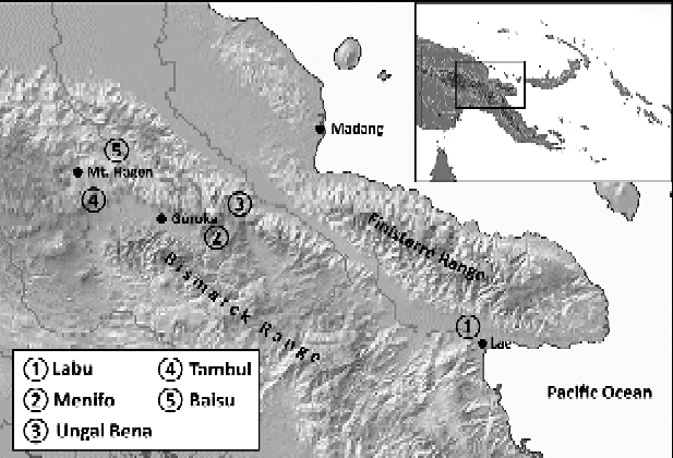
The study was conducted from February to April 2011 in two broad agro-climatic zones, the highlands (specific study sites: Tambul, Baisu, Menifo and Ungai-Bena) and lowlands (specific study site: Labu) in the central region of mainland PNG (fig. 1).
Topographical map of Papua New Guinea (PNG) showing the study sites.

The altitudes of the study sites are 0 m for Labu, 1600–1608 m for Menifo and Ungai-Bena, 1730 m for Baisu and 2320 m for Tambul, with mean annual temperatures of 26°C, 20.1°C, 20.1°C, 18.3°C and 14.7°C, respectively (Bourke, Reference Bourke, Haberle, Stevenson and Prebble2010). The mean annual rainfall for Labu is above 4000 mm and between 2000 and 3500 mm for Ungai-Bena, Baisu and Tambul (Quartermain, Reference Quartermain, Sani, Gray and Baker2004). Menifo is drier than most of the PNG highlands and receives a mean annual rainfall of 1000–1500 mm (Quartermain, Reference Quartermain, Sani, Gray and Baker2004).
The study sites were further characterized by a questionnaire survey, in which 20 farm managers and smallholder farmers were interviewed. It consisted of questions regarding general farm management practices, feeding systems and herd health programmes.
Farm management
The flocks from the three institutional farms (Labu, Baisu and Tambul) were kept together in fenced areas (approximately 20–60 ha), grazed pasture at a high stocking rate at daytime and were kept in houses with wooden, slatted floors at night. At the time of sample collection, the total numbers of sheep and goats in Labu, Baisu and Tambul were 125, 70 and 143, respectively. The subsistence farmers kept few animals (usually fewer than 15) which grazed free range or were tethered and housed at night on slatted floors or on the ground underneath the farmer's house.
Feeding system
Most animals grazed on native grasses and shrubs. Smallholder farmers also fed their animals with starchy vegetables (mostly sweet potatoes). The interviewed farmers also indicated that there were shortages of feed. The animals drank from troughs (sourced from water supply or rainwater tanks), rainwater run-off water or ponds.
Herd health program
The floors of the resting houses were not swept. The animals were penned on dirty floor, ground or on bare concrete floors. The smallholder farmers did not shear their sheep and explained that they did not have the resources for it. Most farm managers reported that the most common signs of illness in their animals were diarrhoea and coughing, followed by itching and hair loss. Most smallholder farmers did not know about causes of diseases in their sheep and goats or the use of anthelmintic drugs for treatment of nematode infections. For instance, a man in Ungai-Bena reported the death of his entire flock (n= 25) and noticed nematode worms in the gut of a dead sheep. The three large institutional flocks were drenched with benzimidazole (Panacur) nominally at bimonthly intervals. At the time of sampling animals had been drenched 2 months previously in Labu and 4 months previously in Baisu and Tambul.
Collection and examination of faecal samples
A total of 110 faecal samples from sheep and 55 faecal samples from goats were obtained from the rectum of randomly selected animals between 07.30 and 09.00 h and kept at 4°C until taken to the laboratory for analysis. Each sample was examined visually for consistency, mucus and macroscopic parasites. Two grams of each faecal sample were examined using a modified McMaster technique (Whitlock, Reference Whitlock1948). Parasites were identified morphologically to genus, or in the case of strongyles, to group level (Soulsby, Reference Soulsby1965). Parasite egg load was expressed as eggs per gram faeces (EPG). The volume of the flotation fluid used in the examinations was 50 ml and the fluid volume examined in the counting chamber was 0.3 ml. Since EPG is calculated using equation 1, the minimum EPG count in our analyses was 83 eggs/g and occurred when only one egg was observed in the counting chamber.
 $$EPG = eggs\,counted\times \frac {total\,volume\,(ml)}{[examined\,volume\,(ml)\times weight\,of\,faeces\,(g)]} $$
Data analysis
Prevalence was calculated as the percentage of positive samples in the total number of samples examined. Apart from the overall prevalence (i.e. the infection with any GI parasite) in each flock, prevalence was also calculated for each parasite type, stratified by animal species and breed, study site, agro-ecological zone and farm management type. Differences in prevalence among groups were compared by Fisher's exact test (FET) (for two groups) or chi-squared analysis (for more than two groups).
EPG counts were transformed to their decadic logarithms to obtain a normal distribution of values prior to statistical testing for significant differences between animal species and breeds, study sites, agro-ecological zones and farm management systems. Differences between/among groups were compared by t-tests or one-way analysis of variance (ANOVA); followed by a post hoc Tukey's HSD test. Statistical analyses were performed using GraphPad Prism version 4 (GraphPad Inc., California, USA).
Results
From the total of 165 small ruminants examined, 128 (78%) were found to be infected with one or more types of GI parasites. A mixed infection with two or more types of GI parasites was found in 39% (50/128) of the infected animals: 33% (26/79) of sheep and 49% (24/49) of goats (not significantly different by FET, P= 0.09).
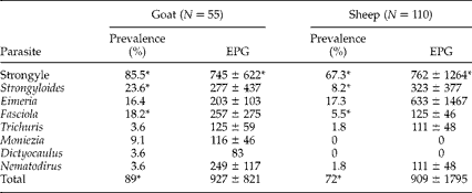
Table 1 shows prevalence and mean EPG counts over all sampling sites, stratified by the type of host animal (sheep or goat) and the type of parasite. The overall prevalence of GI parasite infection was significantly higher (FET; P= 0.017) in goats (89%, 49/55) than in sheep (72%, 79/110). Specifically, prevalence of strongyle (FET; P= 0.0002), Strongyloides (FET; P= 0.013) and Fasciola (FET; P= 0.013) were significantly higher in goats than in sheep. In addition, the mean EPG counts for strongyle were significantly higher in goats than in sheep (t= 2.48, P= 0.014).
GI parasite prevalence and eggs/g (EPG) counts stratified by GI parasite type for the total number of faecal samples collected in the present study. N is the total number of faecal samples. The total prevalence refers to the fraction of animals infected with one or more GI parasite types. EPG counts are given as means ± SD. Where no SD value is given on the mean EPG count, there was only a single or few positive observations with the same values, so that SD could not be calculated. Values with the superscript (*) are significantly different between sheep and goats by either Fisher's exact test (FET) or t-test.
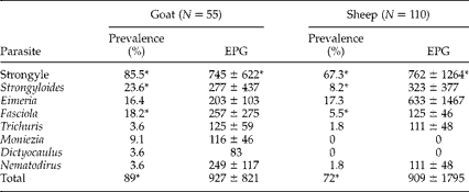
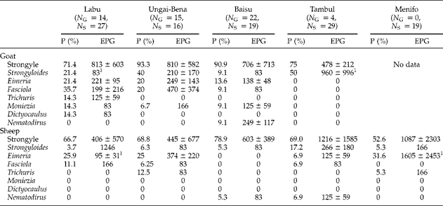
Table 2 summarizes prevalence and mean EPG data grouped by study site. There were no statistically significant differences in the prevalence but there were several significant differences in the EPG counts for different GI parasite types across the study sites.
GI parasite prevalence and eggs/g (EPG) counts for GI parasite type and study site. N G and N S are the total numbers of faecal samples collected at each study site for goats and sheep, respectively. P (%) is the prevalence in per cent. EPG counts are given as means ±SD. Where no SD value is given on the mean EPG count, there was only a single or few positive observations with the same EPG values, so that SD could not be calculated.
1 Values are significantly different by ANOVA/Tukey's HSD testing.
Strongyle and Strongyloides parasites were found in both sheep and goats in all study sites. The mean EPG counts for goats infected with Strongyloides in Tambul were significantly higher than the ones in Labu (ANOVA: F= 3.91, P= 0.05, Tukey's test: P< 0.05). Eimeria was found in sheep from four study sites (Labu, Ungai-Bena, Tambul and Menifo) and in goats from three sites (Labu, Ungai-Bena and Baisu). In sheep, the mean EPG count for Eimeria in Labu was significantly lower than in Menifo (ANOVA: F= 3.41, P= 0.045; Tukey's test: P< 0.05).
No significant differences were observed for the other genera of GI parasites. Fasciola was found in sheep from three study sites (Labu, Ungai-Bena and Tambul) and in goats from three study sites (Labu, Ungai-Bena and Baisu). Trichuris occurred in sheep from two study sites (Ungai-Bena and Menifo) and in goats only from Labu. Nematodirus was found in sheep from two sites (Baisu and Tambul) and in goats only from Baisu. Moniezia was only found in goats from three study sites (Labu, Ungai-Bena and Baisu). Dictyocaulus was only found in goats from Labu.
There was also a trend for goats to be more heavily infected than sheep in all areas. In Labu, mean EPG for strongyle (t= 2.47, P= 0.021) and Eimeria (t= 3.95, P= 0.004) were higher in goats than in sheep. In Ungai-Bena, the prevalence for Strongyloides was higher in goats than in sheep (FET: P= 0.037) and mean EPG for strongyle in goats was higher than in sheep (t= 3.03; P= 0.006).
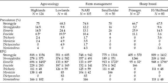
Table 3 shows the prevalence and mean EPG counts for GI parasites with respect to sheep breed, agro-ecology and farm management systems. For comparisons of agro-ecology and farm management systems, the data for sheep and goats were combined. No statistically significant differences were found in either prevalence or mean EPG counts for each parasite type between the two agro-ecological zones, with the exception for Eimeria and Fasciola. The mean EPG for Eimeria was higher in the highlands than in the lowlands (t= 2.1, P= 0.045) and the prevalence of Fasciola was higher in the lowlands than highlands (FET, P= 0.028). There were no significant differences in prevalence or mean EPG between the institutional and smallholder farm management systems with the exception for Eimeria. The mean EPG for Eimeria was higher in the smallholder farms than National Agricultural Research Institute (NARI) farms (t= 3.0, P= 0.004). Similarly, no significant differences were found in both parasite prevalence and mean EPG between sheep breeds, with the exception of Eimeria where the mean EPG in the Highlands Halfbred sheep was higher than in Priangan sheep (t= 2.1, P= 0.045).
GI parasite prevalence and mean eggs/g (EPG) counts stratified for sheep breed, agro-ecology and farm management. N is the number of animals examined and prevalence (%) is the observed percentage of animals infested with one or more GI parasite types. EPG counts are given as mean ± SD. Where no SD value is given for the mean EPG count, there was only a single or few positive observations with the same EPG values, so that SD could not be calculated.
NARI, National Agricultural Research Institute.
H/Halfbred, Highlands Halfbred sheep.
* Values are significantly different by either FET or t-test.
Discussion
The present study observed 78% of the examined farm animals in PNG being infected with one or more types of GI parasites. Similar infection rates for both sheep and goats have been reported in other developing countries, such as Ethiopia and Kenya (Maichomo et al., Reference Maichomo, Kagira and Walker2004; Regassa et al., Reference Regassa, Sori, Dhuguma and Kiros2006). This high parasite prevalence may be attributed to poor farm management, e.g. little pasture rest time, poor nutrition and lack of anthelmintic treatment. At all study sites, mixed-species flocks grazed or browsed on natural forage on the same land for most of the time or were moved to other areas with only short pasture rest times. Studies in Fiji, Tonga and Malaysia found that nematodes have short survival times on pasture and suggested rotational grazing as an effective measure for parasite control (Banks et al., Reference Banks, Singh, Barger, Pratap and Le Jambre1990; Barger et al., Reference Barger, Siale, Banks and Le Jambre1994; Cheah & Rajamanickam, Reference Cheah and Rajamanickam1997).
Natural pasture and shrubs in PNG are often not very nutritious and may contain anti-nutritive factors such as tannin types, which restrict rather than enhance protein availability to the ruminant (Macfarlane, Reference Macfarlane2000). Signal grass, for example, may cause hepatic dysfunction in farm animals (Macfarlane, Reference Macfarlane2000). The low-quality feed may result in subsequent malnutrition which may negatively affect the development of acquired immunity against GI parasites (Kyriazakis & Houdijk, Reference Kyriazakis and Houdijk2006). We did not observe significantly lower GI parasite prevalence at study sites with more frequent anthelminthic treatment.
Strongyles were the most abundant parasites detected in this study. Strongyles, especially Haemonchus species, are highly fecund, laying up to 5000 eggs/day where environmental factors are favourable (Gupta et al., Reference Gupta, Yadav and Chaudhri1987). The pre-parasitic stages of Haemonchus contortus develop and survive better at mean monthly maximum temperatures ≥ 18.3°C (Gupta et al., Reference Gupta, Yadav and Chaudhri1987). Our study sites have mean maximum annual temperatures ranging from 18.9 to 31.1°C with only little variation throughout the year, due to their proximity to the equator. Additionally, most study sites were very humid, which is likely to sustain the survival of the free-living stages of H. contortus, leading to high pasture contamination.
A previous study reported high prevalence (89%) of Eimeria in sheep from three locations in PNG (Varghese & Yayabu, Reference Varghese and Yayabu1985) whereas the present study found a lower prevalence (21–36%) in similar areas. This may be due to the different methodologies used for parasite identification. In the present study a simple flotation procedure was used, while in the previous study a centrifugation/flotation method was used which has been reported to be more sensitive for detection of oocytes in faecal samples (Dryden et al., Reference Dryden, Payne, Ridley and Smith2005). Notwithstanding, this is the first study to report Eimeria in goats in PNG. We found Eimeria in goats in all study sites except Tambul, where only four goats were screened, and Menifo, where no goats were screened.
The trematode Fasciola was present in sheep and/or goats from Labu, Ungai-Bena, Baisu and Tambul. A previous study in PNG found Fasciola in the area of Aiyura and showed that the sheep there are exposed to continual (low-level) pasture contamination leading to chronic fasciolosis at all times (Owen, Reference Owen1989). In PNG, in all areas where the intermediate snail host, Lymnaea species, exists, acute fasciolosis can occur in the wet season, especially in areas where the land is not well drained and the grazing pressure is high (Owen, Reference Owen1989).
This is the first study reporting the prevalence of Dictyocaulus in sheep and goats in PNG. Some studies have found that goats are more susceptible to Dictyocaulus than sheep (Sharma, Reference Sharma1994; Berrag & Urquhart, Reference Berrag and Urquhart1996; Alemu et al., Reference Alemu, Leykun, Ayelet and Zeleke2006). We found Dictyocaulus in 14% of goats examined in Labu (lowland). A similar prevalence has been reported in Ethiopia in areas with an altitude < 1500 m (Alemu et al., Reference Alemu, Leykun, Ayelet and Zeleke2006).
We found higher overall infection levels in goats compared to sheep. Goats were also infected with a wider spectrum of GI parasites. This contradicts some previous studies that found a lower prevalence in goats (Kanyari et al., Reference Kanyari, Kagira and Mhoma2009; Khan et al., Reference Khan, Sajid, Khan, Iqbal and Hussain2010; Abebe et al., Reference Abebe, Gebreyohannes, Mekuria, Abunna and Regassa2011), but is in agreement with a number of other studies, which also reported higher parasite prevalence in goats (Regassa et al., Reference Regassa, Sori, Dhuguma and Kiros2006; Nwosu et al., Reference Nwosu, Madu and Richards2007; Gadahi et al., Reference Gadahi, Arshed, Ali, Javaid and Shah2009; Dagnachew et al., Reference Dagnachew, Amaute and Temesgen2011). Hoste et al. (Reference Hoste, Torres-Acosta and Aguilar-Caballero2008) suggested that goats do not develop resistance as efficiently as sheep and this may be an explanation for our findings.
The EPG counts for Eimeria in PNG Priangan sheep (N= 27) were significantly lower than those in the Highlands Halfbred sheep (N= 83). It is difficult to ascertain the mechanisms behind this difference, especially as, coincidentally, most PNG Priangan sheep samples were collected in Labu, where anthelminthic drug treatment was conducted more regularly. Nevertheless, previous studies have shown that some indigenous sheep breeds exhibit higher levels of resistance against GI parasites and this might partially explain the present findings (Baker & Gray, Reference Baker, Gray, Sani, Gray and Baker2004). PNG Priangan sheep are native to tropical climates, as they originated from South-East Asia and have been exposed to GI parasites in PNG for over a century. In contrast, Highlands Halfbred sheep, which are crossbreeds of the PNG Priangan sheep and the temperate Corriedale and Perendale breeds, have only a fraction of this resistance and will therefore be more susceptible to infection. More detailed studies on immunology and feeding behaviour of the different sheep breeds are required to elucidate this problem further.
The information collected in this study is an important update on GI parasite presence in sheep and goats in PNG. Future investigations should include longitudinal studies and larger cohorts to further assess parasite epidemiology in the diverse agro-climatic zones in the country. Molecular methods should be used to identify the different species of GI parasites infecting sheep and goats in PNG, thus extending previous studies (Varghese & Yayabu, Reference Varghese and Yayabu1985; Owen, Reference Owen1988, Reference Owen1989, Reference Owen1998).
Acknowledgements
The authors would like to sincerely thank the owners of the herds and logistic support by the National Agricultural Research Institute, Department of Agriculture and Livestock in Eastern Highlands Province, and Divine Word University in Papua New Guinea. This research received no specific grant from any funding agency, commercial or not-for-profit sectors. This study was approved by the Murdoch University Animal Ethics Committee (Permit R2368/10).