Introduction
The UK population is ageing; national census data have estimated a 4-fold increase in the population aged over 65 since 1951.1 Cardiovascular disease remains the leading cause of death in the UK,2 and continues to be a significant cause of mortality and morbidity in the ageing population.
Ageing has traditionally been considered a non-modifiable risk factor for cardiovascular disease. Epidemiological data from The Framingham Study suggest that risk factors accumulate with ageing, therefore increasing the risk of developing disease over time.Reference Kannel and Vasan3 Thus ageing remains the biggest non-modifiable risk factor for cardiovascular disease.
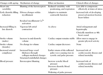
A biological definition of ageing is described as the decline in various physiological body processes culminating in the death of that organism. The relationship between biological and chronological age is not necessarily linear, as environmental impact upon genetics influences the rate and course of ageing. Estimates of arterial function have been found to be more consistent with chronological age. As Thomas SydenhamReference Anon4 remarked: ‘A man is as old as his arteries.’Reference Anon4 Vascular ageing is characterized by mechanical and structural changes within the cardiovascular system (summarized in Table 1) resulting in altered properties of the arterial wall. These changes with ageing are thought to be a contributing factor in the increased incidence of cardiovascular disease amongst the ageing population. These structural and mechanical changes are also affected by modifiable and non-modifiable risk factors that accumulate with ageing, including hypertension, obesity, smoking, lack of exercise, ethnicity and gender.
Changes in the myocardium and vasculature with ageing. Summary of cardiovascular changes with ageing, the mechanisms driving the changes and their effects on function and clinical picture

Literature search
A literature search was conducted using several databases including Embase, Ovid, Wiley, Cambridge Journals, Proquest, Pubmed and Cochrane. Medical Subject Headings (MeSH) terms ageing, aging, cardiovascular, arterial, myocardium, vasculature, elderly, changes, evolution, structure, function, heart, physiology, cellular and senescence were used.
Structural changes in the vascular system with ageing
The arterial system is made up of larger elastic arteries rich in elastin and collagen and smaller muscular peripheral arteries. The arterial wall is made up of three layers: tunica adventitia, tunica media and tunica intima. Between the blood and vessel wall lies a single monolayer of endothelial cells. Larger arteries such as the aorta buffer the flow and pressure variation generated by intermittent cardiac contraction, maintaining oxygen supply to tissues and reducing cardiac pre-load. Loss of elastic recoil impairs coronary flow and may contribute to cardiovascular disease. In the young, blood pressure is determined largely by peripheral vascular resistance but in older people is determined by large vessel stiffness.
In larger arteries such as the aorta the walls thicken with age, elongate and dilate.Reference Gerstenblith, Fredricksen, Yin, Fortuin, Lakatta and Weisfeld5 With ageing aortic wall thickness has been associated with an increase in intima thickness in those with a low incidence of atherosclerosis.Reference Vimani, Avolio, Mergener, Robinowitz, Herderick, Cornhill, Guo, Liu, Ou and O'Rourke6 A study looking at carotid intima thickness post-mortem supports these findingsReference Vimani, Avolio, Mergener, Robinowitz, Herderick, Cornhill, Guo, Liu, Ou and O'Rourke6 with increased intima thickness caused by increased collagen content, covalent cross-linkage of collagen, and elastin fracture, reduction and calcification with ageing. Lifestyle changes and medications such as statins and ACE inhibitors are thought to slow the progression of intima media thickness.
The endothelium plays an essential role in regulating the vascular tone, permeability and response to inflammation by producing vasoactive substances including nitric oxide (a potent vasodilator) and endothelin (a vasoconstrictor and pro-coagulant). Ageing animal studies have demonstrated that endothelial cells become flatter, enlarge and the polypoid nucleus increases.Reference Asai, Kudej, Shen, Yang, Takagi, Kudej, Geng, Sato, Nazareno, Vatner, Natividad, Bishop and Vatner7, Reference Shi, Aida, Vandenberg and Wang8 Structural integrity of the cytoskeleton becomes altered, affecting mobility, migration, proliferation, and proteomic secretions of the endothelial cell.Reference Asai, Kudej, Shen, Yang, Takagi, Kudej, Geng, Sato, Nazareno, Vatner, Natividad, Bishop and Vatner7–Reference Wagner, Hampel, Bernhard, Hala, Zwerschke and Jansen-Durr10 Complex links between endothelial cells in rats decrease with age, causing breaches in the endothelial cell layer.Reference Yeh, Chang, Lu, Lee, Ko, Severs and Tsai11 This may reveal the mechanism in humans which leads to the acceleration of arterial stiffening or atherosclerosis.
Functional changes in the vascular system with ageing
Compliance of any material is defined as the unit change in volume induced by a unit change in pressure. Arterial compliance is a functional property of the artery and it starts to decrease from the age of ten secondary to changes described above, and can be independent of atherosclerosis.Reference Laogun and Gosling12 Reduced arterial compliance causes earlier return of the reflected pulse wave,Reference Safar, Pannier, Laurent and London13, Reference Hodes, Lakatta and McNeil14 causing the wave to return to the heart in systole rather than diastole, instigating a rise in systolic pressure and an increase in pulse pressure.Reference Franklin, Gustin, Wong, Larson, Weber, Kannel and Levy15 This combined with the altered wall properties of reduced arterial compliance reduces coronary blood flow in diastole and increases cardiac work load.Reference Wang, Cheng, Sung, Chuang, Li, Spurgeon, Tring, Najjar, Lakatta, Yin, Chou and Chen16 Arterial stiffness has been identified as an independent predictor of mortality secondary to a cardiovascular event.Reference Laurent, Boutouyrie, Asmar, Gautier, Laloux, Guize, Ducimetiere and Benetos17, Reference London, Blacher, Pannier, Guérin, Marchais and Safar18
Arterial pressure when abnormal results in hypertension; current National Institute of Clinical Excellence (NICE) guidelines are to maintain BP <140/90mmHg.19 The Framingham heart study in the US found that with ageing, systolic blood pressure continues to increase; however, diastolic pressure rises until middle age, balances out, and then begins to decrease after the age of 60 years as demonstrated in Table 2.Reference Franklin, Gustin, Wong, Larson, Weber, Kannel and Levy15 In addition to stroke volume, blood pressure relies on the interplay of peripheral vascular resistance and central arterial stiffness. Peripheral vascular resistance has been identified as a key component in raising systolic and diastolic blood pressure, particularly in young people. Central blood pressure changes affect the blood pressure differently, increasing systolic blood pressure and contributing to a reduction in diastolic pressure, resulting in a widening of pulse pressure. Physiologists have suggested that arterial stiffening may contribute to a resetting of the baroreceptor response within the carotid arteries, resulting in an increase in systolic blood pressure. A blunted neuronal response from the baroreceptors due to an increased resistance to circulating catecholamine as described above may also contribute to a rise in systolic blood pressure in the ageing population.Reference Pan, Hoffman, Pershe and Blaschke20
Blood pressure changes with ageing. Summary of the changes in blood pressure with ageing (adapted from data in Franklin et al.Reference Franklin, Gustin, Wong, Larson, Weber, Kannel and Levy15)

The interplay between the myocardium and vasculature
Changes in the vasculature and myocardium are interlinked and both play a role in the pathogenesis of cardiovascular disease. Bouthier et al.Reference Bouthier, De Luca, Safar and Simon21 found that subjects with altered myocardial structure and function also had altered arterial dispensability.Reference Bouthier, De Luca, Safar and Simon21 Clinically, in those with moderate to large coronary artery disease, arterial stiffness was a key determinant of the ischaemic threshold of the myocardium.Reference Kingwell, Waddell, Medley, Cameron and Dart22
Structural changes of the myocardium with ageing
Macroscopic
Normotensive patients within the Baltimore longitudinal study on ageing demonstrated that left ventricular (LV) cavity size somewhat increased with ageing, but remained within the normal range.Reference Fleg, O'Connor, Gerstenblith, Becker, Clulow, Schulman and Lakatta23 The left ventricle can also become hypertrophic in those with long-standing hypertension in response to increased strain, and studies have suggested that even in those without hypertension LV wall thickness can increase with age.Reference Levy, Anderson, Savage, Kannel, Christiansen and Castelli24 Atrophy can occur, although this is usually confined to disease states. In healthy ageing adults, left atrial enlargement can occur.Reference Lakatta25
Microscopic
Left ventricular myocytes enlarge and decrease in number with ageing.Reference Olivetti, Giordano, Corradi, Melissari, Lagrasta, Gambert and Anversa26 Decreasing myocyte numbers are attributed to drop-out by apoptosis or necrosis, the subsequent outline and valves of the myocardium being replaced by fibrous and calcified tissue.Reference Shirani, Yousefi and Roberts27 Amyloid deposits in the myocardium are a common finding in those aged over 70 years with deposits located mainly in the atria or ventricles. Senile amyloidosis is not associated with cardiomegaly but with atrophied myocardial fibres.Reference Lakatta25 Apoptosis of atrial pacemaker cells occurs, with 50–70% of cells lost by the age of 50;Reference Cheitlin, Zipes, Braunwald, Zipes and Libby28 atrial ventricular node cell numbers are preserved but fibrosis and cellular loss within the bundle of His is thought to contribute to the increasing risk of developing arrhythmia.
Functional changes within the myocardium
Heart rate
With ageing the heart rate in the supine position remains unchanged,Reference Fleg, O'Connor, Gerstenblith, Becker, Clulow, Schulman and Lakatta23 although in the sitting position the heart rate decreases with ageing in both sexes.Reference Rodeheffer, Gerstenblith, Beard, Fleg, Becker, Weisfeldt and Lakatta29 Heart rate variability and adjustments to respiration when measured over a 24-hour period decrease with ageing. This alteration of heart rate is controlled by the sympathetic and parasympathetic nervous system. The activity of the sympathetic nervous system increases with age, resulting in desensitization of the catecholamine receptors driving the sympathetic response, due to the decreasing heart rate with ageing.Reference Lakatta30
Left ventricular systolic and diastolic function
At rest, left ventricular function measured as the ejection fraction is expected to be on average 65%, and is preserved with ageing.Reference Franklin, Gustin, Wong, Larson, Weber, Kannel and Levy15 On exercise the ejection fraction decreases with ageReference Franklin, Gustin, Wong, Larson, Weber, Kannel and Levy15 and this will be discussed later.
The early diastolic filling rate slowly declines from the age of twenty, and at 80 years the diastolic filling rate has reduced by half.Reference Schulman, Lakatta, Fleg, Lakatta, Becker and Gerstenblith31, Reference Swinne, Shapiro, Lima and Fleg32 This has been attributed to structural fibrous changes within the myocardium and residual myofilament calcium (Ca2+) activation from previous myocardial contraction in systole. The length of Ca2+ activation is determined by diastolic stretch and myofilament contraction in systole. Myofilament binding to Ca2+ is thought to combine with structural fibrous alterations of the heart to increase myocardial stiffening; this prolongs early diastolic filling. This is supported by studies undertaken in rats where Ca2+ release from the myoplasm by the sarcoplasmic reticulum resulted in an extended transient rise in Ca2+.Reference Wei, Spurgeon and Lakatta33 These changes are thought to be adaptive to maintain the contractile function of the ageing myocardium.Reference Lakatta30 Despite alterations within the diastolic component of the cardiac cycle, the end-diastolic volume in the supine position remains unchanged in ageing womenReference Fleg, O'Connor, Gerstenblith, Becker, Clulow, Schulman and Lakatta23, Reference Rodeheffer, Gerstenblith, Beard, Fleg, Becker, Weisfeldt and Lakatta29 but increases in men.Reference Swinne, Shapiro, Lima and Fleg32 Vigorous left atrial contraction has been the suggested mechanism leading to increased late diastolic filling.Reference Swinne, Shapiro, Lima and Fleg32, Reference Benjamen, Levy, Anderson, Wolf, Plehn, Evans, Comai, Fuller and Suttton34 The atria are prone to enlargement and hypertrophy, which clinically manifests as a fourth heart sound and giant ‘a’ wave.
In ageing adults free of cardiovascular disease the ejection fraction is not reducedReference Fleg, O'Connor, Gerstenblith, Becker, Clulow, Schulman and Lakatta23 despite common misconceptions. Stroke volume in men increases with age due marginally to the increase in end-diastolic volume, but in women it remains unaltered.Reference Fleg, O'Connor, Gerstenblith, Becker, Clulow, Schulman and Lakatta23 Changes in cardiac output are defined by gender and sedentary lifestyle. Cardiac output in men remains stable with ageing in women; however, cardiac output decreases with ageing. This is thought to be due to the lack of compensation of stroke volume for a lower heart rate. The gender difference is thought to be due to a difference in activity levels between men and women.
Heart failure with preserved ejection fraction in adults
Heart failure is when the heart can no longer pump efficiently enough to meet the body's demands. Of those diagnosed, 40% will die within a year of diagnosis with £635 million of NHS money spent on managing those with heart failure each year.35 It is a clinical syndrome that impairs ventricular filling or ejection.Reference Anderson and Waagstein36 Preserved ejection fraction describes patients with heart failure but normal or near-normal ejection fraction. This contributes to an estimated 50% of the patients with heart failure. Preserved ejection fraction heart failure is thought to be caused by poor left ventricular diastolic filling that in turn requires higher filling pressures in order to maintain left ventricular volume. Elevated pressures increase right-sided pressures and result in increased pulmonary pressures and decreased lung compliance. In advanced disease, low stroke volume may lead to a reduced ejection fraction.
The effects of ageing on the cardiovascular system at a molecular level
Cellular signalling changes
Signalling pathways within the arterial tree include renin, angiotensin I, angiotensin II, angiotensin converting enzyme (ACE), chymase and angiotensin I and II receptors, all of which have been found to be increased in several species.Reference Anderson and Waagstein36–Reference Jiang, Wang, Zhang, Monticone, Telljohann, Spinetti, Pintus and Lakatta40 Studies have shown that all components of the RAS system are up-regulated in the aged arterial wall. Molecules linked to the signalling cascade are also up-regulated; these include calpain-1, matrix metalloproteinase types 2 and 9 (MMP-2 and MMP-9), monocyte chemotactic protein-1 (MCP-1) and transforming growth factor beta-1 (TGF-β1).Reference Anderson and Waagstein36–Reference Jiang, Wang, Zhang, Monticone, Telljohann, Spinetti, Pintus and Lakatta40 Studies on rats to mimic these signalling changes have been shown to cause the structural, functional and biochemical alterations seen in ageing arteries.Reference Wang, Zhang, Spinetti, Jiang, Monticone, Zhao, Cheng, Krawczyk, Talan, Pintus and Lakatta37
Decreasing endothelial cell capacity for replication and repair has been linked to a decrease in nitric oxide production with ageing.Reference Bode-Boger, Muke, Surdacki, Brabant, Böger and Frölich41 It is thought with ageing signalling pathway imbalances disrupt the harmony of oxidase and dismutase. This results in increased levels of superoxide and hydrogen peroxide and a decrease in nitric oxide bioavailability in the coronary and aortic walls of ageing rats, favouring a pro-coagulant state and stimulating new vascular smooth muscle growth. In diseases such as hypertension and diabetes this process is exaggerated and is thought to increase risk of cardiovascular events.Reference McVeigh, Allen, Morgan, Hanratty and Silke42
Telomere biology and cardiovascular ageing
Telomeres make up the ends of eukaryotic chromosomes. They are made up of sequences of DNA laid out in tandem repeats. They are an essential part of the cell cycle, maintaining chromosomal stability.Reference Chan and Blackburn43 As cells divide in mitosis some telomere DNA is lost with each division; the amount that can be lost is variable and can be influenced by oxidative stress.Reference Xu, Neville and Finkel44 The process of losing DNA in each cell division provides a potential mechanism for senescence. Transition to senescence may be when the telomere reaches a critical valueReference Allsop and Harley45 or from cell signalling driving the shortened telomeres.Reference Chan and Blackburn43 Both theories support telomere length as a possible biological marker of ageing, with shorter telomeres indicating increased biological age.Reference Vaziri, Dragowska, Allsop, Thomas, Harley and Lansdorp46 One study showed that subjects over 60 years of age with shorter than average telomere length in circulating leucocytes were more than three times more likely to die of cardiac disease.Reference Cawthon, Smith, O'Brien, Sivatchenko and Kerber47 Shorter telomere length has also been associated with increased likelihood of developing carotid atherosclerosis in individuals with hypertension,Reference Benetos, Gardner, Zureik, Labat, Xiaobin, Adamopoulos, Temmar, Bean, Thomas and Aviv48 decreased left ventricular functionReference Collerton, Martin Ruiz, Kenny, Barrass, von Zglinicki, Kirkwood and Keavney49 type 1 diabetes,Reference Jeancuis, Krolewski, Skurnucj, Kimura, Aviv, Warram and Aviv50 type 2 diabetesReference Sampson, Winterbourne, Hughes, Dozio and Hughes51 and insulin resistance.Reference Demissie, Levy, Benjamin, Cupples, Gardner, Herbert, Kimura, Larson, Meigs, Keaney and Aviv52 It has been inversely correlated with pulse pressure and pulse wave velocity.Reference Benetos, Okuida, Lajemi, Kimura, Thomas, Skurnick, Labat, Bean and Aviv53
Genetics and cardiovascular ageing
Male and females
Androgen receptors are expressed in all vascular tissues, including the endothelium, smooth muscle and myocardium.Reference Wu and von Eckardstein54 The effect of androgens upon the cardiovascular system has long been debated, with oestrogens being considered protective against cardiovascular disease until the recent Women's Health Initiative StudyReference Rossouw, Anderson, Prentice, LaCroix, Kooperberg, Stefanick, Jackson, Beresford, Howard, Johnson, Kotchen and Ockene55 demonstrated the relationship between hormone replacement therapy (HRT) and an increased incidence of non-fatal heart attacks, stroke and thrombo-embolic disease.Reference Rossouw, Anderson, Prentice, LaCroix, Kooperberg, Stefanick, Jackson, Beresford, Howard, Johnson, Kotchen and Ockene55 In men circulating testosterone declines progressively with ageing, but the high levels of testosterone associated with exogenous administration of anabolic steroids are associated with increased cardiovascular morbidity.Reference Sullivan, Martinez, Gennis and Gallagher56 In post-menopausal women on hormone replacement therapy arterial stiffness increasesReference Rajkumar, Kingwell, Cameron, Cameron, Waddell, Mehra, Christophidis, Komesaroff, McGrath, Jennings, Sudhir and Dart57 when HRT is withdrawn, demonstrating that hormonal changes affect vascular compliance.Reference Waddell, Rajkumar, Cameron, Jennings, Dart and Kingwell58 Increased arterial stiffness, reduced systemic arterial compliance and endothelium-dependent dilation have been associated with low androgen levels in men and women.Reference Rajkumar, Kingwell, Cameron, Cameron, Waddell, Mehra, Christophidis, Komesaroff, McGrath, Jennings, Sudhir and Dart57, Reference Dockery, Bulpitt, Donaldson, Fernandez and Rajkumar59 Growing evidence concludes that extremes of androgen excess or deficiency cause an increase in cardiovascular morbidity, but physiological androgen levels may be protective.Reference Getov, Lee, Dockery and Rajkumar60
Race
Afro-Caribbean populations are at a higher risk of developing cardiovascular disease;Reference Chaturvedi61 they are also more likely to have end organ damage from hypertension than anticipated at given levels of blood pressure. Studies have shown that in comparison with European and Asian populations, Afro-Caribbeans have accelerated arterial ageing,Reference Cameron, Bulpitt, Pinto and Rajkumar62 increased aortic stiffness, increased left ventricular wall thicknessReference Chaturvedi, Bulpitt, Leggetter, Schiff, Nihoyannopoulos, Strain, Shore and Rajkumar63 and greater carotid intima thickness.Reference Kalra, Rambaran, Chowienczyk, Goss, Hambleton, Ritter, Shah, Wilks and Forrester64
Lifestyle
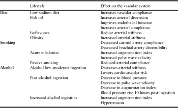
Research suggests that a combination of the following leads to significant premature ageing: smoking, consumption of fruits and vegetables less than 3 times daily, less than 2 hours of physical activity per week, and weekly consumption above the recommended limits for alcohol ingestion in the UK. All-cause mortality data suggested that participants aged 12 years earlier.Reference Kvaavik, Batty, Ursin, Huxley and Gale65 Suggested mechanisms for the effects that lifestyle interactions have, which may accelerate ageing, are summarized in Table 3.
Lifestyle effects upon the vascular system. The interplay of lifestyle and its effects summarized may result in the pathophysiological changes seen in cardiovascular disease more common with ageing

Diet
Evidence and public health messages have associated systolic hypertension and high-salt diets. Low-sodium diets improve vascular compliance in older adults.Reference Gates, Tanaka, Hiatt and Seals66 Fish oil improves arterial distension, endothelial function and arterial compliance.Reference Chin, Gust, Nestel and Dart67 Soy food is associated with a reduced risk of cardiovascular disease,Reference Zhang, Shu, Gao, Yang, Li, Li, Jin and Zheng68 and isoflavones found in soy, tofu and some beans reduce arterial stiffness.Reference Van der Schouw, Pijpe, Lebrun, Bots, Peeters, van Staveren, Lamberts and Grobbee69–Reference Teede, McGrath, DeSilva, Cehun, Fassoulakis and Nestel70
Elderly adults with higher levels of visceral fat have increased aortic stiffness.Reference Sutton-Tyrell, Neman, Simonsick, Havlik, Pahor, Lakatta, Spurgeon and Vaitkevicius71
Smoking and alcohol
Smoking has harmful effects on the arterial wall. Studies have shown that smokers have a decrease in compliance of the carotid artery and distensibility of the brachial artery.Reference Kool, Hoeks, Struijker Boudier, Reneman and Van Bortel72 They also have increased augmentation index and pulse wave velocity in comparison with non-smokers and can be affected acutely post-cigarette.Reference Mahmud and Feely73 Passive smoking is also associated with modified vascular compliance.Reference Fantin and Rajkumar74
Alcohol in low to moderate amounts decreases arterial stiffness and is associated with a lower cardiovascular risk.Reference Sierksma, Muller, van de Schouw, Grobbee, Hendricks and Bots75 In the acute phase post-alcohol ingestion there is a decrease in blood pressure, pulse wave velocity and augmentation index. This gives rise to an increase in blood pressure 10 hours after ingestion.Reference Rosito, Fuchs and Duncan76 In those who ingest high amounts of alcohol (>21 units a week), increases are seen in augmentation index and blood pressure.Reference Mahmud and Feely77
Exercise and ageing
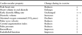
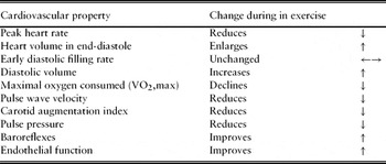
The effect of ageing upon individuals is variable, and this is demonstrated in variability in peak exercise capacity and peak oxygen consumption decline in ageing individuals. This shows that physical fitness as opposed to ageing per se interacts with cardiorespiratory function. The evidence-based effects of exercise upon ageing are summarized in Table 4. Peak heart rate reduces with ageing, and heart volume in end-diastole enlarges. The early diastolic filling rate remains unchanged with age, but the end-diastolic volume in the left ventricle increases as the heart utilizes the Frank-Starling mechanism to maintain stroke volume.Reference Fleg, O'Connor, Gerstenblith, Becker, Clulow, Schulman and Lakatta23 With vigorous exercise the maximal oxygen consumed (VO2, max) declines little when measurements are adjusted to the decrease in lean muscle mass with ageing.Reference Fleg and Lakatta78 Pulse wave velocity, carotid augmentation index and pulse pressure measurements decrease following a period of exercise in previously sedentary subjects.Reference Vaitikevicius, Fleg, Engel, O'Connor, Wright, Lakatta, Yin and Lakatta79, Reference Tanaka, DeSouza and Seals80 Baroreflexes are also improved,Reference Hunt, Farquhar and Taylor81 as is endothelial function,Reference Rywik, Blackman, Yataco, Vaitkevicius, Zink, Cottrell, Wright, Katzel and Fleg82 illustrating that exercise also can illicit intrinsic changes in the arterial wall. With ageing a positive association has been found between increased systemic arterial compliance and exercise tolerance, indicating that either exercise allows acquisition of lower compliance, or that more strenuous exercise is permitted with a more compliant arterial system.Reference Cameron, Rajkumar, Kingwell, Jennings and Dart83
Cardiovascular effects of exercise on a senescent adult. A summary of the effects of ageing on the cardiovascular system during exercise in comparison with a young adultReference Fleg, O'Connor, Gerstenblith, Becker, Clulow, Schulman and Lakatta23, Reference Fleg and Lakatta78–Reference Cameron, Rajkumar, Kingwell, Jennings and Dart83
Conclusions
The biological, structural and mechanical changes in the cardiovascular system in senescence are thought to contribute to the increasing incidence of cardiovascular disease with ageing. Intrinsic genetic factors such as sex and race have been found to influence the ageing myocardial process, demonstrating possible mechanisms of causation for the higher incidence of clinical disease in males versus females, and the implications of race upon the ageing cardiovascular system. Lifestyle factors such as diet, smoking, alcohol ingestion and exercise have also been demonstrated to interact with the ageing cardiovascular system synergistic to the development of cardiovascular disease.
Vascular stiffness has been demonstrated as a useful predictor of biological versus chronological ageing, and independently has been identified as a predictor of mortality secondary to a cardiovascular event.Reference Laurent, Boutouyrie, Asmar, Gautier, Laloux, Guize, Ducimetiere and Benetos17, Reference London, Blacher, Pannier, Guérin, Marchais and Safar18 The value of using vascular stiffness measurements has yet to been transferred into clinical practice. However, with increasing evidence that vascular stiffness is a marker of cardiovascular disease and predictive of mortality, a role for measuring arterial stiffness clinically may assist in the early diagnosis and screening of patients at risk of cardiovascular disease.
Elderly medicine presents great challenges as normal changes meet with pathophysiology to manifest as symptoms often vague and non-specific. Cardiovascular disease in older people is still the leading cause of death in the UK and contributes to significant morbidity and mortality within the elderly population. The identification of those at risk of cardiovascular disease and modification of risk factors may minimize interactions of the ageing process and therefore reduce the incidence of cardiovascular disease within the UK population.
Conflicts of interest
The authors declared no conflicts of interest.