Introduction
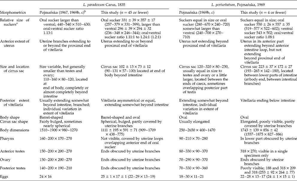
Flukes of the genus Leucochloridium are fairly common avian parasites, two species – L. paradoxum Carus, 1835 and L. perturbatum Pojmańska, 1969 – being particularly frequent in Europe (Casey et al., Reference Casey, Bakke, Harris and Cable2003). Birds become infected by ingesting snails carrying leucochloridiid sporocyst broodsacs which, once in the avian body, give rise to adult worms. Morphological identification of adult leucochloridiids relies on several characteristics, some of those relied upon when identifying L. paradoxum and L. perturbatum being listed in table 1. However, as pointed out by Casey et al. (Reference Casey, Bakke, Harris and Cable2003), species identification of adult leucochloridiids, including the two species mentioned above, is very difficult due to a number of reasons: the parasites possess few autapomorphic characters and lack hard structures, their size ranges overlap, and the internal organs are, in gravid individuals, hidden behind egg-filled uterine loops. In contrast, leucochloridiid broodsacs differ distinctly, the differences being large enough to warrant species descriptions based on colour and banding patterns of sporocyst broodsacs. Specifically, L. paradoxum sporocysts occur in green- and those of L. perturbatum in brown-banded broodsacs (Pojmańska, Reference Pojmańska1967, Reference Pojmańska1969a, Reference Pojmańskab; Bakke, Reference Bakke1978). However, parasitologists frequently face a difficulty of assigning adult specimens they find in birds to sporocysts encountered in organisms preyed upon by those birds (e.g. snails).
Morphometrics of adult Leucochloridium paradoxum and L. perturbatum (measurements are given in μm).

a According to Bakke (Reference Bakke1980), the specific sucker ratio must also be used with caution at the individual level.
b Biometric characters given are those found in L. paradoxum collected from Parus major (Pojmańska, Reference Pojmańska1967).
There are two ways out of this difficulty. First, the life cycle needs to be completed by experimental infection (Pojmańska, Reference Pojmańska1969a, Reference Pojmańska1975, Reference Pojmańska1978; Rietschel, 1972, 1979; Bakke, Reference Bakke1980). This pathway was taken by Pojmańska (Reference Pojmańska1963, Reference Pojmańska1969a) who cultured brown-banded sporocysts of Leucochloridium and experimentally infected birds to produce adult worms. This approach, however, is seldom feasible, as the field-collected material may consist of adult flukes only and no additional sampling for sporocysts is possible. The second option is to use molecular techniques to, first, establish a genetic match between a sporocyst and an adult, and then to apply the molecular characteristics revealed to identify the adults found.
So far, genetic studies on leucochloridiid flukes have focused on their phylogenetic status within the Digenea (Olson et al., Reference Olson, Cribb, Tkach, Bray and Littlewood2003). The first genetic analysis performed on leucochloridiid sporocysts (Casey et al., Reference Casey, Bakke, Harris and Cable2003) allowed comparison of the variability within the sequence of the 5.8S ribosomal RNA gene and internal transcribed spacers (ITS1 and ITS2) of the two species mentioned above. The green-banded L. paradoxum and the brown-banded L. perturbatum sporocysts differed in 6.8% of their ITS sequences. As there were no intraspecific differences, the ITS region was considered a reliable molecular marker with which to differentiate between the two species represented by sporocysts.
However, no corresponding molecular study has been carried out on adult leucochloridiids. Therefore, this study was aimed at finding DNA sequences of adult Leucochloridium specimens matching the sequences described by Casey et al. (Reference Casey, Bakke, Harris and Cable2003) from the two morphologically distinct sporocyst broodsacs (green- and brown-banded). We hypothesized that a match between adult Leucochloridium and sporocysts would be reflected at the molecular level; in other words, the sequences revealed for sporocysts would be found in adults as well. Additionally, we assumed that rDNA sequences would differ between the two adult Leucochloridium morphotypes, and would remain identical within each morphotype.
Materials and methods
Collection and examination of great tits
Adult Leucochloridium were collected from cloaca of the great tit (Parus major) found dead on the Polish Baltic coast. Birds were collected in March 2009 and 2010 at the Hel Peninsula (54°46'N, 18°28'E) (two birds; worm range, 6–9) as well as in October and November 2008 and 2009 near Bukowo-Kopań (54°28'N, 16°25'E) (three birds; worm range, 62–220) and in the city of Szczecin (53°26′N, 14°32′E) (one bird, 132 worms). Digeneans were fixed in 70–75% ethanol, examined under a stereomicroscope and initially assigned to morphotypes using the characteristics given by Pojmańska (Reference Pojmańska1969b), with a due consideration to descriptions provided by Bakke (Reference Bakke1980, Reference Bakke1982).
Morphological and molecular analyses
For the morphometric study, 4, 36 and 9 flukes were collected from the great tits found in Hel, Bukowo-Kopań and Szczecin, respectively. Flukes were stained with alum carmine, mounted in Canada balsam, and examined under a light microscope. For morphological identification, constant (sucker relative size, uterus extension, cirrus sac size and location, vitellaria extension) and additional characters (sensu Pojmańska, Reference Pojmańska1969b) (body shape, cirrus sac shape) were used. In addition, the fluke body was measured (length and width), as were suckers, cirrus sac, anterior and posterior testes, and eggs in those specimens in which the organs were visible.
Molecular analyses were performed on flukes representing the infrapopulations supplying specimens for morphological analyses, although the individuals used were not the same. For sequencing, 11, 75 and 25 specimens from Hel, Bukowo-Kopań and Szczecin were used, respectively. DNA of 70% ethanol-preserved flukes was isolated using the peqGOLD Tissue Mini Kit (PeqLab, Erlangen, Germany) and stored at − 20°C until analysis. The targeted DNA regions (ITS1, partial sequence; 5.8S ribosomal DNA gene and ITS2, complete sequence; and 28S ribosomal DNA gene, partial sequence) were amplified with L18S1 and L28SR1 primers following Casey et al. (Reference Casey, Bakke, Harris and Cable2003). Polymerase chain reaction (PCR) products were identified by their electrophoresis on 1% agarose gel; each PCR product of approximately 1200 bp was sequenced bidirectionally by Genomed (Warszawa, Poland). Sequences were analysed with BioEdit, BLAST and MEGA 4.0 software (Altschul et al., Reference Altschul, Gish, Miller, Myers and Lipman1990; Hall, Reference Hall1999; Tamura et al., Reference Tamura, Dudley, Nei and Kumar2007).
Results
Flukes collected in Hel (oval; 1743 ± 139 × 856 ± 42 μm) were morphologically different from those found in Bukowo-Kopań and Szczecin (table 1). Suckers were almost equal in size; vitellaria extended from the oral sucker centre to below the intestine; the uterus reached beyond the anterior intestinal loop, but did not extend beyond the proximal end of vitellaria; the cirrus sac was located at the lower part of the intestine; and eggs measured 24 ± 1 × 15 ± 1 μm. Hel specimens were tentatively assigned to L. perturbatum (table 1).
Specimens from Bukowo-Kopań and Szczecin were morphologically similar, and differed from those found in Hel by being barrel-shaped, shorter (1111 ± 195 μm), and narrower (591 ± 71 μm); the oral sucker was larger than the ventral; the uterus was substantially enlarged, reaching to the proximal end of vitellaria or extended beyond them; the cirrus sac was located at the posterior end of the body; vitellaria were asymmetrical or identical, ending just beyond the intestine; and eggs measured 25 ± 1 × 17 ± 1 μm. Bukowo-Kopań and Szczecin specimens were tentatively assigned to L. paradoxum (table 1).
Molecular assays of flukes produced characteristic nucleotide sequences deposited in the National Center for Biotechnology Information (NCBI) under accession numbers JF346883 (Szczecin), JF274482 (Bukowo-Kopań) and JF331664 (Hel). Comparison made with the BLAST software showed sequences of Szczecin and Bukowo-Kopań digeneans to be identical. On the other hand, those sequences differed by 1.7% from Hel specimen sequences. Subsequent comparison of sequences obtained in this study with those published by Casey et al. (Reference Casey, Bakke, Harris and Cable2003) showed the Szczecin and Bukowo-Kopań specimen sequences to be identical, except for a single adenine deletion, to those of L. paradoxum (AY258145) of Casey et al. (Reference Casey, Bakke, Harris and Cable2003). Moreover, the Hel specimen sequences were 100% identical to those of L. perturbatum (AY258144). Similar results were obtained by comparing the sequences analysed with other sequences in the NCBI database described as L. paradoxum (JN639012) and L. perturbatum (JN639011). This provides additional confirmation of species identity of the flukes studied. Analysis conducted with the BioEdit software showed the changes (transitions, transversions and deletions) to have been restricted to ITS1 and ITS2 regions. No differences in 5.8S rDNA and in partial 28S rDNA regions were detected. The sequences in question, when analysed with the neighbour joining algorithm of the MEGA 4.0 software, grouped into two clades, one consisting exclusively of specimens representing L. paradoxum and the other, L. perturbatum.
Thus, molecular diagnostics of Hel infrapopulation flukes showed them to represent L. perturbatum. On the other hand, Bukowo-Kopań and Szczecin specimens were diagnosed, with the molecular technique used, as representing L. paradoxum.
Discussion
Tentative morphological identification of leucochloridiid flukes found in dead P. major relied on a number of morphological characteristics which Pojmańska (Reference Pojmańska1967, Reference Pojmańska1969a, Reference Pojmańskab, Reference Pojmańskac) described from adults that had been cultured from distinctly identifiable sporocysts. Those characteristics proved useful for our study, but, unfortunately, their usefulness is not universal. Descriptions, figures and dimensions provided by various authors (Pojmańska, Reference Pojmańska1967; Rutkowska, Reference Rutkowska1973; Machalska, Reference Machalska1980; Sitko, Reference Sitko1993, Reference Sitko2001; Macchioni, Reference Macchioni2003) differ widely. For example, descriptions published by those authors show that adult L. paradoxum and L. perturbatum can be easily distinguished by their relative sucker size. However, Bakke (Reference Bakke1980) reported this character as variable as well. Therefore, we agree with Pojmańska (Reference Pojmańska1969b) that certain characters are manifested sufficiently frequently to be regarded as diagnostic in abundant leucochloridiid collections only. It is conceivable that, in large collections from different localities, multivariate mathematical techniques might be helpful in identifying sets of morphological characters that would eventually assist in morphological diagnosing of adult leucochloridiids.
When a collection is small, uncertainties prevail and can be resolved by using molecular techniques. Molecular analyses to distinguish between morphologically distinct forms of leucochloridiid sporocyst broodsacs representing two unambiguous forms, brown-banded L. perturbatum and green-banded L. paradoxum, were run successfully by Casey et al. (Reference Casey, Bakke, Harris and Cable2003). Not only did they confirm genetic identity of the two species represented by sporocysts, but also provided genetic markers (ITS regions of rDNA) that might be applied to adult flukes of uncertain identity, such as those in our study, where no matching sporocyst broodsacs were available. DNA sequences of adult Leucochloridium flukes proved identical to those from sporocysts of the respective species collected in Denmark and Norway by Casey et al. (Reference Casey, Bakke, Harris and Cable2003). Genetic variation of the flukes we studied was at a level identical to that described by Casey et al. (Reference Casey, Bakke, Harris and Cable2003). Similarity between the genetic make-up of sporocysts and adult flukes confirmed utility of the rDNA region used as marker. Flukes identified as one morphotype (our tentative species) showed an identical level of genetic diversity, whereas the two morphotypes were sufficiently different to be regarded as separate species.
Thus, when there is a need to assess the extent of avian infection with different leucochloridiid species and the actual collection is small, matching sporocyst broodsacs are not available and no experimental infection can be performed, morphological characters can be used as an initial approximation, but the final diagnosis will depend on the results of molecular assays.
Acknowledgements
We are grateful to the ‘Operation Baltic’ team for their help with collecting the materials for this study. We also thank Professor Dariusz Wysocki and PhD students of the Department of Zoology, University of Szczecin, and two anonymous reviewers who provided helpful input with regard to the earlier version of the manuscript. Our thanks go to Dr Teresa Radziejewska for assistance with preparation of the subsequent version of the manuscript.