Introduction
Trichinellosis is a meat-borne zoonosis, which is not only a public health hazard, but also represents a high financial burden in porcine animal production and food safety. Despite concerted control efforts, the disease remains a threat in many countries (Murrell & Pozio, Reference Murrell and Pozio2011) and in 2009 a total of 750 cases were reported in Europe (ECDC, 2011). Moreover, the imminent risk for introducing Trichinella in domestic pigs from non-erasable sylvatic resources stands in full contrast to the concept of Trichinella-free areas (EFSA, 2005) and demands permanent control by means of individual Trichinella examination in terms of the Hazard Analysis and Critical Control Points (HACCP) concept.
Currently, official Trichinella examination is a basic legal principle in meat hygiene and a milestone of preventive consumer protection within the European Union. According to Regulation (EC) No 854/2004 (Anonymous, 2004) all animals intended for human consumption that are susceptible to Trichinella have to be examined post-mortem. Specifications on Trichinella detection in meat are given in Regulation (EC) No 2075/2005 (Anonymous, 2005), which names in its Annex I a reference and several alternative procedures. All these detection methods are based on the artificial digestion of meat samples and comprise the following principal steps: (1) release of Trichinella larvae (L1) by destruction of the surrounding tissues using HCl/pepsin and mechanical treatment; (2) concentration of L1; and (3) visual examination of digests using a trichinoscope or stereomicroscope. The magnetic stirrer method for pooled sample digestion (MSM) is stipulated to be the reference method by the aforementioned legal source and is considered the gold standard (Gamble et al., Reference Gamble, Bessonov, Cuperlovic, Gajadhar, van Knapen, Nöckler, Schenone and Zhu2000).
In the European Union, the estimated annual costs incurred from meat inspection of 167 million pigs (Alban et al., Reference Alban, Pozio, Boes, Boireau, Boué, Claes, Cook, Dorny, Enemark, van der Giessen, Hunt, Howell, Kirjusina, Nöckler, Rossi, Smith, Snow, Taylor, Theodoropoulos, Vallée, Viera-Pinto and Zimmer2011) range from €25 million to €400 million (Kapel, Reference Kapel2005). In view of these considerable economic costs on the one hand, and the importance for preventive consumer protection on the other, it is mandatory that the MSM complies with the highest standards of analytic quality in terms of both qualitative and quantitative analysis.
A thorough analysis of the German national proficiency testing data from 2008 to 2011 shows that MSM cannot meet all demands when performed under daily conditions: despite improvements in qualitative analysis where the sensitivity changed by 6% (89–95%) and specificity by 5% (93–98%) from 2008 to 2011 (Mayer-Scholl, Reference Mayer-Scholl, Reckinger and Nöckler2009, Reference Mayer-Scholl, Reckinger and Nöckler2010, Reference Mayer-Scholl, Reckinger and Nöckler2011, Reference Mayer-Scholl, Reckinger and Nöckler2012), quantitative analysis still provides unsatisfactory results which range from 54.7% L1 recovery in 2008 to 70.2% in 2011, and even the best ten laboratories had an average recovery rate of not more than 89% (2009). These unsatisfactory results strongly indicate that the existing deficits cannot be attributed to insufficient laboratory performance alone, but must also be inherent to the method. Some studies have elucidated analytical errors of the MSM (e.g. Köhler, Reference Köhler1978; Prost & Nowakowski, Reference Prost and Nowakowski1990; Forbes & Gajadhar, Reference Forbes and Gajadhar1999; Gamble, Reference Gamble1999; Forbes et al., Reference Forbes, Parker and Scandrett2005; Rossi & Pozio, Reference Rossi and Pozio2008), and a recent study performed by Riehn et al. (Reference Riehn, Hasenclever, Petroff, Nöckler, Mayer-Scholl, Makrutzki and Lücker2013) indicates that the final step of the MSM reference procedure, the visualization, is the most uncontrollable and thus most error-prone step in the whole protocol.
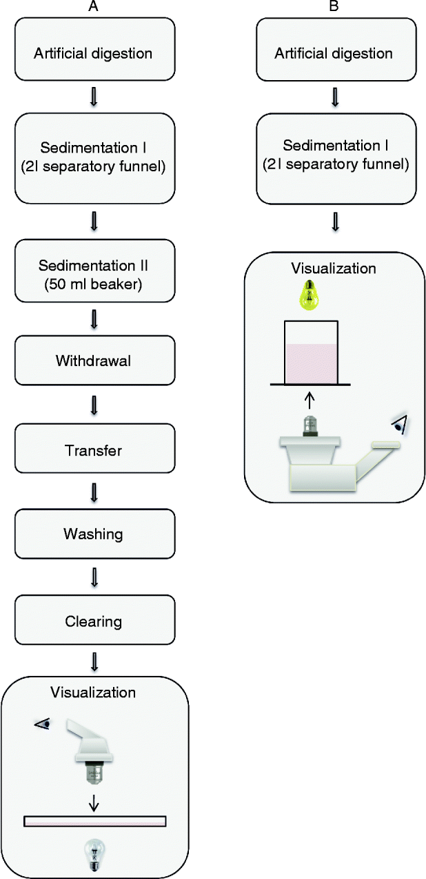
Samples are usually examined in top view using a trichinoscope or stereomicroscope. For this purpose several preparatory steps (fluid transfer, volume reduction, washing) that might cause the loss of L1 are necessary (fig. 1A). In addition, the larval counting basin must have a large surface area in order to reduce the height of the digestion fluid through which L1 have to be detected. Consequently, it is necessary to move the counting basin through the field of vision, a procedure that may lead to increased turbulence and resulting dislocation of the L1.
(colour online) Working sequences of (A) the reference method (MSM) and (B) the modified method (mMSM) using inverse microscopy and the modified larval counting basin (mLCB, ‘Trichoview’).

Here we present an alternative approach that is based on the inversion of the optical path, which enables the direct detection of Trichinella larvae on the bottom of the larval counting basin (fig. 1B). We also redesigned the larval counting basin, which, in combination with inverse microscopy, then offers the possibility to dispense with the aforementioned critical preparatory steps. Furthermore, the extent of the examination area is significantly reduced, which vastly simplifies the assessment of the digest.
Materials and methods
Modified larval counting procedures
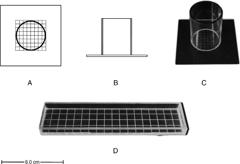
We used a sequential approach for the direct comparison of the modified magnetic stirring method (mMSM) and the conventional magnetic stirring method (MSM). The MSM procedure (fig. 1A) was performed as prescribed in regulation 2075/2005 (Annex I, Chapter I); however, the normal glass beaker for the second sedimentation step was substituted by a modified larval counting basin (mLCB, ‘Trichoview’, fig. 1B) and an additional counting step using inverse microscopy (Axiovert 25, Carl Zeiss AG, Jena, Germany, 50 × magnification) was introduced. Then the normal procedure proceeded as prescribed: 30 ml supernatant withdrawal, transfer of the remaining 10 ml sediment sample to a conventional LCB (inner surface 4 cm × 18 cm, Robert Kind GmbH, Lichtenfels, Germany, fig. 2D), rinsing of the beaker (mLCB) and examination using a stereomicroscope (type SZX 12 Olympus Deutschland, Hamburg, Germany) at 15–20 and 60–100 × magnification.
Schematic drawing of the modified larval counting basin (mLCB, ‘Trichoview’) viewed from above (A) and the side (B) and a complete modified larval counting basin (C) compared with a conventional one (D). Scale bar, 8.0 cm.
The modified larval counting basin (mLCB, ‘Trichoview’) was constructed using transparent acrylic glass (DaBenMo Plexiglas & More, Nordheim, Germany). A hollow cylinder with an inner diameter of 36 mm (10.3 cm2 examination area) and a height of 48 mm (wall thickness 2 mm) was glued to an acrylic plate (8 cm × 8 cm × 0.2 cm) using a special liquid glue (Kallocryl A/C, Speiko-Dr.Speier GmbH, Münster, Germany) in order to prevent formation of bubbles. Prior to that, gridlines were carefully inscribed in 5-mm intervals on the inner surface of the basal plate for easy orientation during visualization. Depth of the gridlines has to be minimal, so that L1 will not get caught in them or be masked by the gridlines. The upper edge of the tube was blackened with acrylic paint (p2 Kosmetik GmbH, Wiener Neudorf, Austria) to prevent stray radiation. The mLCB is shown in fig. 2 schematically (fig. 2A, B) and as used in this study (fig. 2C), in comparison to the conventional larval counting basin (fig. 2D).
Trichinella larvae (L1) were provided by the German Federal Institute for Risk Assessment (BfR, Berlin, Germany). According to the BfR, the infested meat was derived from domestic pigs, which were infected with 40,000 Trichinella spiralis larvae (reference strain ISS 003) and electrically stunned and exsanguined 10 weeks after infection. The meat was stored at +7°C until use. Only viable Trichinella larvae (motile larvae at 37°C) were used in our experiments.
Experimental procedures
The required number of L1 was harvested under the stereomicroscope using a micropipette (Eppendorf reference 20 μl, Eppendorf AG, Hamburg, Germany). L1 were transferred to a watch-glass, counted again, and then transferred to the respective sample. The pipette tip was examined under the stereomicroscope to ensure that no larvae accidentally remained within. All experiments were designed as total blind studies. Different persons conducted spiking and performance of experiments so that the number of L1 was unknown to the respective examiner. The number of spiked and recovered L1 was forwarded to and analysed by a third person who was involved in neither spiking nor performing of the experiments.
For the comparison of mMSM and MSM, we used 60 samples spiked with a total of 1285 Trichinella L1 in varying numbers from 1 to 90. The spiking was performed directly into the modified LCB in order to exclude any further sources of error prior to the second sedimentation, such as larval losses during digestion, filtration or first sedimentation.
In addition, we elucidated potentially relevant experimental influences using the same experimental design for comparing MSM and mMSM, but with different points of spiking: error due to the first sedimentation and the following procedures (73 samples, 1961 L1), spiking error (10 samples, 405 L1), error of supernatant withdrawal and transfer (11 samples, 190 L1) and error due to the substitution of the 50 ml glass beaker by acrylic mLCB (20 samples, 400 L1). We could not observe a significant influence of these experimental aspects on the comparison of MSM and mMSM. However, these data will be presented, along with other sources of error in the context of the estimation of the total analytical variance of MSM and mMSM.
Data analysis
Descriptive statistics were performed using Excel (Microsoft Office 2007 Enterprise, Microsoft Corporation, Redmond, USA); statistical tests (e.g. Fisher's exact test) were performed by use of R (version 2.14.0, R Foundation for Statistical Computing, Vienna, Austria).
Results and discussion
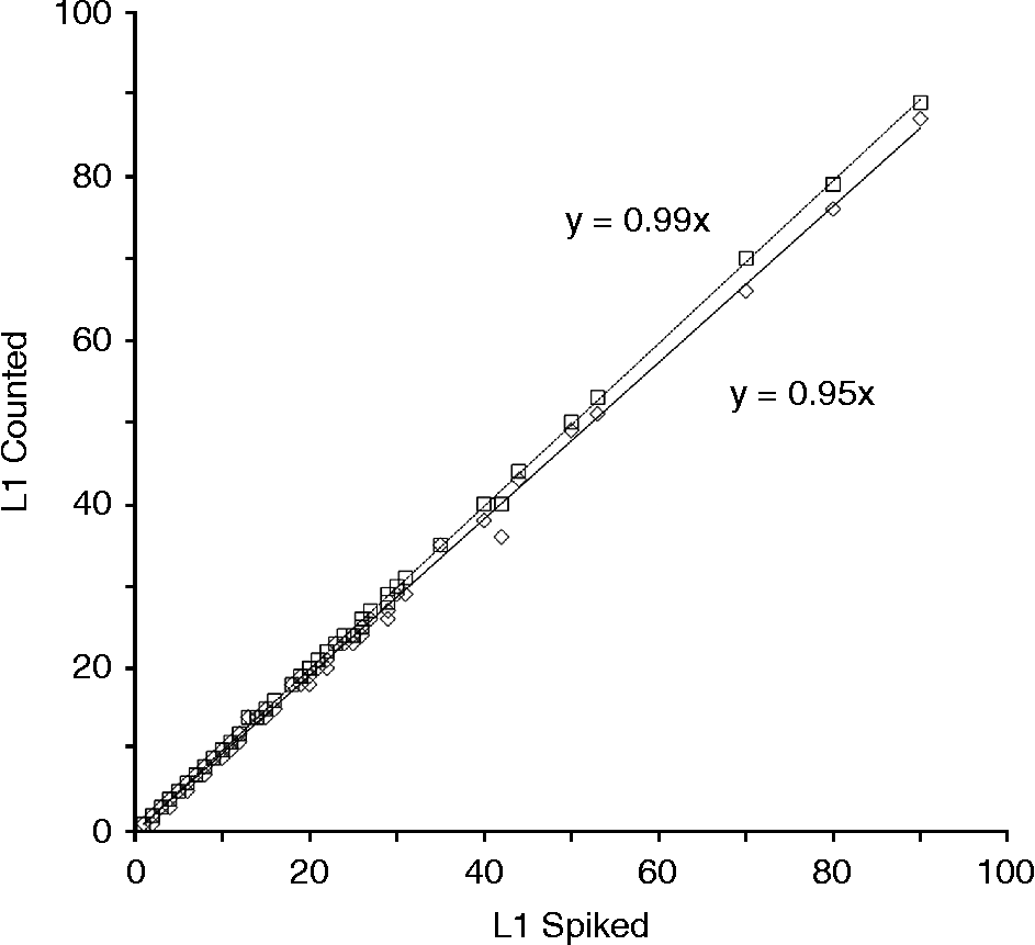
The modified magnetic stirring method (mMSM) introduces a new larval counting basin (fig. 2A–C) and inverse microscopy to the conventional reference method (MSM) as shown in fig. 1. Several analytical steps (supernatant withdrawal, fluid transfer, washing) are superfluous in mMSM; however, new analytical errors could have been introduced. Therefore, we restricted the comparison between MSM and mMSM to the respective analytical steps. Spiking was performed after the first sedimentation into the new LCB followed by a second sedimentation, counting and progressing with the conventional steps (supernatant withdrawal, fluid transfer, washing, counting by means of a conventional counting basin and a stereomicroscope). Sixty samples were used which were spiked with 1–90 L1, giving a total of 1285 L1. The resulting data pairs for spiked/counted L1 are illustrated for conventional MSM and mMSM in fig. 3 by way of a scatter plot and respective regression curves. Overall differences between spiked (x) and counted (y) results were small for both methods, with regression curves close to the bisecting line (y= x). However, the regression coefficient for MSM (y= 0.95x) was distinctly lower than that for mMSM (y= 0.99x). While the number of recovered L1 was correct in 87% of the samples using mMSM, the conventional MSM gave the correct number of spiked L1 only in 37% of the samples. Overall, mMSM recovered 1278 L1 (99.4%) while MSM recovered only 1225 L1 (95.3%). Further results from experiments including MSM/mMSM comparison and spiking at different stages (data not shown) support an approximately 4% higher L1 recovery by mMSM. Thus, we can calculate using Fisher's exact test that the chances of detecting L1 are nearly eight times (7.9) higher using the modified instead of the conventional MSM (95% confidence interval, 3.8–19.3, P= 2 × 10− 11). Furthermore, an analysis of the German national proficiency testing data shows that, interestingly enough, laboratories with a good performance (>80% larva recovery, no false-negative or false-positive test results) also reported samples with an unexpectedly low larval count, and that the average recovery rate, even under highly standardized conditions, was only 84% (Riehn et al., Reference Riehn, Hasenclever, Petroff, Nöckler, Mayer-Scholl, Makrutzki and Lücker2013).
Recovery of Trichinella larvae (L1) using the reference (open diamonds) and modified (open squares) methods with 60 data pairs used for spiked (x) and counted (y) larvae.
Our data suggest that use of the mMSM could improve the performance by about 4 percentage points to 88%. Thus, the chances of a false-negative result in a sample containing five larvae would be reduced by a factor of 4.2: (q 1/q 2)n, where q 1 is the error of conventional MSM (16%), q 2, the error of mMSM (12%) and n the number of L1 in the sample (n= 5).
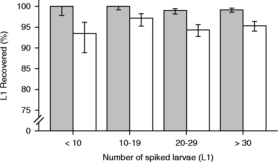
In practical applications, Trichinella examination will generally be faced with relatively low, rather than high, infestation rates and, thus, numbers of L1. As shown in the histogram of fig. 4, where the L1 recovery rate is categorized in four groups, efficiency of mMSM does not decrease for small numbers of L1. In the case of the first and second group (1–19 spiked L1) the recovery rate of mMSM was 100%; we observed no L1 losses with mMSM up to 25 L1 spiked, whereas in conventional MSM the L1 losses already occurred at two spiked L1. With MSM, the recovery in the group of samples spiked with 1–9 L1 was only 93%.
Relative recovery rates (%) of four groups of spiked Trichinella larvae (L1) using the reference (MSM, clear) and modified (mMSM, grey) methods; 60 data pairs were used for each method and errors bars are given with 80% confidence intervals.
The MSM's susceptibility to error has been described in several publications (e.g. Köhler, Reference Köhler1978; Prost & Nowakowski, Reference Prost and Nowakowski1990; Gamble, Reference Gamble1999; Forbes & Gajadhar, Reference Forbes and Gajadhar1999; Forbes et al., Reference Forbes, Parker and Scandrett2005; Rossi & Pozio, Reference Rossi and Pozio2008). It was assumed that insufficient examination results, especially with respect to the quantitative analysis of Trichinella-positive meat samples, cannot only be attributed to a poor performance of the laboratories (Riehn et al., Reference Riehn, Hasenclever, Petroff, Nöckler, Mayer-Scholl, Makrutzki and Lücker2013). Taking into account that several error-prone analytical steps are omitted in the present modified MSM procedure, it is not surprising that the overall larval loss was reduced in this new approach. In fact, the results clearly indicate that no significant new sources of analytical error were introduced by our modification of the MSM.
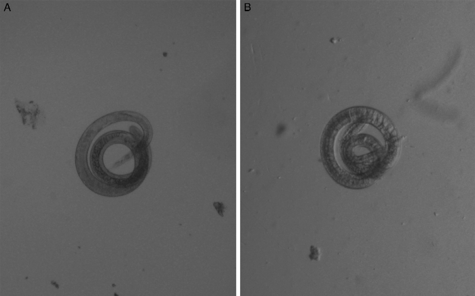
Overall, handling and practicability of the modified MSM were improved when compared to the conventional reference MSM. First, there is a notable reduction of analytical expenditure by the omission of certain analytical steps (supernatant withdrawal, fluid transfer and washing) as well as by the reduced examination time due to the seven times smaller area of the new larval counting basin. The latter, in combination with inverse microscopy, turned out to be easily applicable after only a short period of training. The Trichinella larvae could be clearly identified on the bottom of the mLCB (fig. 5A) and appeared to be less prone to suboptimal digestions with notorious turbidities and undigested particles when compared to the LCB (fig. 5B). This can be attributed to the inversed optical pathway: L1 sediment out faster and are located directly at the bottom of the mLCB, so that other particles, as well as unclear fluid, do not interfere with the optical pathway. It is, however, indispensable to apply a strong light source and blacken the upper fringe of the mLCB in order to achieve clear vision. Moreover, L1 were not generally moved out of the field of vision when the mLCB was moved. Most probably this can be attributed to the drastically higher liquid column of the mLCB which prevents turbulence of the liquid and gives better resistance against acceleration forces compared to the conventional LCBs.
Digital images (50 × ) of Trichinella larvae (L1) in (A) the modified larval counting basin (‘Trichoview’, inverse microscope) and (B) the conventional larval counting basin (stereomicroscope).
Finally, the inversion of the optical path in combination with the new larval counting basin permits depiction of the whole examination area (10.3 cm2) in one single image, using an ordinary digital full-frame camera. This would not only facilitate – for the very first time – full documentation of the examination results, but could also be a starting point for automated digital imaging analysis in Trichinella examination. Altogether we can highly recommend further research in this direction of the modified magnet stirring method.
Acknowledgements
For their valuable technical assistance in the present study, we would like to thank Lia Kieker, Heiko Wellner and Jasem Saffaf. We are particularly thankful to the Federal Institute for Risk Assessment (BfR, Germany) and PD Dr. Nöckler for providing Trichinella-infected meat and giving valuable help. None of the authors of this manuscript has declared any conflict of interest within the past 3 years which may arise from being named as an author on the manuscript.