INTRODUCTION
Many parasites migrate in their hosts towards very distinct habitats. The complex, well-defined paths of migration to the final locations within hosts have been described for many parasites, but little is known about how organisms navigate and locate their destinations within hosts (reviewed by Sukhdeo and Sukhdeo, Reference Sukhdeo and Sukhdeo1994, Reference Sukhdeo and Sukhdeo2002, Reference Sukhdeo and Sukhdeo2004). We studied the orientation of infective larvae of skin-invading parasitic worms after their penetration into agar substrates (Haas et al. Reference Haas, Grabe, Geis, Päch, Stoll, Fuchs, Haberl and Loy2002a, Reference Haas, Haberl, Syafruddin Idris, Kallert, Kersten and Stiegeler2005; Grabe and Haas, Reference Grabe and Haas2004a, Reference Grabe and Haasb). The schistosomula of the human parasite Schistosoma mansoni and the duck parasite and agent of human cercarial dermatitis, Trichobilharzia ocellata, were attracted by sera of their hosts with arginine and glucose as the guiding compounds. We hypothesized that these 2 cues were used by the migrating parasites (1) to orientate towards deeper skin layers, (2) to determine their position during their surface-parallel migration within the epidermis and (3) to locate blood vessels. Arginine and glucose seem to be suitable host signals for these invasion phases, and the question arises whether this navigation strategy may generally be used by digeneans that penetrate skin and seek blood vessels for further migration. Therefore, we studied this topic with Diplostomum spathaceum (syn.: Diplostomum pseudospathaceum, as described by Niewiadomska (Reference Niewiadomska1984) and Niewiadomska and Kiseliene (Reference Niewiadomska and Kiseliene1994)), whose cercariae penetrate fish hosts and migrate towards their eye-lenses. The D. spathaceum – fish host model has the advantages that fish as hosts are more suitable for in vivo experiments than mammals and birds and that much is known about cercarial behaviour and the migration of the parasite within the host.
The cercariae of D. spathaceum attach to a broad spectrum of aquatic animals in response to water currents, touch, and carbon dioxide (Haas, Reference Haas1974a, Reference Haasb, Reference Haas1975), but they remain only on mucus-covered animals in response to small molecular weight carbohydrates. Then, they penetrate their hosts, stimulated by glycoproteins with sialic acids as signalling residues and defined free fatty acids (Haas et al. Reference Haas, Stiegeler, Keating, Kullmann, Rabenau, Schönamsgruber and Haberl2002b). After penetration, the diplostomula migrate towards the head, enter the eyes and then the lenses for further development. Studies on the migratory routes revealed inconsistent results. Histological techniques showed most migrating diplostomula in connective and muscle tissues and, as only few were located in the circulatory system, it was argued that the blood system does not serve as a major migratory route (Erasmus, Reference Erasmus1959; Ratanarat-Brockelman, Reference Ratanarat-Brockelman1974). However, observations in living fish and amphibia and experimental in vivo approaches suggested that diplostomula migrate mainly along blood vessels (Davis, Reference Davis1936; Ferguson, Reference Ferguson1943; Betterton, Reference Betterton1974; White et al. Reference White, Secombes and Chappell1991). Our unpublished observations of diplostomula in fins and tails of guppies and in bodies of the translucent species Chanda ranga and Kryptopterus bicirrhis support the view that blood vessels are important migration routes. It can easily be observed that the parasites enter blood vessels and migrate rapidly within the lumen. But normally they also leave the vessels and burrow their way in a cranial direction in tissues, until they eventually enter blood vessels again. Therefore, most diplostomula are seen digging in tissues, but the few creeping in the vessels cover the main distances. Therefore, we suppose that D. spathaceum is adapted to the location of blood vessels like S. mansoni and T. ocellata. We consider it as a suitable model for analysis of navigation mechanisms, particularly for comparison with what we already know on the navigation strategies of schistosomes.
MATERIALS AND METHODS
Experimental conditions
Cercariae of D. spathaceum were obtained from naturally infected Lymnaea stagnalis snails using methods described by Haas et al. (Reference Haas, Stiegeler, Keating, Kullmann, Rabenau, Schönamsgruber and Haberl2002b). Penetration of cercariae into tail fins and their migration within the fins of female guppy (Poecilia reticulata) was observed with dissecting microscopes and their path of migration was drawn using a camera lucida. Living guppies were fixed with rubber bands in an infection compartment of a Petri dish. The fish were provided with aerated water, free of cercariae, by separating the head and the gills from the infection chamber with a plastic membrane. Groups of 50–200 cercariae were introduced into the infection chamber and the behaviour of individual cercariae recorded.
The in vitro chemo-orientation of the diplostomula was studied with a 2-choice experimental chamber modified after the W-chamber method as described by Haas et al. (Reference Haas, Grabe, Geis, Päch, Stoll, Fuchs, Haberl and Loy2002a) and Grabe and Haas (Reference Grabe and Haas2004a). Briefly, the larvae penetrated agar in the central chamber (5×5 mm, height of agar 6 mm) of a 3-armed cuvet. In typical experiments, the agar (0·5%; pH 7·0, 5 mm sodium phosphate buffer), as well as the test and control substrates contained Ringer solution (composition, see Table 2) or defined salt solutions. Penetration and full transformation into diplostomula (as indicated by lectin staining; see Haas et al. Reference Haas, Grabe, Geis, Päch, Stoll, Fuchs, Haberl and Loy2002a) was stimulated by a 1-mm layer of agar, containing linolenic acid, on the upper surface of the central agar block (0·05 mm linolenic acid in 0·5% agar, pH 7·0, 5 mm sodium phosphate buffer). The central agar block of the chamber was connected with lateral blocks of similarly composed agar, supplemented with test or control substrates, respectively. After placing 70–150 cercariae in 70 μl of water on the central agar blocks (constant numbers per experimental series), the chambers were incubated for 120 min at 22°C in darkness and the number of diplostomula in the agar of the central arm and in the test and control agar was recorded. In experiments with macromolecules (Table 2A, C, D, E), the test and control substrates were integrated in the lateral agar blocks and in experiments with small molecular weight compounds the substrates were applied within buffered Ringer or salt solution (pH 7·0, 5 mm phosphate buffer) on the lateral agar blocks. Concentration gradients of the substrates became established by diffusion across the agar blocks as determined by the diffusion rate of chemicals within the system (Grabe and Haas, Reference Grabe and Haas2004a).
The sensitivity of different cercarial populations (even from the same individual snails) to penetration and orientation stimuli varies from day to day and furthermore depends on the age of the parasites. Therefore, these experiments used cercariae within 3 h after being shed, and all quantitative comparisons were done using the same cercarial populations from 4–15 individual host snails. All results in Table 2 were confirmed by additional experimental series (data not shown).
Chemo-orientation of diplostomula in the W-chambers was evaluated by comparing the numbers of diplostomula leaving the central arm of the chambers in direction to test- and control substrates. More diplostomula left the central arm when there was an attractant in the chamber than in control experiments, but this did not affect the statistical analysis. In Table 2 only the numbers of diplostomula showing orientation (i.e. leaving the central arms) was shown, the numbers of the cercariae penetrating the substrates and the percentage of these showing orientation was omitted for clarity. All in vitro experiments used a blind protocol; the test substrates were given a code unknown to the experimenters.
The effect of melatonin on the photo-orientation of migrating diplostomula was studied with methods modified from Grabe and Haas (Reference Grabe and Haas2004b). Briefly, the agar of the W-chambers contained melatonin in Ringer or Ringer only, and the chambers were illuminated from one side with cold light sources or neon lamps through heat filters at light intensities of 30 000 and 8000 lux respectively.
Preparation and analysis of test substrates
Fish serum was prepared from mixed blood obtained after decapitation of carp (Cyprinus carpio). Separation into molecular size classes was performed by centrifugal filtration with Centricon or Centriprep units (Amicon, Beverley, Massachusetts). Polyanionic glycosaminoglycans in serum were precipitated with Alcian blue by modified methods after Whitehead (Reference Whitehead1978) as described elsewhere (Haas et al. Reference Haas, Granzer and Brockelman1990) and then removed by centrifugation. Proteins were removed from serum by treatment with 10% trichloracetic acid (TCA) solution for 30 min on ice and centrifugation. TCA was removed from the supernatant with diethylether. Proteins in serum were also hydrolysed with trypsin (Type III, Sigma, St Louis, Missouri; 2 successive treatments with 10 units/ml, pH 7·6 for 4 h at 25°C) and then with protease from Streptomyces griseus (pronase E, Sigma; 2 treatments with 2 units/ml, pH 7·5 for 4 h at 37°C). Hydrolysis was verified with the Bradford protein assay. Different incubating conditions modified the stimulating activity of the substrates. Therefore, 3 control substrates had to be run under identical conditions: modifying reagent (e.g. enzyme) without substrate, substrate without reagent (substrate ‘similarly treated’, i.e. positive control), and buffer only.
Amino acid analysis was performed by high-performance liquid chromatography (HPLC) on a RP-18 column applying pre-column derivatization with O-phthaldialdehyde and fluorescence detection as described elsewhere (Haas et al. Reference Haas, Körner, Hutterer, Wegner and Haberl1995). Monosaccharides were detected by gas-chromatography on a ZB-5 column (Phenomenex, Aschaffenburg, Germany) after derivatization with Tri-Sil Z (Pierce, Rockford, Illinois) and incubation at 60°C for 1 h. Osmolarity of the solutions was controlled using a semi-micro osmometer (Type digital, Knauer, Berlin, Germany).
Statistical methods
Replicate percentage data were transformed by arcsine-square root transformation to obtain approximately normally distributed data. Means were compared using a multiple t-test procedure (Tukey-LSD-test) after One-Way Anova and test for homogeneity. Mean values and standard errors (s.e.) were calculated from the transformed data and were retransformed thereafter. For simplification, only the larger of the 2 different s.e. is indicated in the tables. In experiments on orientation of diplostomula within fish fins a χ2-procedure was used to test for statistically significant differences from an equal distribution.
RESULTS
Orientation in the tail fin of fish
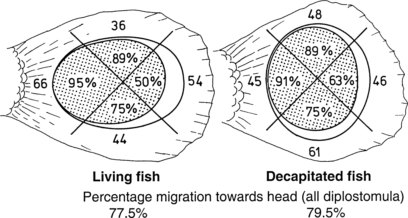
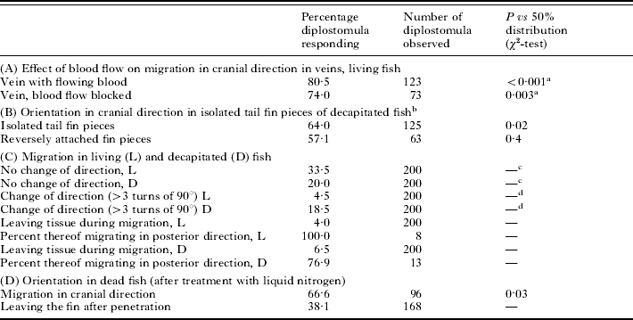
D. spathaceum cercariae readily penetrated tail fins of guppy. Fin rays restricted their path of migration mainly in an antero-posteriad direction. Most diplostomula burrowed directly towards veins and entered them for further migration. Only 1–3% of the diplostomula entered arteries located within the fin rays. During penetration, the cercariae showed no preference for any particular direction, but folds in the living fin forced the attached organisms preferentially in a antero-posteriad direction (Fig. 1). During their further path of migration the diplostomula shifted towards a cranial direction. This was already significant when they had migrated a distance of 1200 μm (approximating 6·5 body lengths) in the fins of living and decapitated fish (Fig. 1). The capability of the parasites to shift to an orientation in a cranial direction was independent from the streaming blood as their orientation was not altered in blocked veins (Table 1A). It was even independent of living cells, as cranial migration occurred also in fins after their treatment with liquid nitrogen (Table 1D). The signalling cues did not originate exclusively from the body of the fish, as the parasites also chose a cranial direction in isolated fins and in pieces of fins, and perhaps even when fin pieces were reversely connected to the fish (Table 1B). The speed of diplostomular migration was below 40 μm/min when they traversed tissues, but in the veins they migrated up to 3–4 mm/min. The mean speed of migration in fins of living guppies over distances of 1198±546 (357–3071) μm was 276±200 μm/min (38–1250 μm/min using data from 51 diplostomula). In the decapitated fish, the parasites migrated slower and showed more turns than in the living fish (Table 1C). In the fins of living or decapitated fish, only few diplostomula left the tissue after penetration, and most of these had migrated in a posterior direction (Table 1C). However, a high percentage of migrating diplostomula left the fins with killed cells (Table 1D).
Choice of cranial direction of Diplostomum spathaceum cercariae after penetration into the tail fin of a live (left) and decapitated guppy (right). The figure shows the orientation of the larvae during penetration (number of cercariae, initial direction) and the percentage of these larvae migrating in cranial direction after 1200 μm of burrowing (spotted). The area of the sectors correlates with the number of cercariae observed. After a longer path of migration all parasites observed shifted to a cranial direction. Data from 200 cercariae and 25 living and 28 decapitated fish individuals.

Orientation of diplostomula in the tail fin of guppies
(Data for 1200 μm distance of migration after penetration.)

a Veins were blocked by rubber bands and by other migrating diplostomula. There was no significant difference between orientation in veins with or without blood flow (P=0·3, χ2-test).
b Direction cranial relative to the original position of the fin pieces. Isolated tail fin pieces were sealed at the cuts with Ringer-agar. Tail fin pieces (3 mm wide) were cut at both ends and fixed at the fin in reverse position (relative to the longitudinal axis of the fish) with Ringer-agar.
c,d Orientation within living and decapitated fish differs significantly (cP=0·002; dP<0·001; χ2-test).
Orientation when migrating in agar
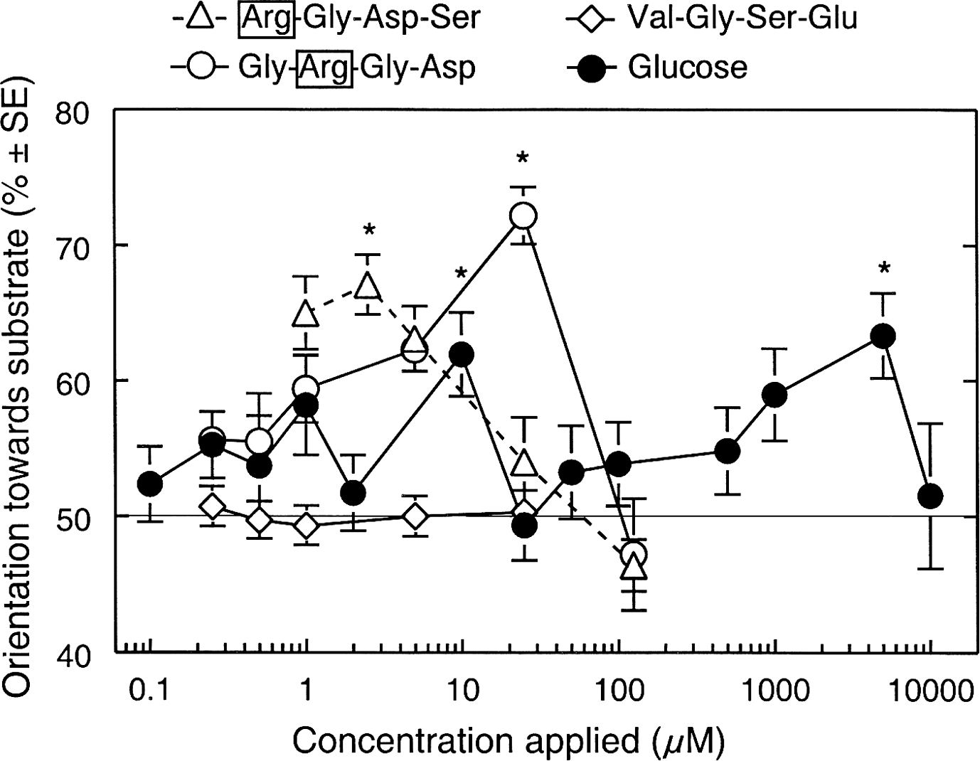
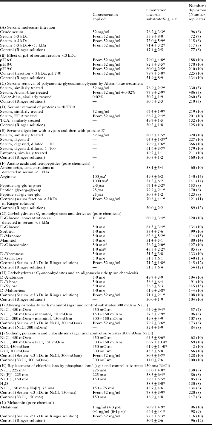
The cercariae penetrated the agar in the W-chambers, and in agar with fish Ringer or other electrolyte solutions, they survived for more than 12 h and showed significant chemotactic orientation. Fish serum was attractive and the stimulating compounds were contained in a fraction with molecular sizes below 3 kDa (Table 2A). This fraction was attractive independent of its pH (Table 2B). The stimuli could not be precipitated with Alcian blue like the stimuli for D. spathaceum cercarial penetration behaviour (Haas et al. Reference Haas, Grabe, Geis, Päch, Stoll, Fuchs, Haberl and Loy2002b) (Table 2C). Also, removal of proteins with TCA had no effect on attractiveness (Table 2D) and protease digestion increased the attraction of serum and also its content of free amino acids (Table 2E, F). However, amino acids were not attractive in concentrations as detected in serum, and arginine, the attractant for S. mansoni and T. ocellata schistosomula (Grabe and Haas, Reference Grabe and Haas2004a), had no effect on the orientation of D. spathaceum larvae at a wide range of concentrations (Table 2F). However, arginine integrated in tetrapeptides attracted the D. spathaceum larvae within narrow concentration ranges. A peptide with terminally positioned arginine attracted at lower concentrations than a peptide with subterminally positioned arginine and a peptide without arginine showed no attraction at all (Table 2F; Fig. 2).
Chemo-orientation of Diplostomum spathaceum diplostomula in the W-chamber. Effect of varying concentration gradients of tetrapeptides and glucose, created by application of different concentrations of the compounds into W-chambers; * significant vs buffered Ringer as control (P<0·001, Tukey multiple t-test); data from 62–295 diplostomula showing chemo-orientation per concentration, 6–10 replicates.

Chemo-orientation of diplostomula in agar towards carp serum, its chemical components and pure chemicals
(The larvae migrated in W-choice-chambers and were exposed to chemical concentration gradients, created by application of defined concentrations of the attractants into W-chambers. The agar and test and control substrates contained Ringer solutionFootnote a unless otherwise noted (I, J, K).)

* P<0·01 vs control (Tukey multiple t-test).
a Fish Ringer according to Haas (Reference Haas1974b), mEq/l: Na+ 120, K+ 4, Ca2+ 5, Mg2+ 4, Cl− 118, SO42− 5, HCO3− 10.
b Total number of diplostomula leaving the central arm of the W-chamber (and number of replicates) per tested substrate.
c The content of amino acids in serum increased after digestion from 11·9 to 92·7 mm and the osmolarity from 269 to 291 mOsm/l.
d Attraction significantly different from that of serum similarly treated, P<0·001 (Tukey multiple t-test).
e Concentration of amino acids, as detected in serum fraction <3 kDa (μm): asp 136, glu 151, asn 128, ser 811, his 412, gly 840, thr 325, arg 127, ala 610, tau 405, tyr 176, val 254, met 62, ile 196, phe 206, leu 203, orn 62, lys 275.
f No significant attraction at other concentrations as well (0·1, 0·25, 0·5, 1·0, 2·0, 10, 25, 50, 500, 5000, 10 000 μm).
g Similar repellent effect at other concentrations (0·01, 0·1, 1·0, 10, 50, 75, 100 μm).
h Na2HPO4 and NaH2PO4, adjusted to the desired Na+-concentration and pH 7·0.
i Weak (not statistically significant) effect at concentrations of 10 and 0·01 ng/ml, no effect at higher concentrations (100, 500, 1000 ng/ml).
Another attractive compound of serum was D-glucose, which stimulated the larvae in a concentration as detected in the serum fraction and in 2 other concentration ranges (Table 2G, Fig. 2). The response to D-glucose was relatively specific, as among other carbohydrates tested, only D-mannose and D-maltotriose were attractive and D-glucosamine had a repellent effect at various concentrations (Table 2G, H).
A concentration gradient of NaCl attracted the larvae (Table 2I). This was not due to the increasing osmolarity of the NaCl gradient as a similar increase of osmolarity with mannitol was not attractive (Table 2I). The chemical stimulus in the increasing gradients of electrolyte solutions was the Cl ion, as a replacement of the Na ions by K ions had no effect (KCl solutions were as attractive as NaCl solutions; Table 2J) and as electrolyte solutions were not attractive, when Cl-ions were replaced by PO4-ions (Table 2K). The cercariae avoided substrates without Cl-ions such as water or sodium phosphate (Table 2K).
The compound to which the diplostomula were most sensitive was melatonin. It was attractive at a very low concentration, but only in a narrow concentration range (Table 2L). The concentration of melatonin in fish retina is higher than in the surrounding tissues and this could lead to an accumulation of the parasites in the retina. However, the parasites leave the retina and migrate towards the eye lens, although they are strongly attracted by melatonin. A cue for their orientation towards the lens could be the direction of light radiation within the eyes. Therefore, we investigated whether the migrating diplostomula shifted to a positive photo-orientation when stimulated with melatonin. However, melatonin failed to stimulate orientation towards light at concentrations of 1 and 100 ng/ml and light intensities of 30 000 and 8000 lux (P vs control 0·7–1·0; 343–706 responding diplostomula; 13–16 replicates).
DISCUSSION
The navigation of D. spathaceum diplostomula along blood vessels and through tissues towards the eye and then to the lens is very complex and our in vivo studies focus only on orientation within the tail fin towards veins and in an anterior direction. Our in vitro experiments revealed information on the chemo- and photo-orientation of the diplostomula, but we can only speculate on the role of these responses during the navigation process. We suggest that the behaviour of migrating diplostomula consists of discrete units, and that they, as in their host-finding behaviour, respond to different host cues in each of these units. The observed responses may have functions in 4 behavioural phases during migration within the fish host, as summarized in Fig. 3 and as presented below.
Hypothetical model of some of the behavioural phases of Diplostomum spathaceum diplostomula when migrating in the fish host (boxes) and supposed cues used for navigation. Diplostomula show chemo-orientation in concentration gradients of glucose and other carbohydrates, peptides with arginine residues, Cl-ions, and melatonin, as well as negative photo-orientation; +, positive, −, negative orientation along increasing concentration gradients or direction of light radiation.

(1) Migration towards deeper skin layers and avoidance of perforation of the covering skin surface layers
To avoid accidental perforation of the skin surface layers is important for the survival of the migrating diplostomula as discussed also for schistosomula of S. mansoni and T. ocellata (Grabe and Haas, Reference Grabe and Haas2004a). In fact, the diplostomula successfully avoided orientation back to the skin surface when the fin tissues were still living (living and decapitated fish). However, when the cells of the fins were killed, many diplostomula left the tissues. So, the cues used to detect the direction towards skin surface or deeper skin layers appear weakened in dead tissue but they still exist. As the diplostomula in vitro migrate along increasing concentration gradients of Cl− and avoid decreasing gradients, Cl− gradients could be the landmark that enables their remaining in the skin. However, little is known about the osmotic conditions and the distribution of electrolytes within fin tissues. In freshwater fish, NaCl uptake seems to be achieved by Na+/H+ and Cl−/HCO3 exchangers, mainly in cells of gills, but the further transport of Na+ and Cl− in the skin tissues is not understood and the published data on concentrations of these ions in fish tissues are controversial, even in studies of gill tissues (reviewed by Marshall, Reference Marshall2002; Perry et al. Reference Perry, Shahsavarani, Georgalis, Bayaa, Furimsky and Thomas2003; Kirschner, Reference Kirschner2004; Evans et al. Reference Evans, Piermarini and Choe2005). There is also a high influx of water (50% of the body water per h), which is compensated by excretion of dilute urine. The water influx occurs presumably mainly across the gills, but it could also occur in the skin. So, there might be concentration gradients of Cl− from the body surface towards deeper tissues, which could be used by the diplostomula as directing cues.
Another strategy of the migrating diplostomula could be to follow an increasing concentration gradient of glucose from the skin surface to deeper tissues. Such a concentration gradient exists in mammals and seems to be used by migrating schistosomula of S. mansoni and T. ocellata (Grabe and Haas, Reference Grabe and Haas2004a). As fish show a persistent hyperglycaemia (Moon, Reference Moon2001; Hemre et al. Reference Hemre, Mommsen and Krogdahl2002), similar conditions can also be expected in fish skin.
An orientation back to the skin surface might also be prevented by the repellent effect of glucosamine on migrating diplostomula. This avoidance behaviour seems to be important for the parasites, as they respond very sensitively to gradients that establish at concentrations as low as 1 nm. It might reflect an adaptation to migration in a fish host, as the schistosomula of S. mansoni and T. ocellata are not repelled by glucosamine (Grabe and Haas, Reference Grabe and Haas2004a). Fish skin contains glycosaminoglycans (GAGs) in surface mucus and epidermal mucus-secreting cells. Degradation products of GAGs from fish skin surface mucus seem to serve as very effective kairomones for shrimps, crabs, and snails (Forward and Rittschof, Reference Forward and Rittschof1999, Reference Forward and Rittschof2000; Rahman et al. Reference Rahman, Forward and Rittschof2000). Disaccharides containing sulfated and acetylated amines were very effective signals among the fish mucus degradation products, but glucosamine was also recognized, at least by nauplius larvae of the brine shrimp (Artemia franciscana) (Forward and Rittschof, Reference Forward and Rittschof1999). Such amino sugars may occur as degradation products of mucus and during synthesis of the polysaccharides in epidermal mucus secreting cells (Lumsden and Ferguson, Reference Lumsden and Ferguson1994); they may then exert their repellent effect on diplostomula. The hypothesis that amino sugars in skin epithelial cells guide diplostomula back to deeper layers requires further study, in particular on the effect of the more abundant galactosamine (Nakagawa et al. Reference Nakagawa, Asakawa and Enomoto1988), as well as sulfated and acetylated amino sugars.
Glucosamine and other amino sugars are major compounds of extracellular matrix. So, the sensitive turning away from these sugars could enable the diplostomula to avoid ineffective migration attempts across extracellular matrix, such as cartilage and basal laminas of epithelial layers. Data on the repellent effect of other amino sugars specific for certain tissues could show which tissue types are avoided during the migration. The D. spathaceum diplostomula are (in contrast to schistosomes) also attracted by carbohydrates other than glucose; they respond to D-mannose and to the trisaccharide maltotriose (and D-glucosamine has a repellent effect). Thus, this fish parasite uses more carbohydrate signals during migration than the parasites of terrestrial vertebrates. The orientation of the diplostomula towards deeper tissue layers could also be enhanced by their negative photo-orientation (Grabe and Haas, Reference Grabe and Haas2004b).
(2) Orientation in direction towards the head of the fish
In the tail fin of fish, the diplostomula navigate precisely in the cranial direction. As they enter veins and use them for further migration in the fin, they could use the direction of the blood flow for navigation. However, their orientation is undisturbed when there is no blood flow, and they migrate in a cranial direction in isolated pieces of the fin, even when the cells in the tissues are dead. The parasites seem to follow a local chemical or electrical gradient the nature of which remains to be explained. It is hypothesized that orientation along increasing concentration gradients of Cl− could be involved in this step of site-finding as well. This assumes that osmolarity (which is mainly determined by NaCl concentration) decreases in a distal direction. However, there seems to be no information on the detailed osmotic conditions in fins, although many studies have dealt with osmoregulation of freshwater fish (reviewed by Karnaky, Reference Karnaky and Evans1998).
(3) Localization of blood vessels
The diplostomula of D. spathaceum enter veins during their further migration. Schistosomula of S. mansoni and T. ocellata also enter veins of their hosts, and have been hypothesized to locate the vessels by following increasing concentration gradients of glucose and arginine (Haas et al. Reference Haas, Grabe, Geis, Päch, Stoll, Fuchs, Haberl and Loy2002a; Grabe and Haas, Reference Grabe and Haas2004a); D. spathaceum diplostomula may use a similar strategy. They are attracted by glucose (Fig. 2) in gradients that establish at concentrations similar to those in fish plasma, i.e., 2–10 mm (Groff and Zinkl, Reference Groff and Zinkl1999; Blasco et al. Reference Blasco, Marimon, Viaplana and Fernandez-Borras2001; Hemre et al. Reference Hemre, Mommsen and Krogdahl2002). This response may guide them to the blood vessels of fish, which are generally hyperglycaemic (Moon, Reference Moon2001). Moreover, the parasites respond to a very flat concentration gradient, which establishes at approximately 10 μm. In addition, schistosomula of S. mansoni respond with a second peak of sensitivity to gradients that establish at 10 μm glucose, suggesting different biological functions of these 2 peaks of sensitivity to glucose gradients. However, as discussed for S. mansoni schistosomula (Grabe and Haas, Reference Grabe and Haas2004a), hypotheses on the adaptive benefits of the responses remain unresolved, as we have no exact information on the nature of glucose concentration gradients the parasites encounter in the host tissues or in our W-chambers. There may be gradients that are useful for navigation and others that should be avoided. For example, the parasites enter mainly veins, which have a lower glucose content than arteries, but probably higher concentrations than the tissues the parasites encounter during migration. However, the responses might also just result from the activity of 2 different glucose receptors. The response to 2 or 3 different concentration gradients is not a general characteristic of blood-vessel invading digeneans, as the schistosomula of T. ocellata respond only to concentration gradients that establish at approximately 1 μm glucose (Grabe and Haas, Reference Grabe and Haas2004a).
In contrast to the schistosomula of S. mansoni and T. ocellata whose response to arginine may enable them to find venules (Grabe and Haas, Reference Grabe and Haas2004a) the diplostomula of D. spathaceum do not respond to arginine. However, like schistosomula, they are attracted by tetrapeptides containing arginine. Further work should demonstrate whether the diplostomula respond similarly as S. mansoni schistosomula to arginine-containing peptides, which occur in endothelial cells, i.e. bradykinin and its fragments, as well as fibronectin. The responses of digenean larvae to peptides with terminal arginine residues is consistent with a variety of other invertebrates that use such molecules as pheromones and kairomones and supports the suggestion that such peptides generally might serve as information molecules in various organisms (Rittschof, Reference Rittschof1993; Rittschof and Cohen, Reference Rittschof and Cohen2004). However, not all blood-vessel invading helminths respond to the same cues. Human hookworms responded differently in our W-chamber bioassay. Ancylostoma duodenale was attracted by human serum, but neither glucose nor arginine were the attractants and Necator americanus showed no chemo-orientation at all (Haas et al. Reference Haas, Haberl, Syafruddin Idris, Kallert, Kersten and Stiegeler2005).
(4) Recognition of the eye
Ferguson (Reference Ferguson1943) concluded from an elaborate series of surgical experiments that the eye tissues, even without a lens, provide a stimulus that attracts the diplostomula. Since fish synthesize melatonin in the retina (Tan et al. Reference Tan, Manchester, Hardeland, Lopez-Burillo, Mayo, Sainz and Reiter2003; Falcon et al. Reference Falcon, Gothilf, Coon, Boeuf and Klein2003), we examined the effect of melatonin on migrating diplostomula. The parasites were attracted by melatonin at gradients, which established at concentrations of 0·1–1·0 ng/ml (0·4–4·3 pm) melatonin. This concentration range occurs also in fish retina, i.e., in Carassius auratus it ranged from 0·5 to 2·5 ng per eye (Iigo et al. Reference Iigo, Furukawa, Hattori, Ohtani-Kaneko, Hara, Suzuki, Tabata and Aida1997) and in Oncorhynchus mykiss from 0·8 to 3·4 ng per eye (Zaunreiter et al. Reference Zaunreiter, Brandstätter and Goldschmid1998). As the melatonin concentration in the fish retina is higher than in plasma, e.g. 9–100 times higher in C. auratus (Iigo et al. Reference Iigo, Furukawa, Hattori, Ohtani-Kaneko, Hara, Suzuki, Tabata and Aida1997), it may be a signal that dipostomula respond to upon contact with the retina. Whether they can also localize the retina following a concentration gradient of melatonin is difficult to evaluate, as ocular melatonin acts in an autocrine or paracrine manner and seems to accumulate in the eye. However, if melatonin reaches the bloodstream (Tan et al. Reference Tan, Manchester, Hardeland, Lopez-Burillo, Mayo, Sainz and Reiter2003), diplostomula could potentially follow the melatonin signal to locate the retina.
Eventually, diplostomula leave the retina and enter the lens. Then, the response to melatonin must be negated by reactions to other cues. We suggested that a shift to positive photo-orientation could guide the parasites towards the lens, but the diplostomula did not migrate towards light after contact with melatonin in physiological concentrations. Future experiments will hopefully indicate whether the diplostomula follow only increasing concentration gradients of melatonin and do not return in decreasing gradients.
Our findings are only a first step in understanding how diplostomula find their way within the fish host. We still have no information on the behaviour patterns of the parasites in the fish body and in the gills, the main penetration sites of the cercariae. Much experimental work must be performed before we are able to understand the navigation of the diplostomula (and parasites in general) over longer distances. The migration of diplostomula in their fish hosts is a suitable model for such studies, as in vitro experiments and manipulations of the host and its organs can easily be performed.
When we consider the orientation behaviour of the migrating larvae of D. spathaceum, S. mansoni, T. ocellata and the human hookworms (Grabe and Haas, Reference Grabe and Haas2004a; Haas et al. Reference Haas, Haberl, Syafruddin Idris, Kallert, Kersten and Stiegeler2005), it is obvious that each of the species has its unique strategy. The species respond to different types of host signals, and when they use similar signal molecules they react to very different concentration gradients, position of arginine in peptides, etc. This diversity of behaviour also occurs when the larvae follow similar routes, for example when entering blood vessels. It seems that the navigation of parasitic worms within hosts is characterized by a similar high diversity of strategies, as seen in their host-finding behaviour (Haas, Reference Haas1994, Reference Haas2003; Haas and Haberl, Reference Haas, Haberl, Fried and Graczyk1997; Sukhdeo and Sukhdeo, Reference Sukhdeo and Sukhdeo2004), and much information will be necessary to understand the adaptive benefits of the individual behavioural responses.
This work was supported by the Deutsche Forschungsgemeinschaft. We thank Renate Landgraf and Christina Loy for their expert technical work and the parasitology group of the University of Erlangen-Nürnberg for snail-collection.