Research aimed to prevent pediatric-onset mood disorders has found that a family history of mood disorders is among the most important risk factors for developing mood disorders. Compared to the general population, offspring of parents with bipolar disorder (BD) and major depressive disorder (MDD) are at increased risk for psychopathology, and are particularly at high risk for mood disorders (Lapalme, Hodgins, & LaRoche, Reference Lapalme, Hodgins and LaRoche1997; Singh, DelBello, Stanford, et al., Reference Singh, DelBello, Soutullo, Stanford, McDonough-Ryan and Strakowski2007). Nevertheless, many youth with a familial risk for BD and MDD do not develop any psychiatric disorders and, instead, go on to lead adaptive and highly functional lives, suggesting there may be important mechanisms that underlie or contribute to resilience from mood disorders (Frangou, Reference Frangou2009; Silk et al., Reference Silk, Vanderbilt-Adriance, Shaw, Forbes, Whalen, Ryan and Dahl2007). Developmental studies have well established that there may be multiple pathways leading to variable outcomes, including the eventual onset of mood and other psychiatric disorders (Cicchetti & Rogosch, Reference Cicchetti and Rogosch1999).
Resilience, the process of positively adapting in spite of adversity, traumatic events, or a negative predisposition (Luthar, Cicchetti, & Becker, Reference Luthar, Cicchetti and Becker2000), can also be defined based on adaptive brain features in the absence of psychiatric symptoms in high-risk youth (Frangou, Reference Frangou2011). Although neural studies of resilience to mood disorders are limited, previous studies have begun to delineate potential neural markers of resilience. Frangou (Reference Frangou2011) found increased connectivity between the ventral and dorsal prefrontal cortex (PFC) in unaffected adult relatives of patients with BD during a cognitive control task, and Fischer, Comacho, Ho, Whitfield-Gabrieli, and Gotlib (Reference Fischer, Camacho, Ho, Whitfield-Gabrieli and Gotlib2018) showed that female adolescents who are resilient to MDD demonstrated compensatory functional connectivity between emotion regulatory regions including the amygdala, orbitofrontal cortex, and dorsolateral PFC during rest. A recent study also identified differences in neural activation among youth and young adults at high and low risk compared to those with BD, demonstrating activation differences during a face emotion labeling task in multiple regions, including the precuneus, superior and inferior frontal gyri, insula, supramarginal gyrus, and fusiform gyrus (Wiggins et al., Reference Wiggins, Brotman, Adleman, Kim, Wambach, Reynolds and Leibenluft2017). Collectively, these studies suggest that functionally specialized neural networks may segregate along resilient versus psychopathological characteristics.
Studies of child offspring of adults with mood disorders are ideally suited to investigate neural substrates of emotion dysregulation. Previous studies have shown that problems with emotion regulation may confer vulnerability to psychopathology (Beauchaine, Reference Beauchaine2015) possibly from environmental as well as heritable factors. For example, youth who spend formative years in families where maladaptive management of emotions is demonstrated are at increased risk for developing psychiatric disorders broadly (Silk et al., Reference Silk, Vanderbilt-Adriance, Shaw, Forbes, Whalen, Ryan and Dahl2007), and mood disorders specifically (Singh, DelBello, Stanford, et al., Reference Singh, DelBello, Soutullo, Stanford, McDonough-Ryan and Strakowski2007). A hallmark feature of mood disorders is a bias toward the negative interpretation of emotional stimuli and emotion dysregulation (Gotlib & Joormann, Reference Gotlib and Joormann2010; Phillips & Swartz, Reference Phillips and Swartz2014), which could in part be learned from caregivers who may struggle with their own emotion regulation.
In addition, neural vulnerability clearly contributes to emotion dysregulation. Prior studies have suggested that emotion dysregulation in the context of psychopathology originates from dysfunction in neural systems that support emotion processing and executive control (Leshin & Lindquist, Reference Leshin and Lindquist2018). In some cases, this neural system dysfunction precedes symptom onset in youth at familial risk for mood disorders (Singh, Chang, et al., Reference Singh, Chang, Kelley, Saggar, Reiss and Gotlib2014; Singh, Kelley, et al., Reference Singh, Kelley, Howe, Reiss, Gotlib and Chang2014). A coordinate-based meta-analysis of neural responses to facial expressions in healthy samples showed consistent activation to emotional faces in the fusiform gyrus, lingual gyrus, cerebellum, inferior parietal lobe (IPL), medial frontal gyrus, anterior cingulate cortex, amygdala, and insula (Fusar-Poli et al., Reference Fusar-Poli, Placentino, Carletti, Landi, Allen, Surguladze and Politi2009). Youth with and at risk for BD and MDD show aberrant function in many of these regions, including the ventromedial PFC and dorsolateral PFC, visual (lingual and fusiform gyri), limbic (amygdala and insula), and striatal (putamen and nucleus accumbens) regions in response to fearful and happy facial expressions (Chang et al., Reference Chang, Garrett, Kelley, Howe, Sanders, Acquaye and Reiss2017; Garrett et al., Reference Garrett, Reiss, Howe, Kelley, Singh, Adleman and Chang2012; Hafeman et al., Reference Hafeman, Bebko, Bertocci, Fournier, Bonar, Perlman and Phillips2014; Hall et al., Reference Hall, Klimes-Dougan, Hunt, Thomas, Houri, Noack and Cullen2014; Olsavsky et al., Reference Olsavsky, Brotman, Rutenberg, Muhrer, Deveney, Fromm and Leibenluft2012; Pavuluri, Passarotti, Harral, & Sweeney, Reference Pavuluri, Passarotti, Harral and Sweeney2009). Further, anomalous functional connectivity between these regions during emotion processing has been demonstrated in youth with and at risk for BD and MDD (Chang et al., Reference Chang, Garrett, Kelley, Howe, Sanders, Acquaye and Reiss2017; Ho et al., Reference Ho, Connolly, Henje Blom, LeWinn, Strigo, Paulus and Yang2015; Kerestes, Davey, Stephanou, Whittle, & Harrison, Reference Kerestes, Davey, Stephanou, Whittle and Harrison2014; Ladouceur et al., Reference Ladouceur, Farchione, Diwadkar, Pruitt, Radwan, Axelson and Phillips2011; Manelis et al., Reference Manelis, Ladouceur, Graur, Monk, Bonar, Hickey and Phillips2015; Rich et al., Reference Rich, Fromm, Berghorst, Dickstein, Brotman, Pine and Leibenluft2008; Zhu et al., Reference Zhu, Wang, Xiao, Liao, Zhong, Wang and Yao2012). These studies suggest that emotion dysregulation is likely principally due to deficient top-down control of limbic regions by executive control regions (Leshin & Lindquist, Reference Leshin and Lindquist2018).
Collectively, these findings suggest potential trait- and disorder-related neural markers of aberrant emotion processing in youth, and neural circuits underlying emotion processing in pediatric mood disorders. Further, these studies suggest that functional abnormalities in neural systems supporting emotion processing may underline the pathophysiology of youth-onset mood disorders. However, most studies of high-risk individuals in the literature are cross-sectional and have included youth who have already developed symptoms (Kerestes et al., Reference Kerestes, Segreti, Pan, Phillips, Birmaher, Brent and Ladouceur2016; Monk et al., Reference Monk, Klein, Telzer, Schroth, Mannuzza, Moulton and Ernst2008; Olsavsky et al., Reference Olsavsky, Brotman, Rutenberg, Muhrer, Deveney, Fromm and Leibenluft2012), making it difficult to evaluate mechanisms of resilience, and to disentangle the causes and effects of psychopathology on emotion processing and emotion dysregulation. Moreover, studies that examined resilience did not investigate functional connectivity during emotion processing and its relation to the emergence of mood symptoms over time. This is an important impetus for elucidating reliable biological factors that can distinguish risk from resilience at the earliest stages of developing psychiatric disorders in high-risk youth who can serve as their own controls with prospective evaluation.
Thus, the aim of this study was to use a multiple levels of analysis approach (Cicchetti & Blender, Reference Cicchetti and Blender2006) to study youth at high risk for mood disorders to determine whether brain function at baseline predicts subsequent resilience versus mood and other psychiatric symptom development 3 years later. At baseline, healthy youth at low and high familial risk for BD or MDD completed functional magnetic resonance imaging (fMRI) scans during an implicit emotion processing task. Three years later, we compared baseline neural activation and connectivity between three groups: low-risk healthy controls (HCL) and high-risk youth who developed (CVT) or did not develop psychopathology (RES). By examining healthy offspring of parents with BD and MDD, we aimed to draw inferences about potential neural markers that differentiate between risk and resilience in this already at-risk group but starting from a stage of health. Based on prior studies of emotion processing in high-risk youth, we hypothesized that youth in the RES group would exhibit functional abnormalities and connectivity of visual, frontal, and limbic regions that reflect adaptive emotion processing strategies compared to CVT and HCL groups. In addition, to confirm that the development of psychiatric symptoms is associated with significant global functional impairment for youth, we also hypothesized that neural differences in emotion processing within the three groups would be correlated with differences in strengths and difficulties, and differences in global functioning.
Method
Participants
The participants in this study were drawn from a longitudinal study of youth with and without a familial risk for BD and MDD. Eighty-four participants who had completed an implicit emotion face processing task at baseline and had been followed clinically over the course of 2.92 ± 1.13 years were included in this analysis. This group included 57 participants with a familial risk for BD or MDD recruited from families receiving treatment at an academic mood disorders program for youth and adults, and from the surrounding community. High-risk youth had at least one parent diagnosed with BD or MDD. In the context of staging models of risk for mood disorders, these high-risk youth were recruited at what would be considered Stage 0, or very early along the risk continuum (Duffy, Vandeleur, Heffer, & Preisig, Reference Duffy, Vandeleur, Heffer and Preisig2017; Raouna, Osam, & MacBeth, Reference Raouna, Osam and MacBeth2018). In addition, 27 healthy youth with no personal or family history of psychopathology (HCL) were recruited from the community by advertisements. Exclusion criteria for all groups at baseline included (a) currently taking medication or receiving psychotherapy for any psychiatric disorder; (b) having a current or lifetime diagnosis of any psychiatric or substance use disorders, or neurological disorders; (c) MRI contraindications including orthodontic braces; or (d) intellectual quotient (IQ) <80 assessed by the Weschler Abbreviated Scale of Intelligence (Weschler, 1999). The institutional review board approved the study, and written informed assent and consent were obtained from all youth and parents, respectively.
Clinical assessment and functioning
At baseline and follow-up, all participants were assessed for psychiatric symptoms by trained interviewers masked to family history status, to evaluate for psychiatric health at baseline and to identify psychiatric diagnoses at longitudinal follow-up. Structured diagnostic interviews were administered separately to participants and their parents (regarding the participants) using the mood sections of the Washington University in St. Louis Kiddie Schedule for Affective Disorders and Schizophrenia (Geller et al., Reference Geller, Zimerman, Williams, Bolhofner, Craney, DelBello and Soutullo2001), and the Kiddie Schedule for Affective Disorders and Schizophrenia for School-Age Children—Present and Lifetime version (Kaufman et al., Reference Kaufman, Birmaher, Brent, Rao, Flynn, Moreci and Ryan1997). The Structural Clinical Interview for the DSM-IV (Williams et al., Reference Williams, Gibbon, First, Spitzer, Davies, Borus and Wittchen1992) was also administered to parents to assess parental family history of mood and other psychiatric disorders. All diagnoses were reviewed and confirmed by a board-certified child and adolescent psychiatrist (M.K.S.).
To ensure that high- and low-risk groups did not differ in levels of mania or depression in the absence of any psychiatric diagnoses, all youth were interviewed using the Children's Depressive Rating Scale—Revised (Poznanski et al., Reference Poznanski, Grossman, Buchsbaum, Banegas, Freeman and Gibbons1984) to confirm low baseline depression symptom severity, and the Young Mania Rating Scale (Young, Biggs, Ziegler, & Meyer, Reference Young, Biggs, Ziegler and Meyer1978) to confirm low baseline mania symptom severity. Finally, the Children's Global Assessment Scale (CGAS; Shaffer et al., Reference Shaffer, Gould, Brasic, Ambrosini, Fisher, Bird and Aluwahlia1983) was administered to assess for overall global functioning.
Parents of participants completed the Strengths and Difficulties Questionnaire (SDQ; Goodman, Reference Goodman1997) to assess their child's psychosocial strengths and psychopathological difficulties. This measure contains scales for behavioral strengths assessed through prosocial behaviors, and delineates behavioral difficulties into emotional problems, peer problems, conduct problems, and hyperactivity subscales. An internalizing score was generated by summing the emotional and peer problems scales, and an externalizing score was generated by summing the conduct and hyperactivity scales. A total difficulties score was generated by summing scores from all the scales except the prosocial scale, which is the primary resilience metric for this scale. At follow-up, some of the assessments were conducted over the phone for participant convenience.
Implicit emotion perception task
In the scanner, participants viewed photographs of young adults with happy, fearful, and calm facial expressions, and scrambled images presented in a block design. Happy and fearful faces were used to probe emotion processing, and calm faces were used as the comparison condition, to subtract activation associated with face processing. Calm faces were considered a better comparison condition than neutral faces because previous studies have suggested that neutral faces activate emotion processing regions such as the amygdala in youth with significant emotion dysregulation (Brotman et al., Reference Brotman, Rich, Guyer, Lunsford, Horsey, Reising and Leibenluft2010). All faces were taken from the MacArthur facial expressions set (“NimStim”; http://www.macbrain.org/resources.htm). Each block contained eight faces of the same expression, and each face was presented for 3 s. Blocks of each expression were alternated throughout the task, and overall, four blocks of each facial expression were shown. Participants were instructed to identify the gender of the faces presented, using a button box to push Button 1 for female faces, Button 2 for male faces, and alternating Buttons 1 and 2 during the scrambled pictures blocks. The entire task lasted 6 min and 24 s. The task was presented using ePrime software (www.pstnet.com), which also collected reaction time and accuracy of behavioral responses.
Neuroimaging data acquisition
Before the MRI scan, participants were familiarized with the scanning environment and trained to minimize head motion using a mock scanner. MRI images were acquired on a 3 Tesla GE Signa scanner (General Electric Co., Milwaukee, WI) using an 8-channel head coil. High resolution anatomical images were acquired to optimize normalization of functional images to a standard template (3D FSPGR pulse sequence; repetition time = 8.5 ms, echo time = 3.32 ms, inversion time = 400 ms, flip angle = 15 degrees, field of view = 25.6 cm, 186 slices in the axial plane, resolution = 1 mm3. Functional images were collected using a spiral in-out pulse sequence (Glover & Law, Reference Glover and Law2001) with the following parameters: repetition time = 2000 ms, echo time = 30 ms, flip angle = 80 degrees, field of view = 22 cm, number of slices = 30 slices in the axial plane, and slice thickness = 4 mm with a gap of 1 mm. A high-order shim was used to improve field homogeneity.
Neuroimaging preprocessing steps
Preprocessing in FSL's FEAT included slice-timing correction, motion correction with MCFLIRT, brain extraction, smoothing with a Gaussian filter (5 mm full width at half-maximum), and volume registration to MNI152 standard space. Subjects were excluded due to motion if after motion correction their absolute or relative mean displacements exceeded 2 mm or more than one-third of volumes had values for spatial standard deviation of successive difference images (DVARS) exceeding the threshold of the 75th percentile plus 1.5 times the interquartile range, as calculated by the fsl_motion_outliers algorithm. Individual subjects’ first-level analyses were performed using FSL's FEAT.
Whole-brain fMRI data analysis
Functional data were processed using FSL (Jenkinson, Beckmann, Behrens, Woolrich, & Smith, Reference Jenkinson, Beckmann, Behrens, Woolrich and Smith2012). For individual subject statistical maps, the time series of each voxel was modeled with a general linear model. Happy, fear, and calm conditions were modeled as regressors of interests. Scrambled blocks and 24 motion correction parameters were included as regressors of noninterest. We also included time points that exceeded the DVARS motion threshold calculated by fsl_motion_outliers (Jenkinson et al., Reference Jenkinson, Beckmann, Behrens, Woolrich and Smith2012) within our general linear model to deweight the effects of these time points in our analyses. Because our primary focus was to examine neural markers of emotion processing associated with resilience to mood disorders, we conducted a priori comparisons using planned t tests to compare resilient to converted and resilient to healthy control groups. Group comparisons were conducted with voxelwise whole-brain t tests using FSL's FLAME (FMRIB's analysis of mixed effects) correcting for mean-centered age and sex for the following contrasts: happy > calm and fear > calm. Statistically significant clusters were identified by thresholding Z statistic images (Z > 2.3) with a cluster probability of p < .05, correcting for multiple comparisons (Worsley, Reference Worsley, Jezzard, Matthews and Smith2001). We extracted mean parameter estimates from significant clusters using fslmeants to determine the direction of activation differences.
Psychophysiological interaction (PPI) analyses
PPI analyses were conducted in FSL's FEAT to examine group differences in context-dependent functional connectivity. Each PPI analysis was conducted using seed regions of interest (ROI) based on significant clusters found in the whole-brain analyses. The ROIs were transformed into individual subject space, and mean temporally filtered time series were extracted. The interaction regressor was computed as the product of the mean extracted time series for the seed ROIs and our contrasts of interest: happy > calm and fear > calm. We also included our original task regressors, and 24 motion correction parameters as regressors of noninterest. Similar to our whole-brain analyses, we included time points that exceeded the DVARS motion threshold calculated to remove the effects of these time points in our analyses. Group comparisons were conducted using FSL's FLAME, correcting for mean-centered age at scan and sex. Significant group differences were identified using a threshold of Z > 2.3 with a corrected cluster significant threshold of p < .05 (Worsley, Reference Worsley, Jezzard, Matthews and Smith2001). Mean parameter estimates from significant PPI clusters were extracted using fslmeants to determine the direction of group differences.
Statistical analyses of clinical and behavioral data
All statistical analyses were conducted using SPSS (version 25) and R. We conducted one-way analyses of variance to test for a main effect of group in clinical and behavioral characteristics. We also conducted repeated measures analyses of covariance with baseline age and sex as covariates to test for group differences in longitudinal changes in SDQ and CGAS scores, followed by post hoc t tests to determine which groups significantly differed from each other. Paired t tests were used to test for within-group differences between baseline and follow-up scores on the SDQ and CGAS. In addition, we also computed Pearson's correlations between parameter estimates extracted from significant clusters from our neuroimaging analyses with change in SDQ and change in CGAS scores within the CVT and RES groups and across all groups combined. Thresholds for significance were corrected for multiple correlations using Bonferroni correction (p = .05 / 8 correlations = .006). We also examined correlations between age, neural activation, and connectivity to investigate age-related developmental differences during positive and negative emotional processing. Finally, given the known predominance of internalizing disorders in girls more than boys especially during adolescence (Martel, Reference Martel2013), we conducted exploratory analyses to examine any sex differences during emotion processing, now as a variable of interest, rather than as a covariate as conducted in our primary analyses.
Results
Participant demographics
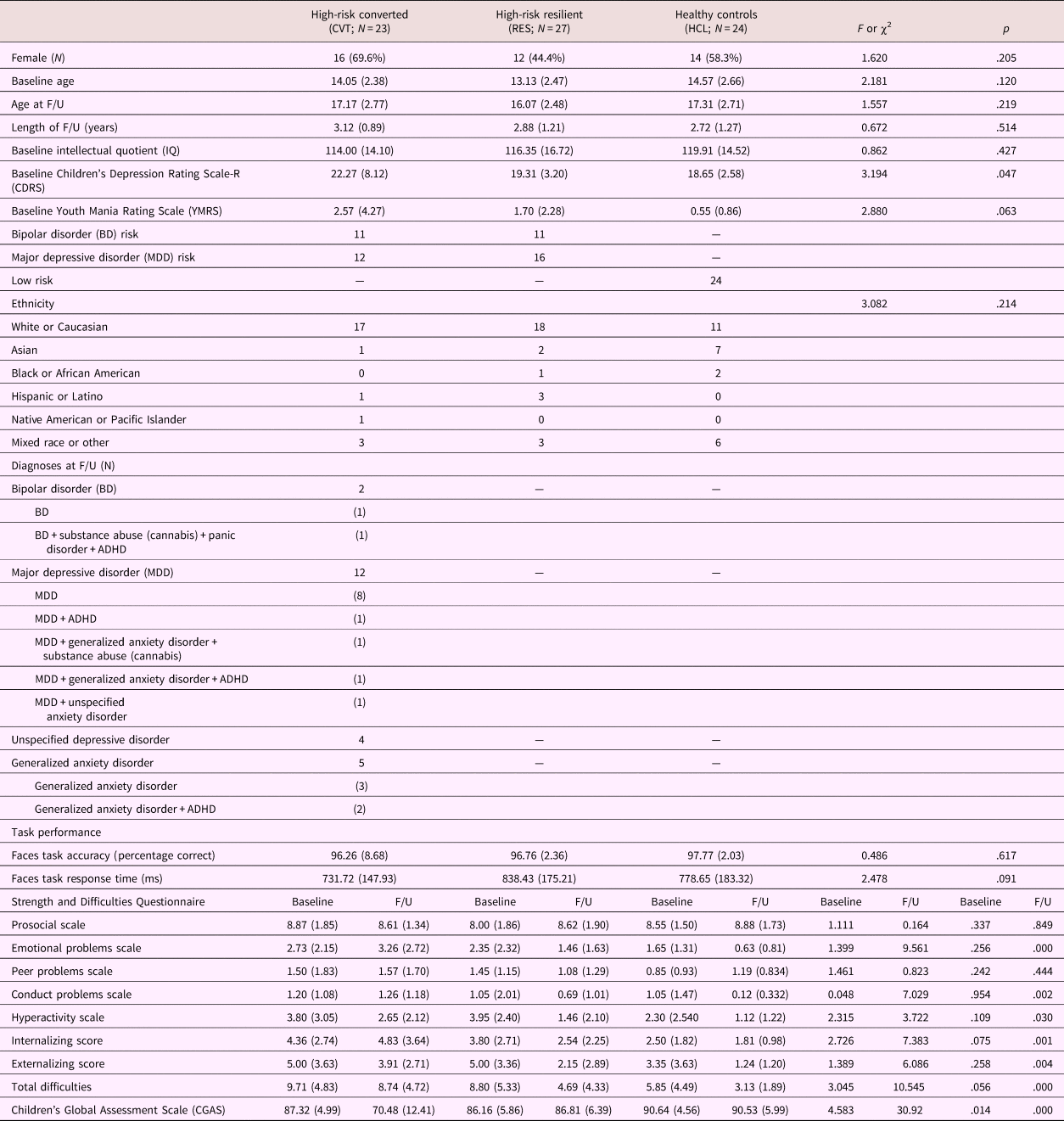
Four participants from the HCL group were excluded for reporting mood symptoms at follow-up, as our investigation was focused on predicting resilience versus symptom development in the high-risk group. Further, the high-risk group was split into two groups: high-risk youth who developed (CVT) psychiatric symptoms and high-risk youth who did not develop psychopathology (RES) at follow-up. Three participants from the RES group and 3 from the HCL group were excluded due to excess head motion in the scanner. Thus, the final sample consisted of 23 CVT, 27 RES, and 24 HCL youth. There were no significant group differences in baseline age, age at follow-up, length of follow-up, IQ, Young Mania Rating Scale, ethnicity, or task accuracy and reaction time. Youth in the CVT group had higher baseline Children's Depressive Rating Scale—Revised scores compared to the RES and HCL groups; however, their scores were not clinically meaningful (below the cutoff of a score of 30). All participant demographic, clinical, and behavioral characteristics, including the frequency of major mood and other psychiatric disorders at follow-up represented in the CVT group, are described in Table 1. Within the CVT group, 2 individuals were later diagnosed with BD (1 comorbid with substance abuse [cannabis], panic disorder, and attention-deficit/hyperactivity disorder [ADHD]), 12 individuals were diagnosed with MDD (1 comorbid with ADHD, 1 comorbid with generalized anxiety disorder [GAD] and substance abuse [cannabis], 1 comorbid with GAD and ADHD, and 1 comorbid with unspecified anxiety disorder), 4 were diagnosed with unspecified depressive disorder, and 5 diagnosed with GAD (2 comorbid with ADHD). No other psychiatric disorders were reported by the CVT group at 3-year follow-up.
Participant characteristics and task performance results

Note: F/U, follow-up. ADHD, attention-deficit/hyperactivity disorder.
Baseline paternal diagnoses
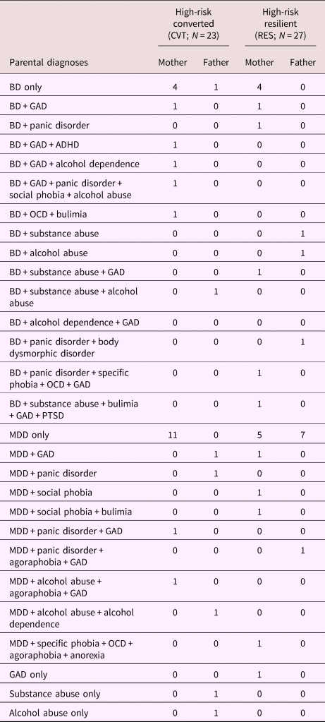
Within the CVT, 9 mothers were diagnosed with BD, 2 fathers were diagnosed with BD, 13 mothers were diagnosed with MDD, 3 fathers were diagnosed with MDD, 1 father was diagnosed with substance abuse, and 1 father was diagnosed with alcohol abuse. Within the RES group, 9 mothers were diagnosed with BD, 3 fathers were diagnosed with BD, 9 mothers were diagnosed with MDD, 8 fathers were diagnosed with MDD, and 1 father was diagnosed with GAD. In both the RES and CVT groups, parents with mood disorders presented with several co-occurring psychiatric disorders. Parental diagnoses and comorbidities are presented in Table 2. It is important to note that both parents had diagnosed mood disorders for 6 subjects. No parent in the HCL group had any current or past diagnosis for psychiatric disorders, as this was an exclusion criterion for HCLs.
Parental diagnoses

Note: BD, bipolar disorder. MDD, major depressive disorder. GAD, generalized anxiety disorder. ADHD, attention-deficit/hyperactivity disorder. OCD, obsessive–compulsive disorder. PTSD, posttraumatic stress disorder.
Task performance
There were no significant differences in task accuracy between CVT, RES, and HCL for happy faces, F (2, 71) = 0.502, p = .607, fearful faces, F (2, 71) = 0.526, p = .593, or calm faces, F (2, 71) = 0.544, p = .583. Similarly, there were no differences in response time among the groups for happy faces, F (2, 71) = 1.356, p = .264, fearful faces, F (2, 71) = 2.270, p = .111, or calm faces, F (2, 71) = 1.900, p = .157.
Assessment of strengths and difficulties, and global functioning
We found significant main effects of group for longitudinal changes in internalizing behaviors, F (2, 40) = 4.181, p = .022, and total difficulties, F (2, 40) = 4.995, p = .012, between baseline and follow-up. Post hoc t tests showed that the CVT group had a greater change in internalizing behaviors and total difficulties compared to the HCL group (ps < .020). There were no other significant between-group differences between baseline and follow-up in regard to scores on the SDQ for emotional problems, conduct problems, hyperactivity, peer problems, prosocial behaviors, and externalizing behaviors (all ps > .05).
There were significant within-group changes on the SDQ between baseline and follow-up. Specifically, the CVT had worsening scores for prosocial behaviors, t (14) = 12.871, p = .000, but improved scores for hyperactivity, t (14) = 4.116, p = .001, and externalizing behaviors, t (14) = 4.076, p = .001, from baseline to follow-up. The RES group had improved scores for emotional problems, t (19) = 2.246, p = .037, total difficulties, t (19) = 3.063, p = .006, hyperactivity, t (19) = 3.932, p = .000, externalizing, t (19) = 3.417, p = .003, and internalizing, t (19) = 2.206, p = .040, behaviors from baseline to follow-up. The RES group also exhibited increased prosocial behaviors, t (18) = 15.720 p = .000, from baseline to follow-up.
There were significant between-group differences in the change between baseline and follow-up global functioning, F (2, 58) = 26.87, p = .000. Post hoc analyses reveal there were significant differences in change in global functioning between CVT and HCL, t (35) = 5.41, p = .000, and between CVT and RES, t (44) = –6.05, p = .000. The CVT group exhibited significantly worse change in global functioning compared to the other groups. There were no significant differences in global functioning between the HCL and RES groups. The CVT group exhibited lower CGAS scores at follow-up compared to baseline, t (21) = 6.716, p = .000, whereas no such change was observed within either the HCL or RES groups.
FMRI whole-brain voxel-wise results
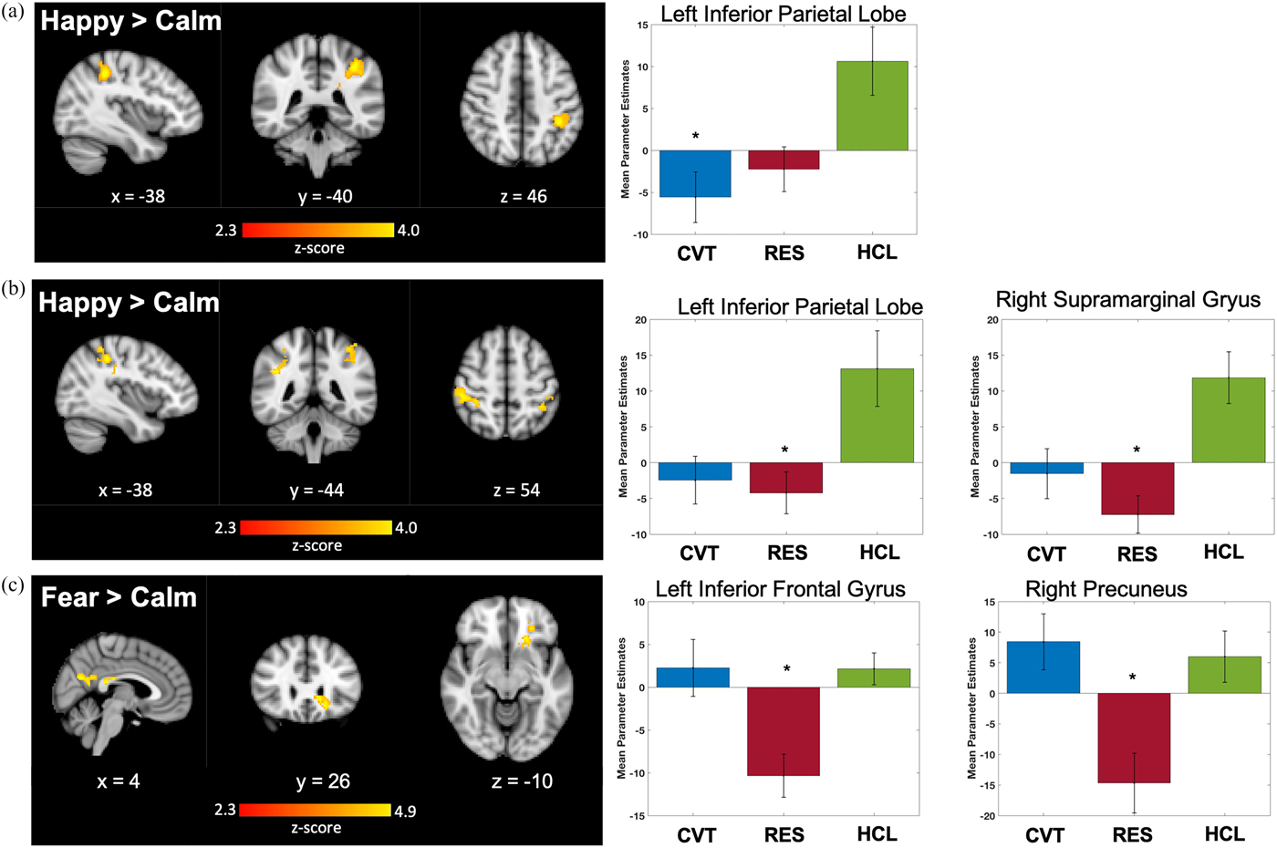
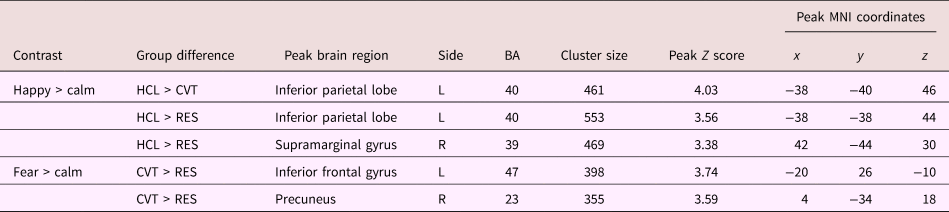
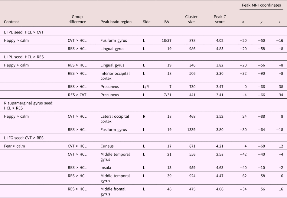
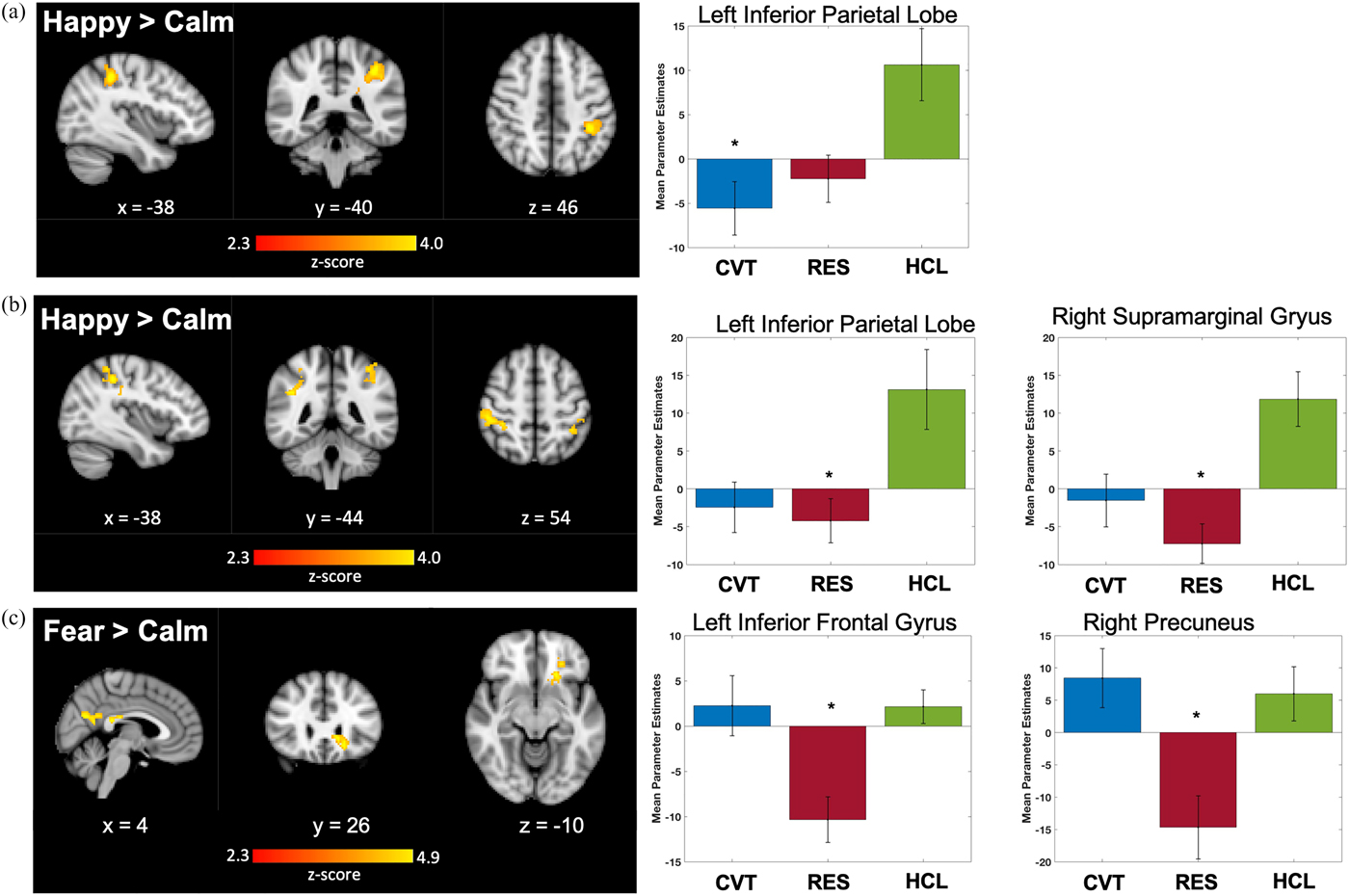
Group differences in activation during happy > calm, and fear > calm contrasts are shown in Figure 1 and Table 3. During happy > calm, the CVT and RES groups had significantly lower activation in the left IPL compared to HCLs. Further, the RES group had significantly lower activation in the right supramarginal gyrus compared to HCLs. During fear > calm, the RES group exhibited significantly lower activation in the left inferior frontal gyrus (IFG) and right precuneus compared to CVT.
Significant clusters of activation and graphs illustrating group differences for each cluster. HCL exhibited significantly greater activation (a) in the left inferior parietal lobe compared to CVT, and (b) in the left IPL and right supramarginal gyrus in RES during happy > calm. (c) RES exhibited significant lower activation in the left inferior frontal gyrus and right precuneus compared to CVT during fear > calm. Z statistics images were thresholded (Z > 2.3) using corrected cluster significance threshold of p < .05. Legend: blue: CVT, red: RES, green: HCL; left side of image corresponds to the right hemisphere.
Significant clusters of whole-brain activation for happy > calm and fear > calm

Note: Coordinates are reported in Montreal Neurological Institute (MNI) space. R, right, L, left. BA, Brodmann area.
Functional connectivity of IPL, supramarginal gyrus, IFG, and precuneus
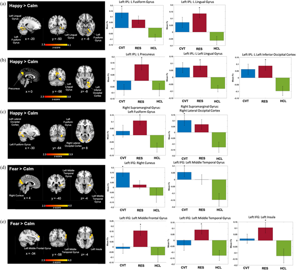
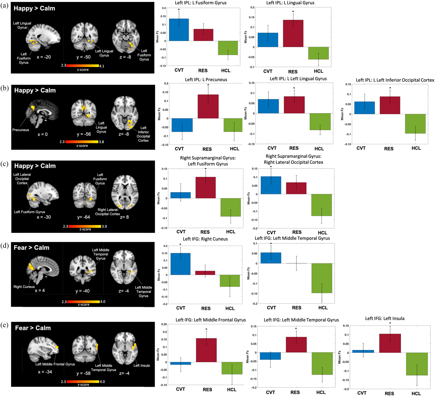
Functional connectivity results are shown in Figure 2 and Table 4. Using the left IPL as a seed region during happy > calm, the CVT group had stronger connectivity with the left fusiform gyrus than the HCL group. The RES group exhibited higher left IPL connectivity with the left lingual gyrus, inferior occipital cortex, and precuneus compared to HCLs. Compared to the CVT group, the RES group had stronger left IPL to precuneus functional connectivity. Using the right supramarginal gyrus seed during happy > calm, the CVT group exhibited stronger connectivity between the right supramarginal gyrus and the right lateral occipital cortex compared to the HCLs. The RES group exhibited stronger connectivity between the right supramarginal gyrus and the left fusiform gyrus compared to HCLs. Using the left IFG seed during fear > calm, the CVT group exhibited stronger connectivity between the left IFG and middle temporal gyrus, and the left IFG and right cuneus, compared to HCLs. The RES group exhibited stronger left IFG connectivity with the insula, middle temporal gyrus, and middle frontal gyrus, compared to HCLs. No regions had significant functional connectivity with the precuneus.
Functional connectivity results. (a) CVT group exhibited higher connectivity than HCL between the left IPL seed and the left fusiform gyrus during happy > calm. RES group exhibited higher left IPL–left lingual gyrus functional connectivity than HCL during happy > calm. (b) RES group exhibited higher left IPL–precuneus connectivity than CVT and HCL, and higher left IPL–left lingual gyrus and left inferior occipital cortex connectivity during happy > calm. (c) RES group exhibited higher left IPL connectivity than HCL between the right supramarginal seed and the left fusiform gyrus during happy > calm. CVT group exhibited higher left IPL–right lateral occipital cortex functional connectivity compared to HCL during happy > calm. (d) CVT exhibited higher left IFG to right cuneus and left middle temporal gyrus functional connectivity compared to HCL during fear > calm. The RES group exhibited higher left IFG–left middle frontal gyrus, middle temporal gyrus, and left insula connectivity compared to HCLs during fear > calm. Mean functional connectivity values are reported as Fisher's z scores (Fz). Z statistics images were thresholded (Z > 2.3) using cluster-corrected significance threshold of p < .05. IPL, inferior parietal lobe. IFG, inferior frontal gyrus. Legend: blue: CVT, red: RES, green: HCL; left side of image corresponds to the right hemisphere.
Clusters showing significant functional connectivity during happy > calm and fear > calm

Note: Coordinates are reported in Montreal Neurological Institute (MNI) space. R, right. L, left. IPL, inferior parietal lobe. IFG, inferior frontal gyrus. BA, Brodmann area.
Brain–behavior correlations
Change in strengths and difficulties
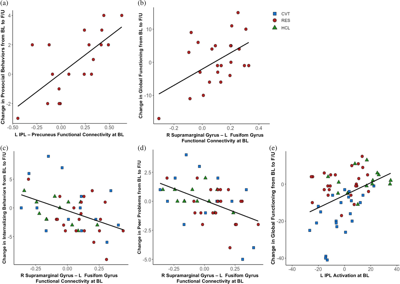
Correlation analyses tested for associations between neural measures at baseline and improved or worsening symptoms at follow-up (compared with baseline). Within the RES group, we found a significant correlation between baseline to follow-up improvement for prosocial behaviors and increased IPL and precuneus functional connectivity, r (16) = .654, p = .004 (Figure 3a). We did not observe any within-group correlations in the CVT group. We also explored correlations between change in scores on the SDQ, and regions identified in our whole-brain analyses and PPI analyses across all groups to generate hypotheses for future research. Across all groups, we found significant negative correlations between worsening internalizing behaviors from baseline to follow-up and the right supramarginal gyrus to left fusiform gyrus functional connectivity, r (42) = –.442, p = .003 (Figure 3c). We also found a negative correlation between worsening peer problems from baseline to follow-up and the right supramarginal gyrus to left fusiform gyrus functional connectivity, r (42) = –.472, p = .001 (Figure 3d). There were no other correlations between change in scores on the SDQ, and activation or functional connectivity during happy > calm or fear > calm across all groups.
Significant correlations between brain activation and functional connectivity, and (a) increased change in prosocial behaviors and (b) improvements in global functioning within the RES group, and (c) worsening internalizing behaviors, (d) worsening peer problems, and (e) increased global functioning across all groups. L, left. R, right. IPL, inferior parietal lobe. BL, baseline. F/U, follow-up.

Change in global functioning
Within the RES group, improvement in global functioning was positively associated with baseline right supramarginal gyrus and left fusiform gyrus functional connectivity, r (18) = .580, p = .005 (Figure 3b). We did not observe any within-group correlations in the CVT group. Upon exploring correlations between change in global functioning, and regions identified in our whole-brain analyses and PPI analyses across all groups, we found a significant positive correlation between increased global functioning from baseline to follow-up and left IPL activation across all groups, r (57) = .424, p = .001 (Figure 3e). There were no other significant correlations between change in global functioning, and activation or functional connectivity during happy > calm or fear > calm across all groups.
Developmental differences
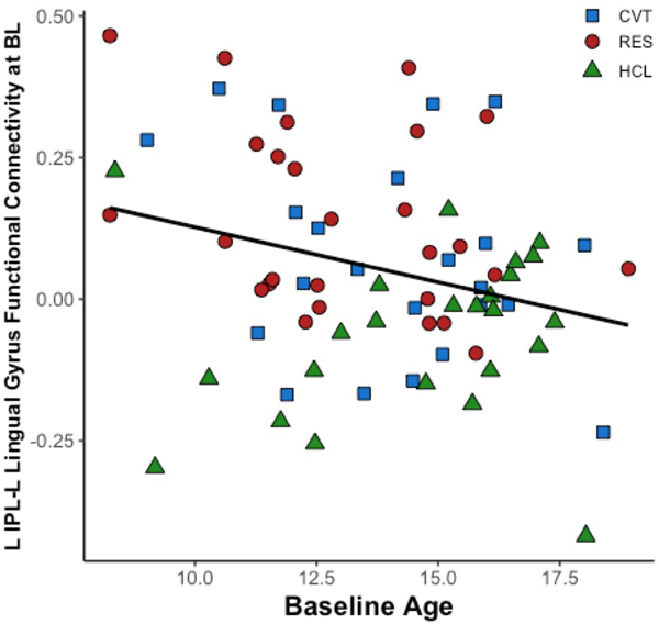
We examined correlations between age, neural activation, and connectivity to investigate age-related developmental differences during positive and negative emotional processing. There was a significant negative correlation between baseline age and increased left IPL and left lingual gyrus functional connectivity during happy > calm across all groups, r (71) = –.255, p = .029 (Figure 4). There were no other correlations between baseline age and activation or functional connectivity of the IPL, supramarginal gyrus, IFG, or precuneus during happy > calm or fear > calm.
Significant correlations between baseline age and left IPL to left lingual gyrus functional connectivity. L, left. IPL, inferior parietal lobe. BL, baseline.

Sex differences
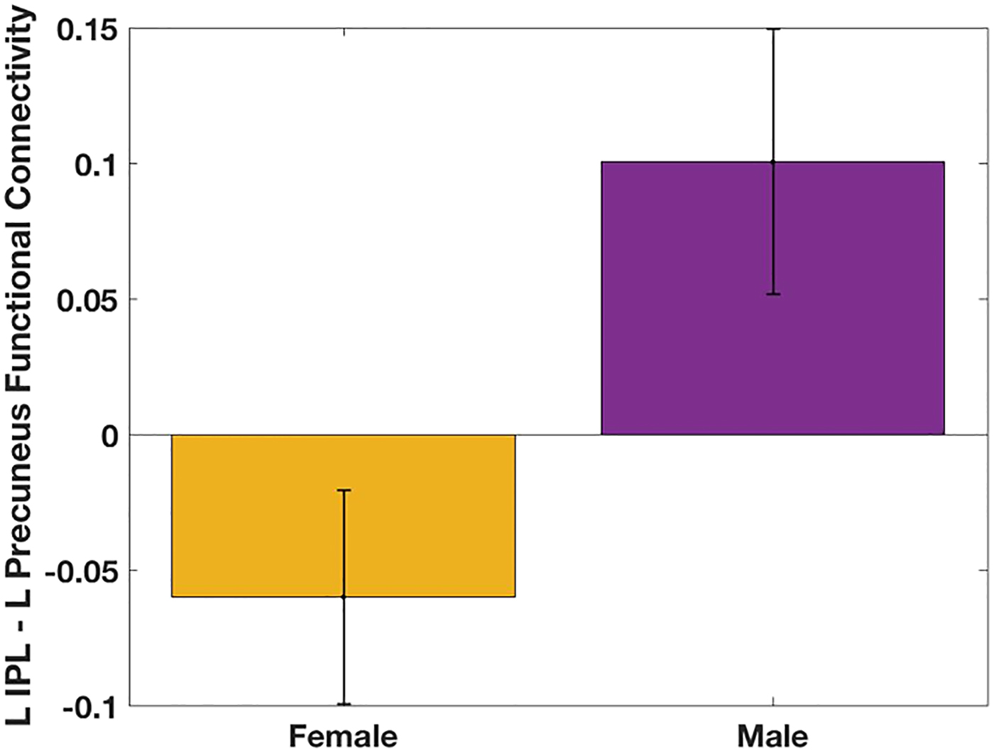
We found significant sex differences for left IPL to left precuneus functional connectivity during happy > calm, t (72) = 2.46 p = .016) (Figure 5). There were no other sex differences found for activation or functional connectivity during the happy > calm or fear > calm contrasts.
Significant sex differences in baseline left IPL to L precuneus functional connectivity. L, left. IPL, inferior parietal lobe. BL, baseline.

Discussion
The aim of this study was to examine whether differences in baseline emotion processing could predict resilience or the development of psychiatric disorders in youth at familial risk for mood disorders. Consistent with our hypotheses, we found differential activation between low-risk youth, high-risk youth who later developed psychopathology, and resilient youth who had no psychiatric symptoms upon follow-up. As predicted, we found differences in brain activation and connectivity in visual, frontal, and limbic regions among the three groups. In particular, we found lower activation in the IPL and right supramarginal gyrus of high-risk youth compared to low-risk youth during positive (happy) facial emotion processing, and lower activation in the left IFG and right precuneus in resilient youth during negative (fearful) facial emotion processing. We also found differential functional connectivity of these regions to other brain regions involved in face processing, emotion processing, and cognition, including the fusiform gyrus, lingual gyrus, precuneus, middle temporal gyrus, middle frontal gyrus, and insula. All of the regions emerging from our voxel-wise analyses have been strongly implicated with face emotion processing in both healthy individuals and individuals with mood disorders (Chen et al., Reference Chen, Lennox, Jacob, Calder, Lupson, Bisbrown-Chippendale and Bullmore2006; Haxby, Hoffman, & Gobbini, Reference Haxby, Hoffman and Gobbini2000; Townsend & Altshuler, Reference Townsend and Altshuler2012). Thus, differences in emotion processing between the three groups could indicate differential neural markers of risk and resilience. Specifically, in the RES youth, stronger IPL and precuneus connectivity was associated with increased prosocial behaviors, and stronger right supramarginal gyrus and left fusiform gyrus connectivity was associated with improved global functioning.
Positive facial emotional processing
During the processing of happy versus calm faces, we found the CVT and RES groups had significantly less activation in the left IPL compared to HCLs, implicating an intrinsic risk shared by CVT and RES groups to this region. Nevertheless, the differences in functional connectivity of this region to other brain regions could differentiate between resilient and converted youth. In our study, we found that whereas the CVT group had stronger IPL connectivity with the left fusiform gyrus, the RES group had stronger IPL connectivity with the left lingual gyrus, inferior occipital cortex, and precuneus compared to HCLs. Stronger IPL connectivity to the precuneus in the RES compared to the CVT youth is of particular importance. The precuneus is involved in the integration of mental processing through its role in cognitive control, including visuospatial imagery and attentional control (Cavanna & Trimble, Reference Cavanna and Trimble2006). Our results suggest that increased IPL to precuneus connectivity in the RES youth may reflect a compensatory mechanism to prevent the onset of mood symptoms through an alteration in the processing of emotional and, in particular, positive stimuli. A previous study found alterations in activation of these regions as candidate resilience markers (Wiggins et al., Reference Wiggins, Brotman, Adleman, Kim, Wambach, Reynolds and Leibenluft2017), further supporting our findings. Similarly, we found increased supramarginal gyrus activation in the RES group, and an increased connectivity between the supramarginal gyrus and the fusiform gyrus. The supramarginal gyrus, similar to the IPL, is involved in attentional control (Igelström & Graziano, Reference Igelström and Graziano2017), and thus its connectivity with the fusiform gyrus, a face-processing region, could indicate increased attentional control to positive stimuli as a potential mechanistic contributor to resilience.
Our study advances our understanding of how these neural systems work differentially in the context of health and psychopathology. Of note, within the RES group, stronger IPL-precuneus connectivity was positively associated with increased prosocial behaviors between baseline and follow-up. Given the RES group had a significant increase in prosocial behaviors at follow-up, we suggest that IPL-precuneus functional connectivity could represent a trait feature or predictor of resilience in youth at high-risk for mood disorders. Prosocial behaviors have been associated with positive outcomes in youth at broad risk for psychopathology (Alarcón & Forbes, Reference Alarcón and Forbes2017; Dang, Reference Dang2014; Silk et al., Reference Silk, Vanderbilt-Adriance, Shaw, Forbes, Whalen, Ryan and Dahl2007). Our data extends this fact to youth at risk for mood disorders. Within the RES group, right supramarginal gyrus connectivity with the left fusiform gyrus was correlated with a more positive change in global functioning, suggesting an adaptive process within the resilient group. As suggested above, supramarginal gyrus connectivity with the fusiform gyrus could indicate increased attentional control to positive stimuli, and thus be related to better functional outcomes in these high-risk youth. Further supporting this notion, across all groups, stronger right supramarginal gyrus to left fusiform gyrus functional connectivity was negatively correlated with worsening internalizing behaviors and worsening peer problems from baseline to follow-up. These findings suggest that right supramarginal gyrus to left fusiform gyrus connectivity may be potentially protective, as increased functional connectivity between these two regions was associated with decreased internalizing behaviors and peer problems. This connectivity may correspond to increased attention to positive emotions, especially given the role of the supramarginal gyrus in the processing of information relating to the self and other (Eddy, Reference Eddy2016; Igelström & Graziano, Reference Igelström and Graziano2017). Thus, this increased functional connectivity could work as a neural mechanism to help individuals process positive emotional information related to both self and others, facilitating the development of healthy peer relationships and minimizing internalizing behaviors. Future studies directly examining the role of supramarginal activation and functional connectivity to enhance peer relationships and reduce internalizing behaviors are warranted. Further, across all groups, stronger IPL activation was associated with increased global functioning, suggesting that IPL activation may be protective against psychopathology in its association with better functional outcomes. Nevertheless, future studies should examine IPL activation before and after the progression of psychiatric disorders in order to examine changes in IPL activation with regard to the onset of psychiatric symptoms.
Negative facial emotional processing
The RES group had significantly less activation in the left IFG during the processing of fearful versus calm faces, and stronger connectivity between the left IFG, and the insula, middle temporal gyrus, and middle frontal gyrus compared to the HCL youth. The IFG is involved in cognitive processes, including emotion regulation, cognitive control, and sustained attention (Liakakis, Nickel, & Seitz, Reference Liakakis, Nickel and Seitz2011). Previous studies have shown abnormal IFG activation during emotion processing in BD, MDD, and high-risk individuals (Breakspear et al., Reference Breakspear, Roberts, Green, Nguyen, Frankland, Levy and Mitchell2015; Hafeman et al., Reference Hafeman, Bebko, Bertocci, Fournier, Bonar, Perlman and Phillips2014; Roberts et al., Reference Roberts, Green, Breakspear, McCormack, Frankland, Wright and Mitchell2013, Reference Roberts, Lord, Frankland, Wright, Lau, Levy and Breakspear2017), with most studies showing an attenuated response of the IFG, especially during response inhibition (Breakspear et al., Reference Breakspear, Roberts, Green, Nguyen, Frankland, Levy and Mitchell2015; Roberts et al., Reference Roberts, Green, Breakspear, McCormack, Frankland, Wright and Mitchell2013). In our study, we found hypoactivation of the IFG in the RES youth but not in the CVT youth. It is possible that our sample of CVT youth are somehow different than those in previous studies, or that emotion-processing tasks yield different activation than the response inhibition tasks used in previous studies. Alternately, it is possible that the processes underlying IFG hypoactivation reported in previous studies are subserved by different prefrontal or limbic regions at this early point in mood disorder progression (Ahmed, Bittencourt-Hewitt, & Sebastian, Reference Ahmed, Bittencourt-Hewitt and Sebastian2015). We found increased IFG functional connectivity with executive functioning regions (middle frontal gyrus) and emotion processing regions (insula) in the RES group compared to HCLs. These findings suggest that increased connectivity between executive and limbic regions in response to negative emotional stimuli could reflect a compensatory or resilience-promoting mechanism in our unaffected high-risk youth during negative emotion processing. It is possible that stronger frontal, and frontal-limbic connectivity in the RES group lessens the biases toward negative emotional stimuli typically seen in high-risk individuals (Sharma et al., Reference Sharma, Barron, Le Couteur, Close, Rushton, Grunze and Le Couteur2017; Whalley et al., Reference Whalley, Sussmann, Romaniuk, Stewart, Kielty, Lawrie and McIntosh2015). Moreover, one recent study found that in BD young adults, the IFG was functionally disconnected from frontal and limbic regions (Roberts et al., Reference Roberts, Lord, Frankland, Wright, Lau, Levy and Breakspear2017). Nevertheless, we did not find any correlations between IFG activation or connectivity and change in global functioning or strengths and difficulties. One possible explanation is that because the IFG is primarily involved with cognitive processes, differences in IFG activation and connectivity at baseline before the development of psychiatric symptoms may not be sensitive enough to changes in prosocial behaviors or global functioning related to emotional processing. Nevertheless, future studies are needed to disentangle neural mechanisms crucial for the integration of emotional processing with executive function in unaffected high-risk youth, especially as common characteristics of pediatric mood disorders involve emotion dysregulation and cognitive control (Beauchaine, Reference Beauchaine2015; Platt, Waters, Schulte-Koerne, Engelmann, & Salemink, Reference Platt, Waters, Schulte-Koerne, Engelmann and Salemink2017).
Further, the RES group exhibited less activation in the right precuneus during the processing of fearful versus calm faces compared to the CVT youth. This finding is supported by a previous finding of altered precuneus activation in high-risk youth compared to BD and low-risk youth (Wiggins et al., Reference Wiggins, Brotman, Adleman, Kim, Wambach, Reynolds and Leibenluft2017), suggesting differential precuneus activation may act as a protective factor. Furthermore, throughout the literature, increased precuneus activation has been associated with depressive symptoms in high-risk individuals (Peterson et al., Reference Peterson, Wang, Horga, Warner, Rutherford, Klahr and Weissman2014). As the precuneus coordinates communication between regions responsible for attentional and cognitive control (Cavanna & Trimble, Reference Cavanna and Trimble2006), deactivation of this region during the processing of negative face stimuli could work to counteract negative cognitive biases seen in high-risk youth (Platt et al., Reference Platt, Waters, Schulte-Koerne, Engelmann and Salemink2017). Nevertheless, future studies would need to disentangle neural mechanisms involved in cognitive control in resilient youth.
Coherence between strengths and difficulties and global functioning
We found both significant group differences in internalizing behaviors and total difficulties and significant within-CVT and RES group changes between follow-up and baseline scores. Whereas the CVT group exhibited lower hyperactivity, prosocial, and externalizing behaviors, the RES group had overall lower difficulties and increased prosocial behaviors. The CVT group had worsened global functioning at follow-up compared to baseline. These results suggest the RES group may have better baseline functioning overall with ceiling effects on certain areas of difficulty observed over development. Increases in prosocial behaviors over time may underlie the observed improvement in global functioning within the RES group, and points to the potentially critical role of prosocial behaviors during this critical stage of emotional development. Previous studies have pointed to the protective factors of prosocial behaviors against the development of psychopathology (Alarcón & Forbes, Reference Alarcón and Forbes2017; Dang, Reference Dang2014; Silk et al., Reference Silk, Vanderbilt-Adriance, Shaw, Forbes, Whalen, Ryan and Dahl2007).
Developmental differences
We examined correlations between age and neural activation and connectivity to investigate age-related developmental differences in positive and negative emotional processing. We found a significant negative correlation between baseline age and left IPL to left lingual gyrus functional connectivity during the processing of positive faces across all groups. This finding suggests that as baseline age increases, functional connectivity between the left IPL and left fusiform gyrus while individuals process positive emotional information decreases. It is interesting to note here that these findings contrast with previous findings suggesting an age-related increase in connectivity between the parietal and visuospatial regions (Solé-Padullés et al., Reference Solé-Padullés, Castro-Fornieles, de la Serna, Calvo, Baeza, Moya and Sugranyes2016). However, this study examined resting-state functional connectivity and not connectivity during emotion processing. It is plausible that related to positive emotion processing, older children and adolescents compared to younger children may have differential functional connectivity between the left IPL and lingual gyrus and rely more on other regions such as the amygdala and prefrontal regions, which has been evidenced in studies examining the development of emotion processing (Del Piero, Saxbe, & Margolin, Reference Del Piero, Saxbe and Margolin2016; Pagliaccio et al., Reference Pagliaccio, Luby, Gaffrey, Belden, Botteron, Harms and Barch2013). Further studies are needed to understand the developmental trajectories of brain regions and networks important for emotion processing and regulation.
Sex differences
We found a significant sex difference for left IPL to left precuneus functional connectivity during the processing of positive faces. Females had significantly lower left IPL to left precuneus functional connectivity compared to males. Our finding suggests that boys exhibit stronger connectivity between the left IPL and left precuneus compared to girls during positive face emotional processing at baseline. These findings are consistent with the literature, which points to potential sex differences in resting-state functional connectivity in healthy (Solé-Padullés et al., Reference Solé-Padullés, Castro-Fornieles, de la Serna, Calvo, Baeza, Moya and Sugranyes2016) and high-risk (Singh, Chang, et al., Reference Singh, Chang, Kelley, Saggar, Reiss and Gotlib2014) youth, and during face emotion processing (Pagliaccio et al., Reference Pagliaccio, Luby, Gaffrey, Belden, Botteron, Harms and Barch2013). A previous study demonstrated that girls show increased activation in face processing regions, such as the fusiform gyrus, and deactivation in the precuneus, and boys exhibit more activation in executive functioning regions in response to face emotional stimuli (Pagliaccio et al., Reference Pagliaccio, Luby, Gaffrey, Belden, Botteron, Harms and Barch2013). Together with the extant literature, our result could indicate important sex differences in attention to or engagement with emotional face stimuli. These sex differences may underlie dissimilarities in risk trajectories for mood disorders seen between girls and boys. Additional studies are clearly needed to understand how sex differences in emotion processing affect brain and behavioral differences in the onset of psychiatric disorders observed between girls and boys (Phillips et al., Reference Phillips, Onopa, Hsu, Ollila, Hillary, Hallmayer and Singh2018).
Transactional model of risk for emotion dysregulation in familial mood disorders
Our study demonstrated that neural differences in emotion processing at baseline could predict resilience versus conversion to a psychiatric disorder in youth at familial risk for mood disorders. It is also likely that there are environmental events that interact with neural susceptibility to psychopathology, representing a transactional model of how mood disorders emerge. Offspring of parents with MDD and BD are often exposed to significant environmental adversity (Oquendo et al., Reference Oquendo, Ellis, Chesin, Birmaher, Zelazny, Tin and Brent2013) and harsh parenting styles (Forbes et al., Reference Forbes, Shaw, Silk, Feng, Cohn, Fox and Kovacs2008; Kopala-Sibley et al., Reference Kopala-Sibley, Jelinek, Kessel, Frost, Allmann and Klein2017; Wiggins, Mitchell, Hyde, & Monk, Reference Wiggins, Mitchell, Hyde and Monk2015), which are potential harbingers of poor outcome. These environmental exposures may further interact with prenatal (Singh, DelBello, Soutullo, et al., Reference Singh, DelBello, Stanford, Soutullo, McDonough-Ryan, McElroy and Strakowski2007; Swales et al., Reference Swales, Winiarski, Smith, Stowe, Newport and Brennan2018) and genetic (Fears et al., Reference Fears, Service, Kremeyer, Araya, Araya, Bejarano and Bearden2014; Park et al., Reference Park, Chang, Hallmayer, Howe, Kim, Hong and Singh2015; Wang et al., Reference Wang, Shen, Guillaume, Chong, Chen, Fortier and Qiu2018) risk factors to increase risk for psychopathology. Prior studies have described the family environments of youth at risk for mood disorders (Daches, Vine, Layendecker, George, & Kovacs, Reference Daches, Vine, Layendecker, George and Kovacs2018; DelBello & Geller, Reference DelBello and Geller2001; Goodman & Gotlib, Reference Goodman and Gotlib1999; Miklowitz et al., Reference Miklowitz, Schneck, Walshaw, Garrett, Singh, Sugar and Chang2019). In families affected by mood disorders, family interactions are typically toxic, and the environments are frequently chaotic and dysfunctional. These are likely major contributors to abnormal cortical and subcortical function (Wackerhagen et al., Reference Wackerhagen, Wüstenberg, Mohnke, Erk, Veer, Kruschwitz and Romanczuk-Seiferth2017), dysconnectivity (Singh, Chang, et al., Reference Singh, Chang, Kelley, Saggar, Reiss and Gotlib2014), and aberrant plasticity (Comasco, Åslund, Oreland, & Nilsson, Reference Comasco, Åslund, Oreland and Nilsson2013) in youth who are also genetically predisposed toward developing mood disorders.
Further, various stressors that children experience, including maltreatment, poverty, and stressful events, can lead to a range of effects on ensuing psychopathology depending on the type, timing, chronicity, and severity of the stressor (Doom & Cicchetti, Reference Doom, Cicchetti, Harkness and Hayden2018). Negative stressful life events commonly precede the first episodes of depression and mania (Horesh, Apter, & Zalsman, Reference Horesh, Apter and Zalsman2011). It is likely that plasticity in cortical and subcortical regions in response to stressful life events could differentiate between youth at risk for mood disorders who do or do not develop stress-related psychopathology (Gröger et al., Reference Gröger, Matas, Gos, Lesse, Poeggel, Braun and Bock2016; VanTieghem & Tottenham, Reference VanTieghem and Tottenham2018). Further, the experience of positive events can be protective. Fischer et al. (Reference Fischer, Camacho, Ho, Whitfield-Gabrieli and Gotlib2018) found a significant association between amygdala to orbitofrontal cortex connectivity and positive life events in resilient female adolescents who were at risk for depression. Thus, it is plausible that differential neurobiological responses to environmental stressors may distinguish high-risk youth who remain resilient from those who develop a mood disorder, as our findings suggest here.
Limitations
Although our findings suggest a potential resilience mechanism that may prevent the onset of mood and other psychiatric disorders, it is important to note some limitations. First, we had a relatively moderate sample size. However, we found consistent results after adjusting for multiple comparisons, and after covarying for age and sex. Second, our CVT group consisted of youth who had converted to a mood diagnosis by, on average, a 3-year follow-up. However, there is a possibility that other participants in our study may convert to a mood or other psychiatric disorders after this follow-up period. It is also possible that the neural markers discovered are not specific to mood disorders, given that youth in the CVT group had mood as well as other co-occurring psychiatric disorders. However, 78% of youth in the CVT group developed some form of a major or subthreshold mood disorder at follow-up, and all had some form of an internalizing disorder, suggesting most of the outcomes for youth at familial risk for mood disorders seem to cluster along the spectrum of internalizing disorders. We examined neural data only at baseline, and although our hypotheses were to investigate whether baseline neural data could predict conversion at follow-up, we cannot make definitive conclusions that aberrant IPL and IFG activation and connectivity during emotion processing is the cause of symptom development. Future studies should evaluate this question longitudinally to examine changes in brain functioning over time.
Conclusions
In this study, we examined whether differences in baseline implicit emotion processing could predict resilience or the development of psychiatric disorders in youth at familial risk for mood disorders. We examined baseline brain activation and connectivity during implicit emotion processing in high-risk youth, some of whom developed psychopathology, and some of whom remained resilient, and low-risk controls. Our results indicate that differential activation and connectivity in the IPL, IFG, and precuneus in response to emotional stimuli may represent differential resilience versus risk markers for pediatric-onset mood disorders as these neural differences are seen before the development of any psychiatric symptoms. Activation and connectivity patterns may provide important clues about risk for or resilience from symptom development and may increase our understanding of means to enhance resilience to psychopathology at the earliest possible time in development.
Acknowledgments
We thank the families who participated in this study and members of the Pediatric Emotion and Resilience Lab who helped with assessment, recruitment, data collection, and data entry.
Financial support
This research was supported by a Ford Foundation Predoctoral Fellowship, the National Science Foundation Graduate Research Fellowship Program, and Stanford's Diversifying Academia, Recruiting Excellence (DARE) Doctoral Fellowship (to A.F.N.); and NIMH Grant K23MH085919 and the Stanford Maternal Child Health Research Institute (to M.K.S.).
Conflict of interest
Manpreet K. Singh receives research support from Stanford's Maternal Child Health Research Institute, the NIMH, National Institute of Aging, Neuronetics, Johnson and Johnson, and the Brain and Behavior Foundation. She is on the advisory board for Sunovion and is a consultant for Google X. No other authors report any biomedical financial interests or potential conflicts of interest.