Introduction
The fertilized oocyte first undergoes a series of early cleavage divisions to produce increasing numbers of progressively smaller cells, known as blastomeres, without changing the overall size of the embryo; it spans compaction and morula formation, and finally, cavitation with formation of a blastocyst. During mouse preimplantation embryo development, the 1-cell embryo develops into a blastocyst, a process that takes 4 days. In the mouse, the first sign of the attachment reaction (apposition stage) in the process of implantation occurs in the evening on day 4 of pregnancy (day 1 = vaginal plug) (Dey et al., Reference Dey, Lim, Das, Reese, Paria, Daikoku and Wang2004; Wang & Dey, Reference Wang and Dey2006). Estrogen secretion around noon on day 4 of pregnancy is essential for on-time blastocyst activation for implantation (Yoshinaga & Adams, Reference Yoshinaga and Adams1966; Paria et al., Reference Paria, Huet-Hudson and Dey1993). However, the mechanisms that regulate preimplantation embryo development are not fully understood.
Tubulointerstitial nephritis antigen-like 1 (TINAGL1; also known as adrenocortical zonation factor 1 [AZ-1] or lipocalin 7) is a secretory protein of 52 kD polypeptides that was cloned from mouse adrenocortical cells and is known to be closely associated with the zonal differentiation of adrenocortical cells (Mukai et al., Reference Mukai, Mitani, Nagasawa, Suzuki, Suzuki, Suematsu and Ishimura2003). Recently, we demonstrated the expression and localization of TINAGL1 in peri-implantation mouse embryos (Igarashi et al., Reference Igarashi, Tajiri, Sakurai, Sato, Li, Mukai, Suematsu, Fukui, Yoshizawa and Matsumoto2009). Just prior to implantation at 23:00 h on day 4 of pregnancy, TINAGL1 was uniquely distributed in the blastocysts. Specifically, TINAGL1 was localized to the blastocoel site surface of the trophectoderm (TE) in implantation-competent (activated) blastocysts. This blastocyst activation can be initiated rapidly by a single injection of estradiol-17β (E2) into ovariectomized and progesterone-primed pregnant mice, and is known as the delayed-implantation model (Yoshinaga & Adams, Reference Yoshinaga and Adams1966; Psychoyos, Reference Psychoyos, Greep, Astwood and Geiger1973). In fact, we have also demonstrated the same localization and increased expression of TINAGL1 in activated blastocysts after E2 treatment using this mouse model (Igarashi et al., Reference Igarashi, Tajiri, Sakurai, Sato, Li, Mukai, Suematsu, Fukui, Yoshizawa and Matsumoto2009). This unique localization may indicate a physiological role for TINAGL1 in the preparation of the blastocyst for successful implantation and/or subsequent pregnancy. We also demonstrated that, at postimplantation, TINAGL1 is a novel component of the Reichert membrane and interacts with laminin (LN) 1, and most likely plays a physical and physiological role in embryo development (Igarashi et al., Reference Igarashi, Tajiri, Sakurai, Sato, Li, Mukai, Suematsu, Fukui, Yoshizawa and Matsumoto2009). However, the relationship between TINAGL1 and other structural extracellular matrix (ECM) molecules in the mouse embryo, including fibronectin (FN) and collagen type IV (ColIV), is not clear.
ECM is present in every tissue but is most highly enriched in connective tissue and basement membrane (BM). ECM provides physical support to tissues and organs by occupying the space between cells. FN is a major constituent of ECM that promotes cell adhesion, spreading migration, and cytoskeletal organization (Hynes, Reference Hynes1990). ColIV is largely considered to be a structural component of BM, where it forms a scaffold with which other BM components, such as LN or FN, can associate (Laurie et al., Reference Laurie, Bing, Kleinman, Hassell, Aumailley, Martin and Feldmann1986), and it also mediates various cell functions directly (Murray et al., Reference Murray, Stingl, Kleinman, Martin and Katz1979; Rubin et al., Reference Rubin, Hook, Obrink and Timpl1981; Aumailley & Timpl, Reference Aumailley and Timpl1986). Some in vitro experiments suggest that FN promotes trophoblast adhesion, which may restrict migration, while other studies indicate that it facilitates motility (Burrows et al., Reference Burrows, King and Loke1993; Damsky et al., Reference Damsky, Librach, Lim, Fitzgerald, McMaster, Janatpour, Zhou, Logan and Fisher1994; Irving et al., Reference Irving, Lysiak, Graham, Hearn, Han and Lala1995; Stephens et al., Reference Stephens, Sutherland, Klimanskaya, Andrieux, Meneses, Pedersen and Damsky1995; Yelian et al., Reference Yelian, Yang, Hirata, Schultz and Armant1995). ColIV can also support the outgrowth of primary trophoblast from the mouse blastocyst (Armant et al., Reference Armant, Kaplan and Lennarz1986; Sutherland et al., Reference Sutherland, Calarco and Damsky1988). Although some investigations into the expression of FN (Zetter & Martin, Reference Zetter and Martin1978; Wartiovaara et al., Reference Wartiovaara, Leivo and Vaheri1979; Yohkaichiya et al., Reference Yohkaichiya, Hoshiai, Uehara and Yajima1988) and ColIV (Leivo et al., Reference Leivo, Vaheri, Timpl and Wartiovaara1980; Sherman et al., Reference Sherman, Gay, Gay and Miller1980) in preimplantation embryos have been performed, detailed analyses of their expression from blastocyst activation to just prior to implantation have not been conducted.
In this study, we compared the immunocytological distributions of TINAGL1, FN, and ColIV during mouse preimplantation development, particularly in blastocysts at three stages: (1) before estrogen secretion; (2) after estrogen secretion; and (3) just prior to implantation. Importantly, it is known that in vitro culture (IVC) of preimplantation embryos alters their global gene expression patterns (Rinaudo & Schultz, Reference Rinaudo and Schultz2004; Rinaudo et al., Reference Rinaudo, Giritharan, Talbi, Dobson and Schultz2006) and affects the behavior of mice after birth (Ecker et al., Reference Ecker, Stein, Xu, Williams, Kopf, Bilker, Abel and Schultz2004; Fernandez-Gonzalez et al., Reference Fernandez-Gonzalez, Moreira, Bilbao, Jimenez, Perez-Crespo, Ramirez, Rodriguez De Fonseca, Pintado and Gutierrez-Adan2004). Therefore, we compared further their expression in in vivo and in vitro fertilized (IVF) blastocysts.
Materials and methods
Animals
All ICR mice were purchased from Japan SLC Inc. (Shizuoka, Japan), and housed under controlled temperatures (22–27°C) with a constant photoperiod (13L–11D). Mice were provided with a pelleted diet (Oriental Yeast Co. Ltd., Japan) and water ad libitum. All investigations were performed in accordance with the Guide for Care and Use of Laboratory Animals of the Graduate School of Agricultural Science, Tohoku University.
In vivo embryo collection
Preimplantation embryos were collected as described previously (Igarashi et al., Reference Igarashi, Tajiri, Sakurai, Sato, Li, Mukai, Suematsu, Fukui, Yoshizawa and Matsumoto2009). In brief, female mature mice were mated with fertile males to induce pregnancy (day 1 [10:00 h] = vaginal plug). Removed oviducts or uteri were flushed with Ca2+- and Mg2+-free Dulbecco's phosphate-buffered saline (PBS, Nissui Pharmaceutical Co., Ltd., Japan) containing 0.1% polyvinyl alcohol (PVA, Sigma). Preimplantation embryos were collected at the following stages during pregnancy: 1-cell, 2-cell, 4-cell, 8-cell, morula, and blastocyst on days 1 (21:00 h), 2 (10:00 h), 3 (01:00 h), 3 (10:00 h), 3 (21:00 h), and 4 (10:00 [before estrogen secretion], 18:00 [after estrogen secretion], and 23:00 h [just prior to implantation]), respectively.
In vitro fertilization and embryo culture
In vitro fertilization and embryo culture were performed as described previously (Matsumoto et al., Reference Matsumoto, Ma, Smalley, Trzaskos, Breyer and Dey2001; Hoshino & Sato, 152 Reference Hoshino and Sato2008). Immature mice were superovulated by subcutaneous injection with 5 IU of pregnant mare serum gonadotropin (PMSG; ASKA Pharmaceutical Co., Ltd, Japan) at 3 weeks of age, followed by injection of 5 IU of human chorionic gonadotropin (hCG; Yell Pharmaceutical Co., Ltd, Japan) 48 h later. Mice were killed by cervical dislocation and oviducts were removed at 14 h post-hCG injection. Mature male mice over 8 weeks of age were killed by cervical dislocation and the epididymis was removed and carefully blotted free of blood and adipose tissues. Cauda epididymis was cut with fine scissors and the sperm droplet was scooped out with a 26-gauge needle (Terumo Co., Japan) and immediately transferred to a 200 μl drop of human tubal fluid (HTF) medium covered with mineral oil (Nacalai Tesque, Japan). Capacitation was allowed to proceed for 2–3 h at 37 °C in 5% CO2 in humidified air. Collected cumulus cell–oocyte complexes (COCs) were moved to the HTF medium; the final concentration was 700 spermatozoa/μl. At 4 h after insemination, oocytes were cultured in a 100 μl drop of potassium simplex optimized medium (KSOM) overlaid with mineral oil in a humidified atmosphere of 5% CO2 in air at 37°C. At 120 and 144 h after embryo culture, blastocysts were collected for immunostaining or western blotting.
Blastocyst stage developed in vivo and in vitro
Animal studies using the mouse model have demonstrated that after blastocoel formation in the morning on day 4 of pregnancy, the blastocysts are activated in utero around noon of day 4 for successful implantation to occur (Paria et al., Reference Paria, Huet-Hudson and Dey1993), when an estrogen secretion takes place (Nilsson, Reference Nilsson1966). Furthermore, just prior to implantation, activated blastocysts have a morphologically distinct structure (reviewed in ref. (McRae & Church, Reference McRae and Church1990)). In this study, the typical stages of in vivo blastocysts were as follows: pre-expansion (at 10:00 h before estrogen secretion); from expanded to hatched (at 18:00 h after estrogen secretion, referred to below as peri-hatching); and implantation-competent (at 23:00 h just prior to implantation, defined below as activated). In contrast, we collected IVF blastocysts following embryo culture for 120 or 144 h, because their most typical stages at each time point were pre-expansion or peri-hatching.
Immunostaining of embryos
Immunostaining of preimplantation embryos was performed as described previously (Matsumoto et al., Reference Matsumoto, Daikoku, Wang, Sato and Dey2004; Li et al., Reference Li, Mukai, Suzuki, Suzuki, Yamashita, Mitani and Suematsu2007; Igarashi et al., Reference Igarashi, Tajiri, Sakurai, Sato, Li, Mukai, Suematsu, Fukui, Yoshizawa and Matsumoto2009), with slight modifications. In brief, preimplantation embryos were fixed in 3.7% formaldehyde (Wako Pure Chemical Industries, Ltd., Japan) in PBS containing 0.1% PVA at room temperature for 30 min, and permeabilized with 0.25% Triton X-100 (Wako) in PBS containing 0.1% PVA for 5 min. After washing three times with PBS containing 0.1% PVA, embryos were incubated with rabbit anti-TINAGL1 polyclonal antibody (diluted 1:200), which was prepared as described previously (Li et al., Reference Li, Mukai, Suzuki, Suzuki, Yamashita, Mitani and Suematsu2007), rabbit anti-fibronectin polyclonal antibody (diluted 1:50; Sigma), or rabbit anti-collagen type IV polyclonal antibody (diluted 1:50; Chemicon) overnight at 4°C. Following washes three times with PBS containing 0.25% Triton X-100 and 0.1% PVA, embryos were incubated with Alexa Fluor 488 goat anti-rabbit IgG (dilution 1:200, Invitrogen) for 1 h at room temperature. Washed three times with PBS containing 0.25% Triton X-100 and 0.1% PVA, nuclei were labelled with 10 μg/ml propidium iodide (Sigma) for 1 h at room temperature. After three washes, embryos were viewed using a Bio-Rad MRC-1024 confocal scanning laser microscope mounted on an Axioplan Zeiss microscope.
Western blotting
Western blot analysis was performed as described previously (Igarashi et al., Reference Igarashi, Tajiri, Sakurai, Sato, Li, Mukai, Suematsu, Fukui, Yoshizawa and Matsumoto2009). In brief, collected blastocysts were solubilized in 2× SDS sample buffer (0.5M Tris–HCl [Sigma] at pH 6.8, 10% β-mercaptoethanol [Wako], and 20% glycerol [Wako]). Electrophoresis was performed with 50 blastocysts in each lane on 12% polyacrylamide gels, and the resolved proteins were transferred to polyvinyli-dene fluoride (PVDF) membranes (Millipore Corporate Headquarters, MA). Thereafter, the membranes were blocked for 1 h at room temperature with Tris-buffered saline (TBS) containing 0.1% Tween 20 (Wako) (TBS-T) and 5% skimmed milk (Wako). Membranes were next incubated with rabbit anti-TINAGL1 polyclonal antibody (diluted 1:2000) overnight at 4 °C. Then, the membranes were reacted with horseradish peroxidase-conjugated goat anti-rabbit IgG (diluted 1:40,000) for 1 h at room temperature. Peroxidase activity was visualized using the ECL Plus western blotting detection system (GE Healthcare, Ltd., UK).
Results
Distribution of TINAGL1, FN, and ColIV in 1-cell embryos to morulae
To compare the cellular localization of TINAGL1 with that of FN and ColIV in preimplantation embryos, immunostaining was performed. In 1-cell to 8-cell embryos before compaction, TINAGL1 was localized in the cytoplasm. However, in compacted morulae, TINAGL1 was primarily localized in the outer cells. FN and ColIV were also expressed in the cytoplasm, but were more strongly distributed on the outer surface of the blastomeres than in the cytoplasm in 1-cell to 8-cell embryos. In morulae, TINAGL1, FN, and ColIV were expressed in the outer cells (Fig. 1).
Distribution of TINAGL1, FN, and ColIV in 1-cell embryo to morula. Collected in vivo embryos (1-, 2-, 4- and 8-cell embryos, and compacted morulae) were immunostained for each protein with Alexa Fluor 488 (green). Red shows nuclei. Upper panel: TINAGL1. Middle panel: FN. Lower panel: ColIV. Bar = 50 μm. (See online for a colour version of this figure.)

Distribution of TINAGL1, FN, and ColIV in blastocysts
In blastocysts, TINAGL1 was expressed in the cytoplasm of TE cells before (10:00 h) and after (18:00 h) estrogen secretion. Importantly, just prior to implantation (23:00 h), the distribution of TINAGL1 changed from the cytoplasm to the inner (blastocoelic) surface of TE cells. Similar to TINAGL1, FN and ColIV were also localized in the cytoplasm of TE in blastocysts before and after estrogen secretion. Just prior to implantation, their distribution patterns changed and they were localized mainly at the inner surface of TE. ColIV was also expressed in the cytoplasm of the inner cell mass (ICM) (Fig. 2).
Distribution of TINAGL1, FN, and ColIV in mouse blastocyst. In vivo blastocysts were collected at 10:00 h (before estrogen secretion), 18:00 h (after estrogen secretion), and 23:00 h (just prior to implantation) on day 4 of pregnancy and immunostained for each protein with Alexa Fluor 488 (green). Red shows nuclei. Upper panel: TINAGL1. Middle panel: FN. Lower panel: ColIV. ICM: inner cell mass. TE: trophectoderm. Bar = 50 μm. (See online for a colour version of this figure.)
Expression level of TINAGL1 in blastocyst developed in vivo and in vitro
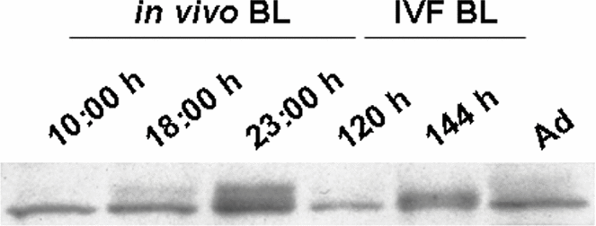
As shown in Fig. 3, the expression of TINAGL1 in IVF blastocysts after 120 h of embryo culture was lower than that in in vivo blastocysts at 10:00 h, indicating that the expression of TINAGL1 in blastocysts at pre-expansion stage was lower in IVF than in in vivo blastocysts. Thereafter, the expression increased in both in vivo and IVF blastocysts (at 23:00 h in in vivo blastocysts and at 144 h in IVF blastocysts).
Expression of TINAGL1 in mouse blastocyst. In vivo blastocysts (BL) were collected at 10:00 h, 18:00 h, and 23:00 h on day 4 of pregnancy. IVF blastocysts were collected at 120 h and 144 h after embryo culture. Ad: adrenal gland as positive control expressing TINAGL1.
Distribution of TINAGL1 and FN in IVF blastocysts
TINAGL1 and FN were localized in the cytoplasm of TE cells in IVF blastocysts (Fig. 4). Immunostaining showed that the expression of FN in IVF blastocysts was lower than that in in vivo blastocysts (refer Fig. 2, middle panel).
Distribution of TINAGL1 and FN in mouse IVF blastocyst. IVF blastocysts collected at 120 or 144 h after embryo culture were immunostained for each protein with Alexa Fluor 488 (green). Red shows nuclei. Upper panel: TINAGL1. Lower panel: FN. Bar = 50 μm. (See online for a colour version of this figure.)
Discussion
In blastocysts, the localizations of FN and ColIV were similar to that of TINAGL1 in the TE, except for the strong expression of ColIV in the ICM. Importantly, these three proteins were distributed at the blastocoelic surface of the TE just prior to implantation. It is known that the TE secretes a BM; several molecules have been localized to the TE-generated BM, including FN, LN, ColIV, and heparin sulfate proteoglycans (Wartiovaara et al., Reference Wartiovaara, Leivo and Vaheri1979; Leivo et al., Reference Leivo, Vaheri, Timpl and Wartiovaara1980; Carnegie, Reference Carnegie1991; Thorsteinsdottir, Reference Thorsteinsdottir1992; Hierck et al., Reference Hierck, Thorsteinsdottir, Niessen, Freund, Iperen, Feyen, Hogervorst, Poelmann, Mummery and Sonnenberg1993; Salamat et al., Reference Salamat, Gotz, Werner and Herken1993). In vitro outgrowth assays have demonstrated that the cellular attachment and outward migration were activated on FN (Bartlett & Menino, Reference Bartlett and Menino1995; Schilperoort-Haun & Menino, Reference Schilperoort-Haun and Menino2002a,Reference Schilperoort-Haun and Meninob) and, to a greater extent, on ColIV (Carnegie & Cabaca, Reference Carnegie and Cabaca1991, Reference Carnegie and Cabaca1993) in several species. It has been suggested that, upon secretion, TINAGL1 immediately binds to the ECM proximal to the secreting cells or their cell surface receptors, and promotes the adhesion of adrenocortical cells in an autocrine or paracrine manner through interaction with cell surface integrin receptors (Li et al., Reference Li, Mukai, Suzuki, Suzuki, Yamashita, Mitani and Suematsu2007). In addition, TINAGL1 immobilized on a substratum or bound to FN or collagen promoted adhesion and spreading of adrenocortical cells (Li et al., Reference Li, Mukai, Suzuki, Suzuki, Yamashita, Mitani and Suematsu2007). In our previous report, we showed that another structural matrix protein, LN1, was not distributed at the blastocoelic surface of the TE and did not colocalize with TINAGL1 at 23:00 h on day 4 of pregnancy (Igarashi et al., Reference Igarashi, Tajiri, Sakurai, Sato, Li, Mukai, Suematsu, Fukui, Yoshizawa and Matsumoto2009). Taken all together, it is conceivable that, just prior to implantation, TINAGL1 secreted from the TE may be involved in some roles of TE-generated BM composed of FN and ColIV, but not LN1, at the blastocoelic surface.
One of the major problems with IVF today is the low pregnancy rate after successful embryo transfer due to implantation failure or early embryonic loss. The fact that growth factor-soaked beads transferred into the uterus of pseudopregnant mice efficiently elicited discrete local implantation-like responses, such as increased vascular permeability, decidualization, and expression of implantation marker genes (Paria et al., Reference Paria, Ma, Tan, Raja, Das, Dey and Hogan2001), indicates that the low pregnancy rate is attributed to the embryos rather than the uterus. Furthermore, different gene expression patterns between in vivo and IVF embryos have been observed (Rinaudo & Schultz, Reference Rinaudo and Schultz2004; Rinaudo et al., Reference Rinaudo, Giritharan, Talbi, Dobson and Schultz2006). In this study, the distributions of TINAGL1 and FN were the same in IVF blastocysts and in vivo blastocysts, except that their expressions were low in IVF blastocysts. These data are in accordance with a previous report showing that in vivo-derived bovine blastocysts exhibited a 2.64-fold increase in FN expression compared with IVF blastocysts (Mohan et al., Reference Mohan, Hurst and Malayer2004). Moreover, in the mouse, it was reported that the expression of procollagen, type IV alpha 1 mRNAs was 0.60-fold lower in IVF blastocysts than in in vivo blastocysts (Giritharan et al., Reference Giritharan, Talbi, Donjacour, Di Sebastiano, Dobson and Rinaudo2007), and immunostaining for ColIV showed that IVF blastocysts contained poorly developed ECM (Summers et al., Reference Summers, McGinnis, Lawitts, Raffin and Biggers2000).
From this study, the following hypothesis is inferred. Low expression levels of ECM proteins, such as FN and ColIV, and supporting molecules, which may include TINAGL1, during blastocyst formation in vitro may affect subsequent postimplantation embryonic development.
Acknowledgements
This work was supported by the Japan Society for the Promotion of Science Grant to E. Sato (No. 21248032).