Introduction
Cancer of unknown primary origin represents a subset of head and neck cancer in 2–3 per cent of patients presenting with cervical lymph node metastases.Reference Muir1 It is defined as biopsy-proven metastatic malignancy the primary site of which is unknown after careful review of the patient's medical history, thorough physical examination and relevant clinical tests. The search for the primary tumour can be very challenging for the clinicians involved, stressful for the patient, time-consuming and expensive. The justification for this exhaustive diagnostic journey is to increase the likelihood of detecting the primary malignancy site, with the aim of offering the patient curative treatment, which may be targeted radiotherapy and/or surgery. It is well known that the identification of the primary malignancy improves survivalReference Wartski, Le Stanc, Gontier, Vilain, Banal and Tainturier2, Reference Pavlidis and Fizazi3 and decreases morbidity, the latter due to targeted loco-regional treatment. In addition, patients with an upper or mid-jugular lymph node metastasis have an 85 per cent chance of the primary being from a head and neck malignancy;Reference Glynne-Jones, Anand, Young and Berry4 hence, the cause is potentially curable in a significant group of patients. The UK Cancer Network has recently reported that the incidence of oropharyngeal cancer is increasing faster than any other tumour type in the UK. This has partly been attributed to the increase in human papillomavirus (HPV) mediated head and neck squamous cell carcinoma,Reference Schache, Liloglou, Risk, Filia, Jones and Shaw5–7 which frequently presents with bulky neck disease and a tiny or occult primary in the oropharynx. This situation clearly makes clinical examination difficult, and highlights the importance of imaging.
Malignancy-induced metabolic changes can precede structural alterations. Positron emission tomography (PET) is a non-invasive diagnostic tool that provides information about metabolic events in tissues. There are several radiopharmaceutical agents employed in PET imaging, but Reference Gutzeit, Antoch, Kühl, Egelhof, Fischer and Hauth18F-fluorodeoxyglucose (FDG), an analogue of glucose, remains by far the most commonly used PET radiopharmaceutical. It is well known that malignant cells utilise glucose at greater rates than normal tissue, mainly by glycolysis for energy production, regardless of the availability of oxygen, a phenomenon known as the ‘Otto Warburg effect'.Reference Warburg, Posener and Negelein8 Fluorodeoxyglucose is taken up by normal and tumour cells during the first stages of the normal glucose metabolic pathway. However, unlike glucose, after initial phosphorylation into FDG-6-phosphate, FDG cannot undergo further metabolism and is trapped in metabolically active tumour cells. This trapping phenomenon is exploited for FDG PET imaging, which can often detect early-stage malignant disease before any structural abnormality is evident. It can also help to exclude the presence of malignancy in an anatomically altered structure. This added role of FDG PET imaging has been well documented in a variety of malignancies for more than 25 years.Reference Gambhir, Czernin, Schwimmer, Silverman, Coleman and Phelps9 The introduction of hybrid molecular imaging, whereby PET and computed tomography (CT) are combined in a single system (PET-CT), has significantly increased the accuracy with which areas of increased glucose metabolism can be localised to their correct anatomical sites, subsequently improving the sensitivity and the specificity of this novel imaging technique.
The primary aim of the present study was to determine retrospectively the diagnostic performance of FDG PET-CT in a cohort of patients who were referred to our tertiary head and neck specialist centre with cervical lymph node metastases for which no primary malignancy could be identified after a thorough physical examination and (negative) relevant clinical tests. In addition, a systematic review of the literature was undertaken in order to compare the present study's findings with those of previous reports, and to combine findings.
Materials and methods
We reviewed retrospectively the referral indications of all patients who presented to our regional head and neck cancer multidisciplinary team between April 2008 and July 2009. At our institution, the multidisciplinary team tumour board meetings were attended by head and neck surgeons, radiologists, radiotherapists, pathologists and oncologists.
Sixty-two patients were referred with cervical lymph node metastases from an unknown primary malignancy. Of these 62 patients, 32 were included in the present study as they had undergone FDG PET-CT after negative clinical investigation, which had consisted of clinical examination, fibre-optic endoscopy, and routine contrast-enhanced magnetic resonance imaging (MRI) following a dedicated head and neck imaging protocol. The remaining 30 patients had a primary detected following referral to our tertiary centre, without the need for PET-CT.
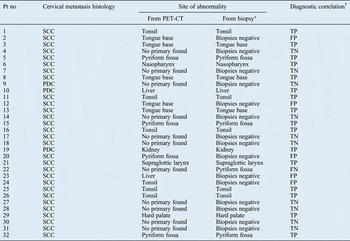
The 32 included patients comprised 23 men and 9 women. The patient population mean and median ages were both 61 years (age range, 39–86 years). In 29 patients, histological analysis of the lymph nodes showed squamous cell carcinoma; the remaining 3 patients showed poorly differentiated tumour infiltration (Table I). The imaging results were compared to the ultimate histological diagnosis, where known, to calculate the diagnostic performance of FDG PET-CT in the detection of a primary tumour. In patients with FDG-PET abnormality outside of the tonsils, a targeted biopsy was performed; patients with focal tonsillar activity underwent tonsillectomy. In FDG-PET negative cases, non-directed biopsy specimens were taken from the post-nasal space, tonsil and base of the tongue.
Patient clinical data

*Site of pathological findings on ultimate biopsy. †Comparing positron emission tomography computed tomography (PET-CT) findings to biopsy findings. Pt no = patient number; SCC = squamous cell carcinoma; TP = true positive; FP = false positive; TN = true negative; PDC = poorly differentiated carcinoma; FN = false negative
As this study was based on retrospective data collection, ethical approval was not required. The review was conducted in full accordance with the approving local institutional audit review board. Multidisciplinary team discussion was the primary mode of referral for PET-CT.
Combined positron emission tomography computed tomography acquisition and analysis
All patients were fasted for at least 6 hours prior to scanning, and their blood glucose level was checked routinely prior to injection of FDG. Patients weighing less than 100 kg were injected intravenously with 400 MBq (±40 MBq depending on body mass index), while patients weighing more than 100 kg received 500 MBq. Following a 60-minute uptake period, patients were scanned using a two-dimensional acquisition technique (as performed on all head and neck cancer patients), proceeding from vertex to mid-thigh, with the patient's head resting in a cradle and their arms by their side, spending five minutes in each of the six to seven bed positions required (per scan) to cover the body. For attenuation correction and localisation purposes, a non-contrast CT was performed prior to PET image acquisition (CT attenuation correction scan parameters: 140 kV, 80 mA). All patients were scanned using a mobile PET-CT unit.
Literature review
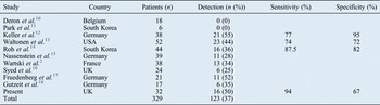
An extensive Medline search identified 10 studies (2005–2011) which specifically assessed patients receiving combined PET-CT for malignant cervical lymph nodes of unknown primary origin (Table II). Unfortunately, not all studies presented data amenable to calculating sensitivity or specificity values.
Primary tumour detection rates in patients with cervical lymph node metastases from unknown primary: published findings

Results
Primary tumour site detection
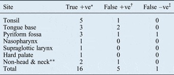
The commonest sites of primary tumour detected on PET-CT and confirmed by histology were the tonsil, tongue base and pyriform fossa (see Table III). False positives (i.e. a suspicious PET-CT but normal histological results) most commonly involved the tongue base. The only false negative site (i.e. negative PET-CT but positive histology) was the pyriform fossa (see Table III).
Primary tumour sites: true positives, false positives & false negatives

Data represent patient numbers. *Positron emission tomography computed tomography (PET-CT) positive and histology positive; †PET-CT positive but histology negative; ‡PET-CT negative but histology positive. **Kidney or liver. +ve = positive; –ve = negative
Diagnostic performance of imaging modalities
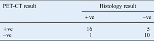
The diagnostic performance of PET-CT can be seen in Table IV. Of the 32 patients, the primary tumour was correctly detected in 16 (50 per cent detection rate). Positron emission tomography computed tomography had a sensitivity of 94 per cent (16 of 17) and a specificity of 67 per cent (10 of 15) in detecting the primary tumour. This diagnostic performance is compared with previously published data, as shown in Table II.
Diagnostic performance of PET-CT
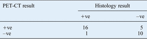
Data represent patient numbers. PET-CT = positron emission tomography computed tomography; +ve = positive; –ve = negative
Distant lesions
One or more distant lesions were identified in eight patients. These were either further metastases or a synchronous tumour, and consisted of bowel (n = 2), bone (n = 1), oesophagus (n = 1), liver (n = 2), kidney (n = 1) and lung (n = 1) tumour tissue.
Discussion
Determining the primary site of malignancy in head and neck cancer is important, as it may offer the potential for curative treatment with subsequent improved prognosis. Haas et al. Reference Haas, Hoffmann, Engers and Ganzer19 found that the three-year survival rate for patients with identified head and neck primary tumours was 100 per cent, compared with 58 per cent for patients in whom the primary tumour was not identified.
In the detection of unknown primary tumours, distant metastases and synchronous primary tumours, FDG PET-CT is a more sensitive imaging technique than either CT or MRI alone.Reference Adams, Baum, Stuckensen, Bitter and Hör20–Reference Goerres, Schmid, Bandhauer, Huguenin, von Schulthess and Schmid23 The diagnostic performance of PET alone in patients with cervical lymph node metastases from an unknown primary ranges from 20 to 35 per cent.Reference Kole, Nieweg, Pruim, Hoekstra, Koops and Roodenburg24–Reference Rusthoven, Koshy and Paulino31 However, when used alone PET has lower spatial resolution than CT or MRI. The fusion of PET and CT, first described by Beyer et al.,Reference Beyer, Townsend, Brun, Kinahan, Charron and Roddy32 enables the anatomical detail provided by CT to be combined with the functional information provided by FDG PET, and has been shown to increase the specificity and accuracy of tumour detection and to improve the diagnostic yield from targeted biopsy.Reference Wartski, Le Stanc, Gontier, Vilain, Banal and Tainturier2, Reference Deron, Bonte, Vermeersch and Van de Wiele10–Reference Gutzeit, Antoch, Kühl, Egelhof, Fischer and Hauth18
In our cohort, the diagnostic performance of PET-CT in primary tumour detection was 50 per cent; combining this result with those of 10 previous studies gave an overall primary tumour detection rate of 37 per cent. These figures highlight the added value of PET-CT in the diagnostic investigation of patients with cervical lymph node metastases from an unknown primary.
We emphasise the positive influence of a specialised head and neck cancer multidisciplinary team or tumour board on the diagnostic yield of PET-CT guided, targeted biopsy. Such a multidisciplinary team serves as a conduit for eliciting further clinical information, and allows direct communication between the radiologist interpreting the PET-CT and the surgeons (who may have performed fibre-optic examination and may be proceeding to directed biopsy).
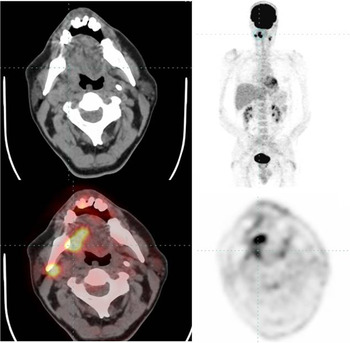
The most common primary sites found in our cohort were the tonsil, tongue base and pharyngeal wall, all parts of the oropharynx. This finding correlates with the results of other studies reviewing patients presenting with cervical lymph node metastases from an unknown primary.Reference Mendenhall, Mancuso, Parsons, Stringer and Cassisi33 This finding also fits with the rise in incidence, and significant impact, of HPV in head and neck cancer.Reference Schache, Liloglou, Risk, Filia, Jones and Shaw5–7 Examples of two confirmed PET-CT positive primaries are shown in Figures 1 and 2. Positron emission tomography computed tomography examination includes most of the body, and can result in the identification of disease outside of the head and neck region. In our study, two patients had biopsy-proven primary malignancy arising from distant primary sites, while six patients were identified with distant metastases or synchronous tumours elsewhere. This additional information can have a significant impact on treatment options, and may also prevent unnecessary surgery and influence the patient's overall prognosis.
Positron emission tomography computed tomography images for case one, showing avid 18F-fluorodeoxyglucose uptake in the left tonsillar bed; this was confirmed as squamous cell carcinoma on targeted biopsy. A left level 2 neck node is also shown to be involved.

Positron emission tomography computed tomography images for case two, showing avid 18F-fluorodeoxyglucose uptake in the right tongue base; this was confirmed as squamous cell carcinoma on targeted biopsy. Bilateral involved neck nodes are also seen.

We acknowledge that this study was limited by retrospective data collection and a small sample size. All our patients were scanned using a mobile PET-CT scanner, a suboptimal environment as regards adherence to the conventional protocol of silence during the FDG uptake period, which is essential to prevent complex physiological muscle FDG activity in the head and neck region. This may have contributed to our false positive rate (5 of 32), which is slightly higher than that reported in the literature. We emphasise that some of our ‘false positive’ cases had an inadequately short clinical follow-up period, and may well prove to be malignant in the longer term.
• This study evaluated positron emission tomography computed tomography (PET-CT) detection of occult primary tumours in patients with neck lymph node metastasis
• The primary tumour detection rate was 50 per cent
• The sensitivity was 94 per cent and the specificity 67 per cent
• Pooling with previous, comparable study findings gave an overall detection rate of 37 per cent
• To date, PET-CT has the highest detection rate for head and neck cancer patients with neck lymph node metastasis of unknown origin
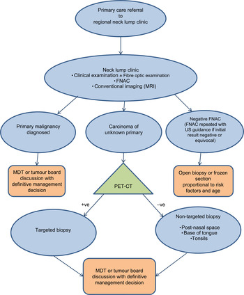
From our experience, we have devised a diagnostic algorithm for the management of patients presenting with cervical lymph node metastasis of unknown primary origin (Figure 3). In all cases, PET-CT is advised prior to directed biopsy.
Flow diagram illustrating a diagnostic algorithm for the management of patients presenting with a cervical lymph node of unknown primary origin. FNAC = fine needle aspiration cytology; US = ultrasound; MDT = multidisciplinary team; PET-CT = positron emission tomography computed tomography; +ve = positive; –ve = negative

Conclusion
This study adds further weight to the developing body of evidence advocating the use of PET-CT in the detection of primary tumour in head and neck cancer patients presenting with cervical lymph node metastases from a primary malignancy the site of which is unknown after careful review of the patient's medical history, thorough physical examination and relevant clinical tests.