Introduction
Insects use humoral and cellular defences against invading pathogens and parasites. Humoral defence responses include the production of antimicrobial peptides (cecropins, attacins, defensins, lysozymes, haemolins, lectins) (Lowenberger Reference Lowenberger2001), reactive intermediates of oxygen or nitrogen (Bogdan et al. Reference Bogdan, Rollinghoff and Diefenbach2000), and the prophenoloxidase (proPO) activating system that regulates melanisation of haemolymph (Kanost et al. Reference Kanost, Jiang and Yu2004). Cellular defence responses include haemocyte-mediated immune responses like phagocytosis, nodulation, and encapsulation (Lavine and Strand Reference Lavine and Strand2002). Small pathogens such as bacteria and fungi are generally killed by humoral defence responses or phagocytosed by haemocytes, whereas parasites such as parasitoids and nematodes are destroyed by haemocytic encapsulation and melanisation (Schmidt et al. Reference Schmidt, Theopold and Strand2001). Cellular reactions are significantly lowered in parasitised insect hosts by parasitic factors such as polydnavirus (PDV), venom, ovarian proteins, and teratocytes (cells derived from the serosal membrane of parasitoid eggs) (Pennacchio and Strand Reference Pennacchio and Strand2006; Pruissjers et al. Reference Pruissjers, Falabella, Eum, Pennacchio, Brown and Strand2009). In Cotesia kariyai (Watanabe), the laid eggs can be protected from the encapsulation reaction by an ovarian immuno-evasive protein encoded in the wasp genome (Tanaka et al. Reference Tanaka, Matsumoto and Hayakawa2002). Venom, as another host immune-suppressive factor, acts alone in Cotesia melanoscela (Ratzeburg) or synergistically in Cotesia glomerata (Linnaeus) (Bae and Kim Reference Bae and Kim2004). A PDV plays a significant role in blocking host plasma melanisation and haemocyte-spreading behaviour (Shelby et al. Reference Shelby, Ade yeye., Okot-Kotber. and Webb.2000). PDVs are symbiotic double-stranded, circular DNA viruses found in some Ichneumonoidea families, being classified as bracovirus (BV) or ichnovirus (IV) according to the host parasitoid and viral morphology (Webb et al. Reference Webb, Beckage, Hayakawa, Krell, Lanzrein and Strand2000). PDVs are present as proviruses in host chromosomes in which they are integrated and form viral particles following replication in the wasp's female reproductive organs (Wyler and Lanzrein Reference Wyler and Lanzrein2003; Pennacchio and Strand Reference Pennacchio and Strand2006). The viral particles are injected into the haemocoel of the parasitised host by the parasitoid along with the eggs (Stoltz Reference Stoltz1993). In the parasitised host, PDV genes are expressed, which induce several important parasitic roles, causing abrogation of host immunity and alteration of developmental programme (Webb and Strand Reference Webb and Strand2005).
Cotesia flavipes is a gregarious wasp that parasitises larvae of the sugarcane borer, Diatraea saccharalis (Fabricius) (Lepidoptera: Crambidae). Parasitisation of D. saccharalis larvae by the wasp causes an acute and chronic suppression of the host's cellular immune response (encapsulation and nodulation), and a prolonged larval period, which result in failure of pupal metamorphosis (Mahmoud et al. Reference Mahmoud, De Luna-Santillana and Rodríguez-Pérez2011). We have observed that parasitised larvae are susceptible to opportunistic infections (e.g., bacterial septicaemia) (Mahmoud et al. Reference Mahmoud, De Luna-Santillana and Rodríguez-Pérez2011), suggesting that the humoral antibacterial defences of these insects are also compromised. It is well known that different haemocyte types are involved in cellular immune processes. This research was aimed to analyse cellular immunosuppression of D. saccharalis parasitised by C. flavipes in terms of haemocyte composition and we measured the change of the total haemocyte population in both parasitised and unparasitised larvae. We also showed how parasitism of C. flavipes influenced the humoral immunity in terms of phenoloxidase (PO) activity of its host, D. saccharalis.
Methods
The parasitic wasps used in this study were originally imported from Africa. Cotesia flavipes were reared on its habitual hosts, D. saccharalis and have been maintained in our laboratory since 2009. Adult parasitoids were fed on honey solution and the host larvae were reared on an artificial diet, while the adults were fed with 10% sucrose as described by Mahmoud et al. (Reference Mahmoud, De Luna-Santillana and Rodríguez-Pérez2011). Diatraea saccharalis were maintained at 28 ± 1°C with a 16:8, light:dark photoperiod. Parasitism by C. flavipes was carried out by hand exposing third to sixth D. saccharalis instar to a female wasp one day after parasitoid mating. The parasitoid was allowed to oviposit one time in a single larva, and then the parasitised larvae were transferred to a new diet in a new Petri dish. After 10 days of parasitisation, the parasitoid larvae exited from the host larvae and spun cocoons (Mahmoud et al. Reference Mahmoud, De Luna-Santillana and Rodríguez-Pérez2011). Mature cocoons were collected and kept in a centrifuge tube containing a small piece of moist cotton at the bottom and a drop of honey in the inner wall for feeding the parasitoids. All solutions, glassware, and plastic materials used in the study were sterilised.
Fourth instar D. saccharalis larvae were divided into two groups at the beginning of this experiment. One group was used as a control (unparasitised), and the second group was parasitised by C. flavipes. Haemolymph was collected after 1, 3, 6, and 9 days postparasitoid oviposition and unparasitised larvae of the same age were used as control. For haemocyte counts, the larvae were chilled in ice for 5 minutes, sterilised in 70% ethanol, and then bled onto a small sterilised Petri dish by cutting off one of the abdominal prolegs with sterile scissors. Ten microlitres of haemolymph were collected using a pipette in an Eppendorf tube containing 50 μl of an anticoagulant buffer (98 mM NaOH, 186 mM NaCl, 17 mM Na2EDTA, and 41 mM citric acid, pH 4.5) to avoid haemocyte aggregation after collection. The diluted haemolymph samples were kept in ice, and the haemocytes were counted immediately after mounting using a haemocytometer under a phase contrast microscope (Olympus BX51, Tokyo, Japan). Haemocyte types were classified into granular cells, plasmatocytes, and others by distinguishing morphological characters (Gardiner and Strand Reference Gardiner and Strand1999; Ibrahim and Kim Reference Ibrahim and Kim2006). Granular cells were round cells with granules in their cytoplasm, while plasmatocytes assumed a spindle shape in the unspread state and exhibited a bilateral cytoplasmic extension when spread. Other types of haemocytes including prohaemocytes, spherules, and oenocytoids were observed but collectively named as “others” in this study.
In order to assess the capacity of whole haemolymph to display a melanisation reaction, haemolymph was collected in the same way as explained above by using the parasitised and unparasitised fifth instar larvae of the same age. The undiluted haemolymph was left on a sterilised small Petri dish for 30 minutes at ambient temperature. A change in coloration of the haemolymph, from emerald green to brown-black, was recorded as normal melanisation, whereas the maintenance of the original green colour was considered as reflecting an inhibition of melanisation (Doucet and Cusson Reference Doucet and Cusson1996). Darkening of the haemolymph (indicating that melanisation was occurring) was assessed by eye after 1, 2, 4, 6, and 7 days after parasitisation.
The fifth instar larvae of D. saccharalis were divided onto two groups at the time of haemolymph collection: larvae parasitised 1, 2, 4, 6, and 7 days earlier and unparasitised larvae of the same age which were used as control. Haemolymph PO activity was determined with a substrate, L-3,4-dihydroxyphenylalanine (DOPA), as described by Kim et al. (Reference Kim, Park, Kim and Lee2001). The larvae were sterilised in 70% ethanol and kept for 5 minutes on ice to anaesthetise the larvae before cutting the abdominal proleg for haemolymph collection. PO activity was measured in haemocytes and plasma. Haemocytes and plasma were separated by centrifugation at 8000 rpm for 10 minutes at 4°C. The haemocytes were then resuspended in 20 μl of 50 mM phosphate buffer saline (PBS, pH 7.4). For plasma, 2 μl were mixed with 10 μl PBS. To measure the activity of PO in each component, 2-μl laminarin (100 μg) was added to each reaction mixture and the mixture was incubated at room temperature for 10 minutes. The reaction mixture was then added to 900-μl substrate, 10 mM L-3,4-DOPA (Sigma Company, St. Louis, Missouri, United States of America) in PBS. The absorbance was measured at 490 nm every 5 minutes for an hour.
All data were analysed by using the SPSS11.0.0 software (Statistical Package for the Social Sciences (SPSS) 2001). The differences between means were analysed by independent samples t-test.
Results
Effect of parasitism on the total (THC) and differentiated (DHC) haemocyte counts
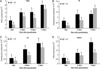
Data of THC and DHC are summarised in Figure 1. Experiments were conducted in order to assess the effect of parasitism by C. flavipes on the abundance of haemocytes in D. saccharalis haemolymph. Morphologically, the haemocytes of D. saccharalis could be classified into granular cells, plasmatocytes, and other types. As can be seen in Figure 1A, THCs were between 2 × l05 and 1.9 × l05 cells/ml of haemolymph up to one day after treatment in unparasitised and parasitised larvae, respectively. Between 3 and 9 days posttreatment, the total cell counts rose to 6 × l05 and 5 × l05 cells/ml in control and parasitised larvae, respectively. By using the t-test, the THC changed through the parasitism period and there was a significant reduction in THC counts in parasitised larvae at 3 and 6 days postparasitoid oviposition (t = 2.3457; df = 34; P = 0.025 and t = 2.0827; df = 26; P = 0.0473), respectively. However, no significant difference in THC counts was found in parasitised and unparasitised larvae after 1 and 9 days postparasitoid oviposition (t = 1.7594; df = 16; P = 0.0976 and t = 1.1851; df = 27; P = 0.2463), respectively (Fig. 1A). Regarding the differentiated haemocytes, in nonparasitised D. saccharalis, the major haemocyte type was the granule cells, which consisted of 39% in the total haemocyte population. Plasmatocytes were the next most frequent haemocyte type and composed ∼35% of the total numbers of haemocytes. Together, granular cells and plasmatocytes composed 74% of the total haemocyte population. There was a significant reduction in the plasmatocyte population in the parasitised larvae after 6 days of parasitoid oviposition (t = 2.4452; df = 26; P = 0.0216), however, no significant differences were found between parasitised and unparasitised larvae after 1 and 9 days of parasitism (t = 0.7261; df = 16; P = 0.4783 and t = 1.8317; df = 12; P = 0.0919), respectively (Fig. 1B). The population reduction of granular cells in parasitised larvae was significant only after 6 days of parasitism (t = 2.2010; df = 26; P = 0.0368), and there were no significant differences in granular cells after 1 and 9 days of parasitism (t = 0.2264; df = 16; P = 0.8237 and t = 0.6079; df = 11; P = 0.5556), respectively, in both parasitised and unparasitised larvae (Fig. 1C). The other types of haemocytes did not differ significantly after 1 and 6 days of parasitism (t = 0.3132; df = 16; 0.7582 and t = 0.9788; df = 26; P = 0.3367), respectively. However, the other types decreased significantly after 9 days of parasitism in parasitised larvae (t = 2.6970; df = 11; P = 0.0208) (Fig. 1D). At 1 and 3 days postparasitoid oviposition, there were no obvious differences found in haemocyte morphology from parasitised and unparasitised larvae. However, the haemocytes from parasitised larvae at 6 and 9 days after parasitisation showed pronounced morphological alterations. The most obvious feature was the appearance of large clumps of rounded shape haemocytes. These results suggest that parasitism by C. flavipes resulted in a significant reduction in immunocompetent cells of D. saccharalis by targeting all classes of haemocytes at potentially different times.
Effect of parasitism by Cotesia flavipes on total haemocyte counts (THC) (A), and differentiated haemocyte counts (DHC), (B) for plasmatocytes (Pl), (C) for granulocytes (Gr), and (D) for other types (others) of haemocytes in the haemolymph of parasitised (P) and unparasitised (NP) Diatraea saccharalis larvae 1 (n = 10 for NP and 8 for P), 3 (n = 8 for NP and 10 for P), 6 (n = 10 for NP and 16 for P), and 9 days (n = 8 for NP and 5 for P) following parasitisation by C. flavipes. Different letters above error bar indicate significant difference of means (±SE) at a = 0.05 (t-test).

Effect of parasitism on melanisation of haemolymph and PO activity
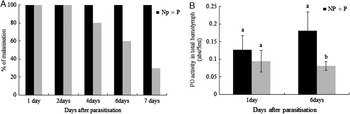
Haemolymph drawn from each larva was exposed to air and checked for evidence of melanisation after 30 minutes. As indicated in Figure 2, haemolymph from unparasitised larvae darkened progressively with time once exposed to air. By contrast, haemolymph from parasitised larvae melanised more slowly and to a lesser extent than that from the unparasitised larvae. Nearly every haemolymph sample obtained from unparasitised and parasitised larvae melanised at all five sampling times. Melanisation was also observed in 100% of samples collected from parasitised and unparasitised insects after 1 and 2 days of oviposition, but only 80%, 60%, and 30% of such samples showed evidence of melanisation 4, 6, and 7 days after parasitisation, respectively (Fig. 2A). In order to determine if the inhibition of melanisation described here was the result of an inactive PO system, PO activity was measured in whole haemolymph of unparasitised and parasitised larvae. Results presented in Figure 2B indicate that PO activity in parasitised larvae was significantly lower than in control animals 6 days after parasitisation (t = 2.4785; df = 824; P = 0.0382), and there was no significant difference in PO after 24 hours of parasitism (t = 0.6272; df = 4; P = 0.5646) (Fig. 2B).
(A) Percentage of haemolymph samples showing melanisation 1, 2, 4, 6, and 7 days following parasitisation by Cotesia flavipes (n = 3). (B) Phenoloxidase (PO) activity in whole haemolymph in parasitised (P) (n = 5) and unparasitised (NP) (n = 4), Diatraea saccharalis, 1 and 6 days postparasitoid oviposition. Different letters above error bar indicate significant difference of means (±SE) at a = 0.05 (t-test).

Effect of parasitisation on PO activity in different fractions of D. saccharalis haemolymph
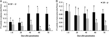
In order to characterise the localisation of active PO in the haemolymph, PO activity was compared between the plasma and haemocytes (Figs. 3A, 3B). Although both showed significant levels of PO activity, the plasma possessed higher level of PO activity (Fig. 3A). The PO activity of the plasma decreased significantly after 6 and 7 days of parasitisation (t = 2.8883; df = 4; P = 0.0446 and t = 2.8818; df = 4; P = 0.0449), respectively. However, no significant differences were found in plasma samples from parasitised and unparasitised larvae after 1, 2, and 4 days of parasitism (t = 0.7238, df = 6; P = 0.4965; t = 0.9535; df = 6; P = 0.3772 and t = 1.6048; df = 6; P = 0.1597), respectively (Fig. 3A). As shown in Figure 3B, on day 6 and 7 of parasitisation the PO activity of the haemocytes decreased significantly in parasitised larvae (t = 4.9421; df = 4; P = 0.0078 and t = 2.9982; df = 4; P = 0.0400), respectively. Moreover, there were no significant differences in the PO activity of the haemocytes between parasitised and unparasitised larvae after 1, 2, and 4 days of parasitism (t = 0.0389; df = 4; P = 0.9708; t = 0.0272; df = 6; P = 0.9792 and t = 0.5473; df = 6; P = 0.6046), respectively. PO activity was found to be present in all compartments of the haemolymph tested; haemocytes and plasma, but the majority of PO activity detected in haemolymph of D. saccharalis larvae was associated with the plasma. The most interesting finding, however, was that PO activity in both plasma and haemocytes decreased significantly by 6 days postparasitisation in parasitised larvae compared with unparasitised controls.
Phenoloxidase (PO) activity in plasma (A) and haemocytes (B) in parasitised (P) (n = 4) and unparasitised (NP) (n = 4), Diatraea saccharalis. Different letters above error bar indicate significant difference of means (±SE) at a = 0.05 (t-test).

Discussion
Decrease of the haemocyte population in the parasitised D. saccharalis
Haemocytes are functional elements involved in recognition and encapsulation of foreign objects by suppressing the host's immune system. Most parasitoids suppress the host's immune reactions by interfering with the function of haemocytes (Ibrahim and Kim Reference Ibrahim and Kim2006). It has been mentioned that the primary physiological functions of parasitoid-associated BVs and IVs is to suppress the host immune response, either by altering host haemocyte behaviour and adhesiveness (Beck and Strand Reference Beck and Strand2005), or by inducing apoptosis of the cells so they are no longer able to mobilise an effective encapsulation response (Amaya et al. Reference Amaya, Asgari, Jung, Hongskula and Beckage2005). Our study showed that parasitism of D. saccharalis by C. flavipes clearly affected the haemocyte load, and reduced the haemocyte counts of D. saccharalis (either THC or DHC) (Fig. 1). The reductions were most evident in the numbers of the granular cells and plasmatocytes. Moreover, this study suggests that these two types of haemocytes are the major haemocytes in D. saccharalis, and play the most significant roles in cellular immune reactions in D. saccharalis. In general our data were similar to the results reported in many other parasitoid/host systems such as Cotesia vestalis (Haliday) (as Cotesia plutellae Kurdjumov)/Plutella xylostella (Linnaeus) (Lepidoptera: Plutellidae) (Ibrahim and Kim Reference Ibrahim and Kim2006; Yu et al. Reference Yu, Chen, Chen, Huang, Lou and Liu2007), Macrocentrus cingulum Brischke (Hymenoptera: Braconidae)/Ostrinia furnacalis (Guenée) (Lepidoptera: Crambidae) (Hu et al. Reference Hu, Xiong, Wen and Fu2003), Tranosema rostrale (Brischke) (Hymenoptera: Ichneumonidae)/Choristoneura fumiferana (Clemens) (Lepidoptera: Torticidiae) (Doucet and Cusson Reference Doucet and Cusson1996), and Microplitis demolitor Wilkinson (Hymenoptera: Braconidae)/Pseudoplusia includens (Walker) (Lepidoptera: Noctuidae) (Strand and Pech Reference Strand and Pech1995), in which parasitism variously increased or reduced the host's haemocyte load. It has also been demonstrated that changes of haemocytes can be induced by the injection of calyx fluid, or PDVs alone or in the presence of venom (Yu et al. Reference Yu, Chen, Chen, Huang, Lou and Liu2007). This indicates that these maternal factors may actively interfere with the haemocyte immune reaction. In several endoparasitoids, the parasitism results in a significant reduction of host haemocyte population, presumably due to directly induced immunosuppression (Ibrahim and Kim Reference Ibrahim and Kim2006). Haemocyte reduction in the parasitism of C. kariyai was caused by apoptosis of the circulating haemocytes and by histolysis of the haematopoietic organ (Teramoto and Tanaka Reference Teramoto and Tanaka2004). The fact that the plasmatocytes are produced from the granular cells by mitotic division and differentiation in some Lepidoptera species (Nakahara et al. Reference Nakahara, Kanamori, Kiuchi and Kamimura2003) suggests that the reduction of the haemocyte populations in the parasitised D. saccharalis is induced by cytotoxic effects of the parasitism on the circulating granular cells and by a shutdown of plasmatocyte production from the haematopoietic organ of D. saccharalis. The latter may result in a delayed decrease of the plasmatocyte population until the natural death of the circulating plasmatocytes. In fact, the granular cell population decreased faster than that of plasmatocytes (Lavine and Strand Reference Lavine and Strand2002). In the parasitism of a braconid wasp, M. demolitor, the PDV has been shown to induce apoptosis in only the granular cells of its host, P. includens (Strand and Pech Reference Strand and Pech1995). The significant reduction of granular cells and plasmatocytes following parasitisation suggests that these two haemocytes play significant roles in the cellular immune responses against C. flavipes parasitism. Recent genome approaches to studying the C. plutellae BV suggest that some viral candidate genes are implicated in modifying cell signalling such as protein tyrosine phosphatases (Ibrahim and Kim Reference Ibrahim and Kim2006) and IkB-like (Kim et al. Reference Kim, Basio, Ibrahim and Bae2006), which may disrupt the activation of actin-polymerisation in haemocytes (Baldwin and Barbieri Reference Baldwin and Barbieri2005; Walzer et al. Reference Walzer, Galibert and De Smedt2005). Further, the recovery of the total haemocyte number within the hosts 9 days after parasitism suggests additional factors exist and maintain the haemocyte numbers in the hosts at a low level, possibly substances secreted from teratocytes or the larva elicit effects on host hematopoiesis. Also, it may be more likely that the combinatorial immunosuppressive effects of parasitism (ovarian proteins + venoms + PDV + teratocytes + larval secretions, etc…) just begin to wear off in the latent stages as the parasitoid larvae near emergence. Many systems have a “peak” of immunosuppressive activities that wear off over time.
Effect of parasitism by C. flavipes on haemolymph melanisation and PO activity
Parasitisation of Lepidoptera larvae by endoparasitoids is usually associated with suppression of host cellular and humoral immunity (Glatz et al. Reference Glatz, Asgari and Schmidt2004). Our results show that parasitisation by C. flavipes reduces haemocytes and total haemolymph melanisation response of D. saccharalis larvae. When PO activity in haemocytes and plasma from unparasitised and parasitised D. saccharalis larvae were compared, two important observations were made. First, in all cases, PO activity was present in each of the haemolymph fractions. Second, there was less activity in fractions from parasitised larvae compared with those from unparasitised insects. The reduced level of haemolymph defensive melanisation in the parasitised host has been accompanied by reduced activity of PO (Lavine and Beckage Reference Lavine and Beckage1995). Phenoloxidase is an essential enzyme for melanisation, catalysing the early steps of a cascade of reactions leading to deposition of eumelanin onto foreign particles (Ashida and Brey Reference Ashida and Brey1995). Reduced plasma melanisation, attributed to reduced PO activity, is characteristic of infection by several PDVs (both BV and IV) (Shelby and Webb Reference Shelby and Webb1999). For example, the Cotesia congregata (Say)/Manduca sexta (Linnaeus) (Lepidoptera: Sphingidae) (Beckage et al. Reference Beckage, Metcalf, Nesbit, Schleifer, Zetlan and DeBuron1990), and C. plutellae/P. xylostella (Bae and Kim Reference Bae and Kim2004) systems exhibited significant reductions of haemolymph melanisation and monophenoloxidase activity following parasitisation. Similar effects were observed in Trichoplusia ni (Hübner) (Lepidoptera: Noctuidae) parasitised by Hyposoter exiguae (Viereck) (Hymenoptera: Ichneumonidae) (Stoltz and Cook Reference Stoltz and Cook1983). Stoltz and Guzo (Reference Stoltz and Guzo1986) observed markedly slower rates of melanisation in haemolymph from host Malacosoma disstria Hübner (Lepidoptera: Lasiocampidae) parasitised by Hyposoter fugitivus (Say), and correlated this phenomenon with the presence of PDV and enhanced stability of the normally labile haemocytes (oenocytoids), which are haemocytes that are involved in the synthesis and storage of proPO (Gupta Reference Gupta1991). Moreover, Stoltz and Cook (Reference Stoltz and Cook1983) found that Heliothis virescens plasma melanisation is greatly reduced by IV infection (CsIV). This was the first indication that PDV infection compromises humoral elements of the host immune response; however, the specific mode of action of any PDV gene in this regard remains unknown.
With regard to the distribution of PO activity, haemolymph of D. saccharalis resembles that of other Lepidoptera such as Lacanobia oleracea (Linnaeus) (Noctuidae) and Bombyx mori (Linnaeus) (Bombycidae), where the enzyme is present in both haemocytes and plasma (Huxham and Lackie Reference Huxham and Lackie1988; Richards and Edwards Reference Richards and Edwards2000). Furthermore, in B. mori oenocytoids synthesise and store the enzyme for release. It has been shown that specific haemocyte subpopulations synthesise proPO, which is released into the plasma via cell lysis, as proPO evidences no signal secretion sequence (Ashida and Brey Reference Ashida and Brey1998). The activation of proPO in the plasma is mediated by a serine protease cascade, and the resulting active PO has a tendency to adhere to foreign surfaces, including the haemocyte surface (Ling and Yu Reference Ling and Yu2005; Mavrouli et al. Reference Mavrouli, Tsakas, Theodorou, Lampropoulou and Marmaras2005). In addition, Hilgarth (Reference Hilgarth1997) reported that Campoletis sonorensis (Cameron) (Hymenoptera: Ichneumonidae) calyx fluid reduces the concentrations of PO substrates tyrosine and tyrosine glucoside. In summary, this study shows that parasitism of C. flavipes down-regulates the haemocyte population of D. saccharalis and suppresses both cellular and humoral immune reactions. In the future, we need to identify the immunosuppressive genes from the parasitoid and use these genes to make a biopesticide as an effective and save method to control D. saccharalis.
Acknowledgements
We thank the Secretaria de Investigación y Posgrado del Instituto Politécnico Nacional for financial support (grant number 20101198). Mario A. Rodríguez-Pérez holds a scholarship from the Comisión de Operación y Fomento de Actividades Académicas of Instituto Politécnico Nacional (IPN). Ali Mohammed holds a doctoral scholarship from the Consejo Nacional de Ciencia y Tecnología – México (CONACYT). We also acknowledge the Centro de Biotecnología Genómica and Centro de Investigación en Ciencia Aplicada y Tecnología Avanzada (Unidad Altamira) of IPN. Ali Mohammed is also grateful to Esraa Gamal for her support and encouragement. We thank Drs. Nancy Beckage and Robert Meagher for providing the parasitoid-host system.