Introduction
In the latter part of the nineteenth century, there was much debate and excitement over the use of cotton wool and other objects to improve hearing, by applying them to the middle-ear structures.Reference Chu and Jackler1 However, the process was for the most part abandoned by the medical profession, because of the culture of quackery surrounding such devices. That there was some merit to their use is highlighted by cases such as the one presented here.
Case report
A 56-year-old Russian woman presented with longstanding, bilateral middle-ear disease and worsening hearing. She had undergone at least one surgical procedure on her right ear in Russia, the details of which were unavailable. Despite her ear disease, she continued to perform as an accomplished violinist.
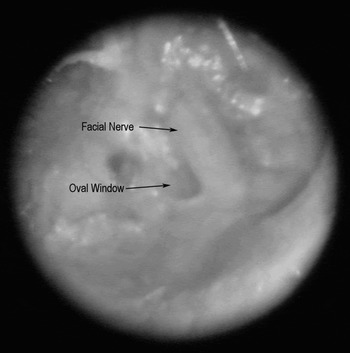
On examination, she was noted to have cotton wool in both ears. When this was removed, she was seen to have extensive atelectasis of both tympanic membranes, with bilateral scutal erosion or removal. Both drums were adherent to the medial wall of the middle ear, with no ossicles visible on either side (Figure 1).
Otoscopic view of medial wall of left tympanic cavity.

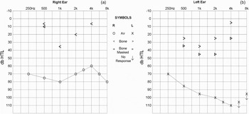
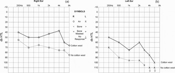
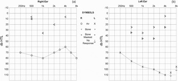
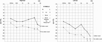
Pure tone audiometry demonstrated a severe mixed loss on the right and a severe to profound mixed loss on the left. There were large conductive components to the hearing loss bilaterally (Figure 2). Unusually, the patient was observed to have a significant improvement in her hearing when she inserted cotton wool into her ears, up to 40 dB difference (Figure 3).
Baseline audiogram.
Audiogram showing hearing improvement following cotton wool insertion.
This technique had been taught to her by a Russian doctor prior to her emigration. The patient would stand beside a running tap and twist moist cotton wool into her ear, stopping when the tap was perceived loudest.
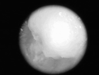
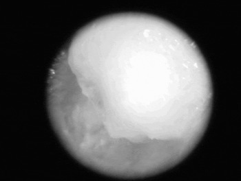
Photographs were taken of the smallest amount of cotton wool able to achieve this effect, in an attempt to identify the position of the cotton wool in the ear (Figure 4).
Otoscopic view of cotton wool pellet in the middle ear, at the site inducing maximum hearing improvement.
The patient had a satisfactory result from a trial of hearing aids, and failed to attend any further follow up.
Discussion
Cotton wool applied to the ear is well known to decrease sound intensity.Reference Veale2 The application of moist cotton wool to a particular part of the ear to increase sound intensity seems counterintuitive. Historically however, cotton wool has been used to improve hearing in patients with a tympanic membrane perforation.Reference Yearsley3–Reference Graf7 A pellet of wool or other device would be inserted to rest on the small portion of the tympanic membrane which remained, although the exact mechanism of how this produced benefit was unknown. Raut and Kerr reported a case of hearing improvement resulting from a patient inserting a saliva-moistened paper towel into his ear.Reference Raut and Kerr8 This patient had a postero-superior perforation the anterior edge of which was adherent to the promontory. They postulated the mechanism to be a temporary ossiculoplasty rather than occlusion of the perforation. In our case, the tympanic membrane was also atelectatic but was without perforation.
We postulate that the surface area difference between a relatively large pellet of cotton wool coupled to a smaller area on the stapes footplate produced an amplification effect. The in vivo effect of an intact tympanic membrane and ossicular chain accounts for approximately 40 dB of hearing amplification.Reference von Bekesy9 Interestingly, this is about the degree of improvement our patient experienced with her cotton wool insertion.
Alternatively, the cotton wool may perhaps create a sound pressure phase difference so that, in the absence of ossicles, sound does not hit the round and oval windows simultaneously. Theoretically, this could dampen the flow of pressure being transmitted in a unilateral direction from the vibrating window through the perilymph.
A baffle effect may be a further possible explanation of our patient's hearing improvement.Reference Pierce10 Sound intensity within the perilymph is increased if the pressure wave is unable to escape back to the middle ear via the occluded round window. This potentially turns the cochlea into a blind-ended sac, acting as a baffle for the oval window which is still free to vibrate. While our patient had a very atelectatic tympanic membrane, it did not appear to be retracted into the round window niche (Figure 4), thereby suggesting that occlusion of the round window was an unlikely cause of improved hearing in our case.
• Unusually, the reported patient's hearing was improved by insertion of cotton wool onto a retracted tympanic membrane
• In the latter part of the ninetheenth century, there was much debate over the use of cotton wool and other objects applied to middle-ear structures to improve hearing
• The mechanism of hearing improvement in this case is unknown, but is likely to relate to sound amplification from the relatively large cotton wool surface area to the smaller oval window
The baffle effect and phase difference theories assume that cotton wool only has an insulating or dampening effect on sound. The amplification theory, however, implies that moist cotton wool is sufficiently rigid to act as a sound conductor.
Foreign body insertion into the ear is not advocated by modern otologists. Nevertheless, there are still intricacies of sound conduction and sound-conducting materials which are difficult to explain. Our patient's significant improvement in hearing on inserting moist cotton wool in her ear is such a point. Whereas Raut and Kerr's patient did not tolerate the distortion his hearing aid produced, our patient had an excellent functional improvement from her more modern aid, and continued to use this in her role as an accomplished professional musician.Reference Raut and Kerr8