INTRODUCTION
Members of the heat shock protein 40 (Hsp40) and heat shock protein 70 (Hsp70) families, many of which are molecular chaperones, are proposed to play a significant role in the establishment and development of the malaria parasite within host cells. Plasmodium falciparum Hsp40s (PfHsp40s) and Hsp70s (PfHsp70s) display, in addition to the features typical of the heat shock proteins of these classes, unique characteristics, which are likely to have arisen for the distinctive lifestyle of the parasite (reviewed in Njunge et al. Reference Njunge, Ludewig, Boshoff, Pesce and Blatch2013).
All Hsp40s have a signature J-domain, and are classified into types I to IV based on the presence of various other domains (reviewed by Botha et al. Reference Botha, Pesce and Blatch2007). The canonical type I and II Hsp40s have some independent chaperone properties. However, most Hsp40s act as co-chaperones of Hsp70s, using their J-domain to interact with Hsp70 when a client (substrate) protein is present, stimulating its ATP hydrolysis rate and thereby activating its chaperone activity. Hsp40s provide client protein specificity to the Hsp70 chaperone system, either by interacting with and delivering client proteins to Hsp70 or by activating an Hsp70 in the proximity of a client protein. We will not attempt to fully describe the structure and function of the Hsp40 and Hsp70 chaperones here, as this system has been extensively reviewed in the literature (reviewed in Kampinga and Craig, Reference Kampinga and Craig2010).
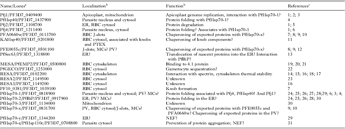
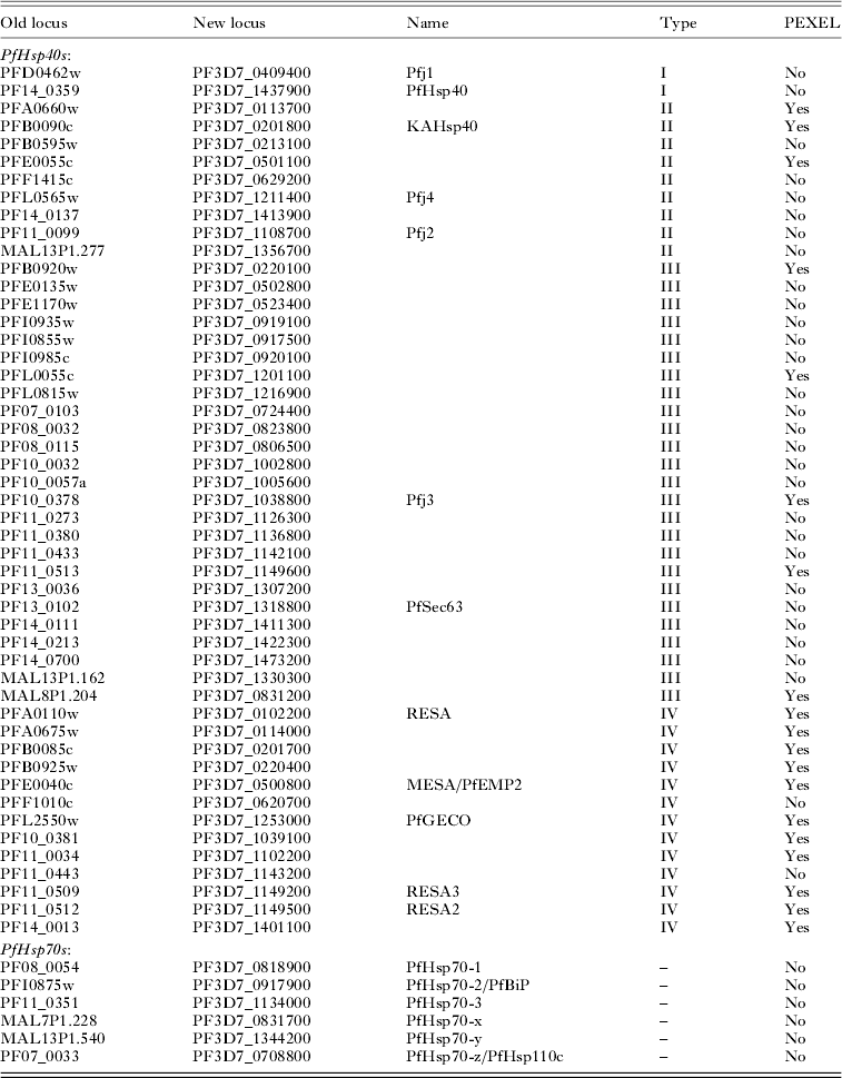
Up to now, as many as 49 PfHsp40s and 6 PfHsp70s have been identified in P. falciparum (Appendix Table A1; Shonhai et al. Reference Shonhai, Boshoff and Blatch2007; Njunge et al. Reference Njunge, Ludewig, Boshoff, Pesce and Blatch2013); however, only a few of them have been investigated (Table 1). Several PfHsp40s are predicted to be part of the ‘exportome’ as they bear the Plasmodium export element (PEXEL) or vacuolar translocation signal (VTS) (Hiller et al. Reference Hiller, Bhattacharjee, van Ooij, Liolios, Harrison, Lopez-Estraño and Haldar2004; Marti et al. Reference Marti, Good, Rug, Knuepfer and Cowman2004; Sargeant et al. Reference Sargeant, Marti, Caler, Carlton, Simpson, Speed and Cowman2006; Boddey et al. Reference Boddey, Carvalho, Hodder, Sargeant, Sleebs, Marapana, Lopaticki, Nebl and Cowman2013) with the type IV Hsp40s being the majority (Botha et al. Reference Botha, Pesce and Blatch2007; Njunge et al. Reference Njunge, Ludewig, Boshoff, Pesce and Blatch2013). Conversely, only one PfHsp70, PfHsp70-x, has been experimentally located in the parasitophorous vacuole (PV) and the infected erythrocyte cytosol (Külzer et al. Reference Külzer, Charnaud, Dagan, Riedel, Mandal, Pesce, Blatch, Crabb, Gilson and Przyborski2012; Grover et al. Reference Grover, Chaubey, Ranade and Tatu2013).
Localization and function of some P. falciparum Hsp40s and Hsp70s

a If a common name exists this is given; if a common name does not exist the commonly used old gene locus is given; the new gene locus is provided for all proteins.
b Abbreviations: ER, endoplasmic reticulum; RBC, red blood cell or erythrocyte; MCs, Maurer's clefts; PV, parasitophorous vacuole.
c References: 1. Watanabe (Reference Watanabe1997); 2. Kumar et al. (Reference Kumar, Tanveer, Biswas, Ram, Gupta, Kumar and Habib2010); 3. Misra and Ramachandran (Reference Misra and Ramachandran2009); 4. Botha et al. (Reference Botha, Chiang, Needham, Stephens, Hoppe, Külzer, Przyborski, Lingelbach, Wipf, Brodsky, Shonhai and Blatch2011); 5. Botha et al. (Reference Botha, Pesce and Blatch2007); 6. Pesce et al. (Reference Pesce, Acharya, Tatu, Nicoll, Shonhai, Hoppe and Blatch2008); 7. Maier et al. (Reference Maier, Rug, O'Neill, Brown, Chakravorty, Szestak, Chesson, Wu, Hughes, Coppel, Newbold, Beeson, Craig, Crabb and Cowman2008); 8. Külzer et al. (Reference Külzer, Rug, Brinkmann, Cannon, Cowman, Lingelbach, Blatch, Maier and Przyborski2010); 9. Külzer et al. (Reference Külzer, Charnaud, Dagan, Riedel, Mandal, Pesce, Blatch, Crabb, Gilson and Przyborski2012); 10. Grover et al. (Reference Grover, Chaubey, Ranade and Tatu2013); 11. Acharya et al. (Reference Acharya, Chaubey, Grover and Tatu2012); 12. Bhattacharjee et al. (Reference Bhattacharjee, van Ooij, Balu, Adams and Haldar2008); 13. Tuteja (Reference Tuteja2007); 14. Foley et al. (Reference Foley, Murray and Anders1990); 15. Foley et al. (Reference Foley, Corcoran, Tilley and Anders1994); 16. Silva et al. (Reference Silva, Cooke, Guillotte, Buckingham, Sauzet, Le Scanf, Contamin, David, Mercereau-Puijalon and Bonnefoy2005); 17. Pei et al. (Reference Pei, Guo, Coppel, Bhattacharjee, Haldar, Gratzer, Mohandas and An2007); 18. Diez-Silva et al. (Reference Diez-Silva, Park, Huang, Bow, Mercereau-Puijalon, Deplaine, Lavazec, Perrot, Bonnefoy, Feld, Han, Dao and Suresh2012); 19. Lustigman et al. (Reference Lustigman, Anders, Brown and Coppel1990); 20. Bennett et al. (Reference Bennett, Mohandas and Coppel1997); 21. Waller et al. (Reference Waller, Nunomura, An, Cooke, Mohandas and Coppel2003); 22. Morahan et al. (2011); 23. Durand et al. (Reference Durand, Migot-Nabias, Andriantsoanirina, Seringe, Viwami, Sagbo, Lalya, Deloron, Mercereau-Puijalon and Bonnefoy2012); 24. Kumar et al. (Reference Kumar, Koski, Harada, Aikawa and Zheng1991); 25. Shonhai et al. (Reference Shonhai, Boshoff and Blatch2008); 26. Vincensini et al. (Reference Vincensini, Richert, Blisnick, Van Dorsselaer, Leize-Wagner, Rabilloud and Braun Breton2005); 27. Nyalwidhe and Lingelbach (Reference Nyalwidhe and Lingelbach2006); 28. Ramya et al. (Reference Ramya, Surolia and Surolia2006); 29. Shonhai et al. (Reference Shonhai, Boshoff and Blatch2007); 30. Akide-Ndunge et al. (Reference Akide-Ndunge, Tambini, Giribaldi, McMillan, Müller, Arese and Turrini2009); 31. Muralidharan et al. (Reference Muralidharan, Oksman, Pal, Lindquist and Goldberg2012).
PARASITE RESIDENT Hsp70S AND Hsp40S: HOW MUCH DO WE KNOW?
Experimental data are available for about a third of the PfHsp40s with the putative exported ones having received much attention as they may be implicated more directly in the remodelling of the host cell and therefore pathogenesis and virulence of the malaria parasite. In contrast, the parasite resident PfHsp70-1 has been extensively studied especially with regards to its molecular chaperone properties (Matambo et al. Reference Matambo, Odunuga, Boshoff and Blatch2004; Shonhai et al. Reference Shonhai, Boshoff and Blatch2005, Reference Shonhai, Botha, De Beer, Boshoff and Blatch2008; Ramya et al. Reference Ramya, Surolia and Surolia2006; Bell et al. Reference Bell, Chiang and Brodsky2011). PfHsp70-1 is highly abundant, with high levels throughout the parasite asexual life cycle (Acharya et al. Reference Acharya, Pallavi, Chandran, Chakravarti, Middha, Acharya, Kochar, Kochar, Subudhi, Boopathi, Garg, Da and Tatu2009), which further increase at febrile temperatures (Kumar et al. Reference Kumar, Koski, Harada, Aikawa and Zheng1991; Joshi et al. Reference Joshi, Biswas and Sharma1992; Pesce et al. Reference Pesce, Acharya, Tatu, Nicoll, Shonhai, Hoppe and Blatch2008). It localizes in the parasite nucleus and cytosol (Kumar et al. Reference Kumar, Koski, Harada, Aikawa and Zheng1991; Pesce et al. Reference Pesce, Acharya, Tatu, Nicoll, Shonhai, Hoppe and Blatch2008) and is believed to function as a typical Hsp70 involved in protein homoeostasis as well as cytoprotection during stress conditions. PfHsp70-1 could also be important in keeping certain proteins in an unfolded translocation-competent state; such as proteins destined for the apicoplast (Foth et al. Reference Foth, Ralph, Tonkin, Struck, Fraunholz, Roos, Cowman and McFadden2003) or for secretion (Gehde et al. Reference Gehde, Hinrichs, Montilla, Charpian, Lingelbach and Przyborski2009). In this regard, proteomic studies have reported the presence of PfHsp70-1 in the PV (Nyalwidhe and Lingelbach, Reference Nyalwidhe and Lingelbach2006), and so it is tempting to propose an interaction between PfHsp70-1 and components of the Plasmodium translocon of exported proteins (PTEX) complex (de Koning-Ward et al. Reference de Koning-Ward, Gilson, Boddey, Rug, Smith, Papenfuss, Sanders, Lundie, Maier, Cowman and Crabb2009). However, the localization of this chaperone in the PV has not been confirmed via immunofluorescence and EM analyses (Kumar et al. Reference Kumar, Koski, Harada, Aikawa and Zheng1991; Pesce et al. Reference Pesce, Acharya, Tatu, Nicoll, Shonhai, Hoppe and Blatch2008) and therefore it is plausible that the PV localization resulted from contamination of the PV cellular fraction with the parasite fraction. Moreover, another member of the Hsp70 protein family, PfHsp70-x, has been recently identified in the PV (Külzer et al. Reference Külzer, Charnaud, Dagan, Riedel, Mandal, Pesce, Blatch, Crabb, Gilson and Przyborski2012; Grover et al. Reference Grover, Chaubey, Ranade and Tatu2013) making it a more suitable candidate for a role in protein trafficking across the PV membrane (PVM) (Fig. 1). PfHsp70-1 was also identified in the proteome of P. falciparum-infected erythrocyte membrane ghost fractions inferring its presence in the Maurer's clefts (Vincensini et al. Reference Vincensini, Richert, Blisnick, Van Dorsselaer, Leize-Wagner, Rabilloud and Braun Breton2005). Also in this case, though, the lack of further evidence suggests cross contamination due to the high abundance of this protein in the parasite. As it has been found for other eukaryotic chaperone systems, potential PfHsp40 partners of PfHsp70s are starting to be identified. The canonical type I PfHsp40 (called PfHsp40) represents a likely co-chaperone of PfHsp70-1 since it is similarly heat inducible and localizes mainly in the parasite cytosol (Botha et al. Reference Botha, Chiang, Needham, Stephens, Hoppe, Külzer, Przyborski, Lingelbach, Wipf, Brodsky, Shonhai and Blatch2011). Although a functional interaction between PfHsp70-1 and PfHsp40 was shown by ATPase and aggregation assays in vitro, a direct association of the two proteins in vivo is yet to be validated (Botha et al. Reference Botha, Chiang, Needham, Stephens, Hoppe, Külzer, Przyborski, Lingelbach, Wipf, Brodsky, Shonhai and Blatch2011). In contrast, there is evidence for a partnership in vivo between PfHsp70-1 and Pfj4, but proof of a co-chaperone–chaperone interaction is still missing (Pesce et al. Reference Pesce, Acharya, Tatu, Nicoll, Shonhai, Hoppe and Blatch2008). A third PfHsp40, Pfj1, was shown to functionally interact with PfHsp70-1 when using in vitro protein refolding and ATPase assays (Misra and Ramachandran, Reference Misra and Ramachandran2009). However, the subsequent report of Pfj1 as an apicoplast Hsp40 (Kumar et al. 2010) is not consistent with a Pfj1-PfHsp70-1 partnership. In addition, Kumar and co-authors suggested that Pfj1 binds to the apicoplast genome, and in their experimental conditions they excluded it from the mitochondrion even though a putative mitochondrial import signal has been previously proposed (Watanabe, Reference Watanabe1997). Based on these contrasting data we propose that the localization and function of Pfj1 warrants deeper investigation.
A proposed role for Hsp40 and Hsp70 chaperones in protein export in P. falciparum-infected erythrocytes. A section through a parasite-infected erythrocyte is schematically depicted, showing the parasite with its nucleus (N), endoplasmic reticulum (ER) and plasma membrane (PPM), enclosed in the parasitophorous vacuole (PV) and its membrane (PVM). The infected erythrocyte is shown with Maurer's clefts (MC), knobs (K) and J-dots (J). This model addresses the role for chaperones in the export of soluble proteins which may be Plasmodium export element (PEXEL)-containing proteins or PEXEL-negative exported proteins (PNEPs) (Spielmann and Gilberger, Reference Spielmann and Gilberger2010). Similar roles may also apply for membrane proteins, and these are discussed in the text. (1) Proteins destined for export emerge from the ribosome (R) in the parasite cytosol, and may associate with Hsp40 and Hsp70 chaperones before being transported into the ER as occurs with post-translational transport; however, it has been proposed that signal-recognition particle-based co-translational transport into the ER predominates in P. falciparum (Tuteja, Reference Tuteja2007; Zimmermann and Blatch, Reference Zimmermann and Blatch2009). The unfolded protein is translocated through an ER translocon (e.g. a Sec61-based translocon) and captured by Hsp70 and Hsp40 chaperones in the lumen of the ER (e.g. PfBiP and PfSec63). (2) The Hsp40 and Hsp70 chaperones or other unidentified chaperones (plasmepsin V involved in cleaving the PEXEL motif, may also act as a dedicated ‘chaperone’; Bullen et al. Reference Bullen, Crabb and Gilson2012) may be involved in maintaining the exported protein in an unfolded but translocation competent state as the protein is trafficked through the ER and subsequent vesicular compartments before being released into the PV. (3) The PV is rich in chaperones, including Hsp70s (e.g. PfHsp70-x; Külzer et al. Reference Külzer, Charnaud, Dagan, Riedel, Mandal, Pesce, Blatch, Crabb, Gilson and Przyborski2012; Grover et al. Reference Grover, Chaubey, Ranade and Tatu2013) and potentially Hsp40s, which may be involved in delivering the unfolded exported protein to a translocon in the PVM (e.g. Plasmodium translocon of exported proteins, PTEX; de Koning-Ward et al. Reference de Koning-Ward, Gilson, Boddey, Rug, Smith, Papenfuss, Sanders, Lundie, Maier, Cowman and Crabb2009). (4) The exported protein is transported into the infected erythrocyte cytosol where it is bound by parasite or host Hsp40 and Hsp70 chaperones (e.g. KAHsp40, Acharya et al. Reference Acharya, Chaubey, Grover and Tatu2012). Parasite Hsp40 and Hsp70 chaperones associated with J-dots (e.g. PfHsp70-x, PFA0660w and PFE0055c) may be involved (Külzer et al. Reference Külzer, Rug, Brinkmann, Cannon, Cowman, Lingelbach, Blatch, Maier and Przyborski2010, Reference Külzer, Charnaud, Dagan, Riedel, Mandal, Pesce, Blatch, Crabb, Gilson and Przyborski2012). Finally the exported protein is released at its final location and allowed to fold into its three-dimensional functional state.

Although three putative co-chaperones of PfHsp70-1, PfHsp40, Pfj4 and PfHOP (see Gitau et al. Reference Gitau, Mandal, Blatch, Przyborski and Shonhai2012) have been suggested, the client proteins of PfHsp70-1 are yet to be described. The identification of client proteins for PfHsp70-1 and their discrimination from co-chaperones is a challenging task; co-chaperone–chaperone interactions tend to be transient with electrostatic contacts predominating, while unfolded or partially folded client proteins interact more stably with chaperones through mainly hydrophobic contacts.
Of the remaining parasite resident PfHsp40s, Pfj2 and PfSec63 are notable members that are predicted to be localized to the endoplasmic reticulum (ER). Pfj2 contains a putative ER retrieval sequence as well as a putative PEXEL motif within the J-domain. The processing of the PEXEL sequence in the ER for transport into the infected erythrocyte would give rise to a protein lacking a functional J-domain. We therefore favour the idea that this is not a genuine export signal and the protein is retained in the ER (Botha et al. Reference Botha, Pesce and Blatch2007). However, Pfj2 could be exported to the infected erythrocyte cytosol, where its thioredoxin and thioredoxin-like domains could have a role independent of the J-domain. In fact, until the location of Pfj2 is clearly established we cannot exclude the possibility that it could function in both the ER and the infected host cell. Its similarity to human ERdj5 also suggests that it may act as a disulphide bond reductase and together with PfBiP be involved in ER-associated degradation (ERAD) (reviewed in Hagiwara and Nagata, Reference Hagiwara and Nagata2012). PfSec63 might be part of the Sec complex (Fig. 1) and therefore participate in the translocation of nascent proteins into the ER by interacting with PfBiP (Tuteja, Reference Tuteja2007). The silencing of other Sec63 homologues of parasitic origin (e.g. Trypanosoma brucei Sec63) results in dramatic effects on protein translocation (Goldshmidt et al. Reference Goldshmidt, Sheiner, Bütikofer, Roditi, Uliel, Günzel, Engstler and Michaeli2008). Thus, while PfSec63 has not been experimentally investigated, its putative role in the translocation of a large number of proteins destined for the infected erythrocyte makes it a potential drug target of interest.
PfBiP (also called PfHsp70-2) is an ER-inducible Hsp70 (Kumar et al. Reference Kumar, Koski, Harada, Aikawa and Zheng1991) which is routinely used as a marker for ER localization. It is generally accepted that it would be involved in Sec-based protein translocation into the ER, protein folding and ERAD. Similarly to PfHsp70-1, proteomics studies found PfBiP in the Maurer's clefts and PV (Vincensini et al. Reference Vincensini, Richert, Blisnick, Van Dorsselaer, Leize-Wagner, Rabilloud and Braun Breton2005; Nyalwidhe and Lingelbach, Reference Nyalwidhe and Lingelbach2006), but also in this case contamination with other cellular fractions might be responsible for these findings. Although the in vivo client proteins and co-chaperones of PfBiP are largely unknown, Ramya and co-workers showed that PfBiP was able to prevent the thermal aggregation of certain protein substrates in vitro and its activity was enhanced by PfHip (PfHsp70 interacting protein) (Ramya et al. Reference Ramya, Surolia and Surolia2006). In another study, the expression of the genes encoding for PfBiP and PfHsp70-3 (mitochondrial PfHsp70) was shown to correlate with the expression of genes coding for antioxidant enzymes and was enhanced in response to oxidative stress; this is of particular importance because several antimalarial agents produce oxidative stress (Akide-Ndunge et al. Reference Akide-Ndunge, Tambini, Giribaldi, McMillan, Müller, Arese and Turrini2009).
PfHsp110c, also known as PfHsp70-z, belongs to the Hsp110 family together with the putative ER PfHsp70-y, and as such is proposed to function as a nucleotide exchange factor (NEF) for PfHsp70s (Shonhai et al. Reference Shonhai, Boshoff and Blatch2007). PfHsp110c is cytosolic and was recently shown by Muralidharan et al. (Reference Muralidharan, Oksman, Pal, Lindquist and Goldberg2012) to prevent the formation of aggregates at normal and heat shock temperatures. In addition, results from attempts to knock out PfHsp110c and from conditional knock-down strongly indicate that this protein is essential. Its role appears to be particularly important during febrile episodes as conditional knock-down parasites lost viability upon exposure to 40 °C when the activity of PfHsp110c was disrupted and proteostasis was thus disturbed (Muralidharan et al. Reference Muralidharan, Oksman, Pal, Lindquist and Goldberg2012).
Except for the proteins we have described, very little is known about the other parasite resident PfHsp40s. We also have to remember that several proteins such as the so-called PNEPs (PEXEL-negative exported proteins) are transported into the infected erythrocyte and therefore the lack of a PEXEL sequence does not necessarily exclude the possibility that some of the Hsp40s we currently consider as parasite resident are actually exported.
EXPORTED PARASITE Hsp40S AND Hsp70S
As mentioned in the previous section, a number of putative exported PfHsp40s have been investigated. Although PFE0055c, PFA0660w and KAHsp40 (knob associated Hsp40) are all type II Hsp40s with considerable sequence conservation, suggesting redundant functions (Botha et al. Reference Botha, Pesce and Blatch2007), recent studies have shown that they are quite distinct. Both PFA0660w and PFE0055c have been localized to the J-dots within the infected erythrocyte cytosol (Külzer et al. Reference Külzer, Rug, Brinkmann, Cannon, Cowman, Lingelbach, Blatch, Maier and Przyborski2010); however, PFE0055c knocked-out parasite lines could be obtained while the gene encoding for PFA0660w could not be disrupted (Maier et al. Reference Maier, Rug, O'Neill, Brown, Chakravorty, Szestak, Chesson, Wu, Hughes, Coppel, Newbold, Beeson, Craig, Crabb and Cowman2008). It would appear that the two proteins might fulfil different tasks despite both being closely associated with J-dots. Külzer et al. revealed that the two Hsp40s did not co-localize with Maurer's clefts markers excluding their presence in these structures (Külzer et al. Reference Külzer, Rug, Brinkmann, Cannon, Cowman, Lingelbach, Blatch, Maier and Przyborski2010). In contrast, a previous study found that PFE0055c partially co-localized with PfSBP1 (P. falciparum skeleton-binding protein 1), a protein residing in the Maurer's clefts (Bhattacharjee et al. 2008). The Maurer's clefts localization would be in accordance with proteomics data reported by Vincensini et al. (Reference Vincensini, Richert, Blisnick, Van Dorsselaer, Leize-Wagner, Rabilloud and Braun Breton2005). The study by Külzer et al. (Reference Külzer, Rug, Brinkmann, Cannon, Cowman, Lingelbach, Blatch, Maier and Przyborski2010), which thoroughly analysed the potential co-localization of PFE0055c and PFA0660w with PfSBP1, STEVOR and MAHRP1 (membrane associated histidine-rich protein 1) and could not find a positive association with any of them, brings into question the previous Maurer's cleft association of PFE0055c. PFA0660w and PFE0055c partially co-localized with PfEMP1 (P. falciparum erythrocyte membrane protein 1) suggesting that they may assist its transport through the infected erythrocyte. PFA0660w and PFE0055c may achieve their tasks by modulating PfHsp70-x. The latter is the only known exported PfHsp70, which was found to closely associate with PFA0660w, PFE0055c, and, though not consistently, with PfEMP1. PfHsp70-x was also detected in the PV and not in the Maurer's clefts as no co-localization with PfSBP1 and STEVOR could be observed (Külzer et al. Reference Külzer, Charnaud, Dagan, Riedel, Mandal, Pesce, Blatch, Crabb, Gilson and Przyborski2012). A subsequent study carried out by another group showed that in their experimental conditions PfHsp70-x was expressed predominantly in the PV with the infected erythrocyte cytosol showing a punctate signal. In contrast to Külzer et al. (Reference Külzer, Charnaud, Dagan, Riedel, Mandal, Pesce, Blatch, Crabb, Gilson and Przyborski2012), the authors of this second study could identify a partial association of PfHsp70-x with a different Maurer's clefts marker, MAHRP1 (Grover et al. Reference Grover, Chaubey, Ranade and Tatu2013). Despite these differences, the presence of PfHsp70-x in the PV suggests that this chaperone may be important in keeping the proteins destined to cross the PVM in an unfolded state (Fig. 1). Many questions about PfHsp70-x, PFA0660w and PFE0055c are still unanswered. What exactly are the J-dots and why are there two Hsp40s associated with them? Do they form heterodimers? Is the activity of PfHsp70-x modulated by these two potential co-chaperones? And finally, which proteins are chaperoned by the J-dots complex?
Though very similar to PFA0660w and PFE0055c, KAHsp40 associates with components of the knobs such as KAHRP (knob associated histidine-rich protein), PfEMP3 and PfEMP1 (Acharya et al. Reference Acharya, Chaubey, Grover and Tatu2012). It also partially co-localizes with constituents of the PTEX complex (Acharya et al. Reference Acharya, Chaubey, Grover and Tatu2012), therefore possibly representing the Hsp40 that ‘captures’ the proteins exported through the translocon (Fig. 1).
The most studied exported PfHsp40 is RESA (ring-infected surface antigen). This protein accumulates within the dense granules and is released as the PV forms around the merozoite upon erythrocyte invasion (Aikawa et al. Reference Aikawa, Torii, Sjölander, Berzins, Perlmann and Miller1990; Culvenor et al. Reference Culvenor, Day and Anders1991). Once in the host cell, RESA is phosphorylated and binds to spectrin stabilizing its tetrameric structure (Foley et al. Reference Foley, Murray and Anders1990, Reference Foley, Corcoran, Tilley and Anders1994; Pei et al. Reference Pei, Guo, Coppel, Bhattacharjee, Haldar, Gratzer, Mohandas and An2007). The influence of RESA on the cytoskeleton makes the infected erythrocytes less deformable in particular at early developmental stages and at febrile temperatures (Mills et al. Reference Mills, Diez-Silva, Quinn, Dao, Lang, Tan, Lim, Milon, David, Mercereau-Puijalon, Bonnefoy and Suresh2007). Additionally, the growth of RESA knock-out parasites was inhibited upon exposure to 41 °C (Silva et al. Reference Silva, Cooke, Guillotte, Buckingham, Sauzet, Le Scanf, Contamin, David, Mercereau-Puijalon and Bonnefoy2005). A change in the physical properties of infected erythrocytes can have an impact on their circulation in blood capillaries and within the spleen (Safeukui et al. Reference Safeukui, Correas, Brousse, Hirt, Deplaine, Mulé, Lesurtel, Goasguen, Sauvanet, Couvelard, Kerneis, Khun, Vigan-Womas, Ottone, Molina, Tréluyer, Mercereau-Puijalon, Milon, David and Buffet2008). A recent study by Diez-Silva et al. investigated the effect of RESA on the circulation of ring-infected erythrocytes in experimental settings that reproduced the physical conditions present in the vascular and splenic microcirculation (Diez-Silva et al. Reference Diez-Silva, Park, Huang, Bow, Mercereau-Puijalon, Deplaine, Lavazec, Perrot, Bonnefoy, Feld, Han, Dao and Suresh2012). It was found that the presence of RESA reduced the transit velocity of infected erythrocytes compared with uninfected erythrocytes at 37 and at 41 °C. When tested in the ‘spleen-like’ system, expression of RESA increased the filterability of infected erythrocytes at febrile temperatures. It has been proposed that RESA is a key factor in determining the circulatory behaviour of ring-stage infected erythrocyte and protects the parasites during febrile episodes. The fact that exposure to fever temperatures increased the RESA-mediated augmented filterability of infected erythrocytes suggests an increased retention and therefore spleen clearance (Diez-Silva et al. Reference Diez-Silva, Park, Huang, Bow, Mercereau-Puijalon, Deplaine, Lavazec, Perrot, Bonnefoy, Feld, Han, Dao and Suresh2012). The biological significance of this particular finding is not clear.
Two other RESA-like proteins, RESA2 and RESA3, have not been significantly investigated. The effect of RESA3 knock-out parasites on some properties of infected erythrocyte was examined in parallel to RESA knock-outs. From this study it is evident that RESA3 fulfils distinct functions from RESA as it did not influence the rigidity or the response to heat shock conditions of infected erythrocytes (Silva et al. Reference Silva, Cooke, Guillotte, Buckingham, Sauzet, Le Scanf, Contamin, David, Mercereau-Puijalon and Bonnefoy2005). The gene encoding for RESA2 was also knocked out in a different study in which characteristics such as erythrocyte rigidity and cytoadherence were tested. Absence of RESA2 did not have any significant effect (Maier et al. Reference Maier, Rug, O'Neill, Brown, Chakravorty, Szestak, Chesson, Wu, Hughes, Coppel, Newbold, Beeson, Craig, Crabb and Cowman2008). Taken together, these studies suggest that the role of RESA2 (and RESA3) may be important in vivo (Durand et al. Reference Durand, Migot-Nabias, Andriantsoanirina, Seringe, Viwami, Sagbo, Lalya, Deloron, Mercereau-Puijalon and Bonnefoy2012). During the large-scale knock-out analysis described above, Maier and co-authors identified a type IV Hsp40, PF10_0381, that seems to be involved in knob formation. Erythrocytes infected with PF10_0381-negative parasites displayed reduced cytoadherence and under-developed knobs suggesting that proper knob formation requires PF10_0381 (Maier et al. Reference Maier, Rug, O'Neill, Brown, Chakravorty, Szestak, Chesson, Wu, Hughes, Coppel, Newbold, Beeson, Craig, Crabb and Cowman2008). The exact mechanism of action of this protein as well as interacting proteins are still unknown but it is reasonable to propose that PF10_0381, as a chaperone, may be involved in protein complex assembly by the knobs.
MESA (mature parasite-infected erythrocyte surface antigen) has also been linked to the erythrocyte cytoskeleton, as it binds the cytoskeletal 4.1R (Lustigman et al. Reference Lustigman, Anders, Brown and Coppel1990; Bennett et al. Reference Bennett, Mohandas and Coppel1997; Waller et al. Reference Waller, Nunomura, An, Cooke, Mohandas and Coppel2003). The precise role of the binding of MESA to 4.1R is still not understood; nevertheless their interaction appears fundamental as the accumulation of unbound MESA in the cytosol of infected erythrocytes lacking 4.1R is lethal (Magowan et al. Reference Magowan, Coppel, Lau, Moronne, Tchernia and Mohandas1995). Interestingly, the absence of MESA did not affect either growth or cytoadherence in cultures (Magowan et al. Reference Magowan, Coppel, Lau, Moronne, Tchernia and Mohandas1995).
Most PfHsp40s have been analysed in the asexual stages of the parasite life cycle with the exception of PfGECO. It is primarily expressed in gametocytes although it does not seem to play a role in heat protection or in gametocyte production and maintenance. Lack of PfGECO did not disrupt the export of a number of gametocyte proteins. It has been suggested that PfGECO could be important in gametocyte sequestration (Morahan et al. Reference Morahan, Strobel, Hasan, Czesny, Mantel, Marti, Eksi and Williamson2011).
It should be noted that most of the exported PfHsp40s, including many discussed in this section (MESA, RESA, RESA2, RESA3, PF10_0381 and PfGECO), are type IV Hsp40s containing a J-domain in which the normally invariant HPD motif is not conserved (Botha et al. Reference Botha, Pesce and Blatch2007). We still do not know the function of these J-like domains. In other organisms, mutations in the HPD motif are proposed to deregulate the co-chaperone–chaperone partnership (reviewed by Walsh et al. Reference Walsh, Bursać, Law, Cyr and Lithgow2004). However, it is not clear if such deregulation occurs within the malaria parasite system and whether it is part of an Hsp40 specialized mechanism of action. Is it possible that these PfHsp40s may not interact with Hsp70s but with other ATPases? It is certainly remarkable that P. falciparum contains so many type IV Hsp40s and that nearly all of them contain the PEXEL sequence (Table A1).
WHAT IS THE ROLE OF PFHsp40S AND PFHsp70S IN PROTEIN EXPORT?
The malaria parasite significantly relies on protein trafficking to modify the host cell in which it grows and develops. The intra-erythrocytic phase of the life cycle of P. falciparum has been primarily investigated because of its importance in the pathology of the disease. Nevertheless, protein transport to the infected host cell also occurs in the hepatocytes. Recently, protein and membrane trafficking in infected erythrocytes has been comprehensively reviewed (Bullen et al. Reference Bullen, Crabb and Gilson2012; Deponte et al. Reference Deponte, Hoppe, Lee, Maier, Richard, Rug, Spielmann and Przyborski2012; Grüring et al. Reference Grüring, Heiber, Kruse, Flemming, Franci, Colombo, Fasana, Schoeler, Borgese, Stunnenberg, Przyborski, Gilberger and Spielmann2012) and therefore in this section we will mainly focus on possible roles of Hsp40s and Hsp70s.
As molecular chaperones, Hsp40s and Hsp70s are involved in prevention of protein aggregation, protein folding, and assembly and disassembly of protein complexes. All these functions are crucial during the extensive export of proteins to the infected erythrocyte cytosol and plasma membrane. We have mentioned earlier that proteins harbouring the PEXEL motif are targeted to the infected erythrocyte. These proteins enter the secretory pathway via the ER and after processing (e.g. cleavage at the PEXEL motif) they are delivered to the PV via vesicular transport. There is a growing body of evidence, which indicates that soluble PEXEL-proteins cross the PVM through a translocon that is most likely PTEX (Gehde et al. Reference Gehde, Hinrichs, Montilla, Charpian, Lingelbach and Przyborski2009; de Koning-Ward et al. Reference de Koning-Ward, Gilson, Boddey, Rug, Smith, Papenfuss, Sanders, Lundie, Maier, Cowman and Crabb2009; Riglar et al. Reference Riglar, Rogers, Hanssen, Turnbull, Bullen, Charnaud, Przyborski, Gilson, Whitchurch, Crabb, Baum and Cowman2013). To be able to move across a translocon, proteins need to be in an unfolded translocation-competent status (Gehde et al. Reference Gehde, Hinrichs, Montilla, Charpian, Lingelbach and Przyborski2009). One component of PTEX is Hsp101, which was proposed to harness the energy of ATP to unfold exported proteins before they enter the translocon (Bullen et al. Reference Bullen, Crabb and Gilson2012). It was also previously proposed that unfolding prior to PVM crossing may be mediated by chaperones residing in the PV (Charpian and Przyborski, Reference Charpian and Przyborski2008). In accordance with this model, PfHsp70-x may be fulfilling such a task (Fig. 1). Alternatively, PEXEL-proteins could be bound by chaperones already in the ER and be kept in an unfolded state until they are translocated (Fig. 1). We favour the latter hypothesis, as maintenance of an unfolded state until being released to fold at the final destination is energetically more efficient than folding in the ER, unfolding in the PV to translocate across the PVM and then refolding. However, experimental validation is needed. Once the PEXEL-proteins have passed through the PVM, it is likely that they are captured by chaperones present in the infected erythrocyte (Fig. 1). The recent finding of a possible association between PTEX components and KAHsp40 (Acharya et al. Reference Acharya, Chaubey, Grover and Tatu2012) makes this Hsp40 a likely candidate for chaperoning exported proteins; however its interaction with a PEXEL-protein is still to be experimentally proven. In addition, several PfHsp40s are potentially present in the erythrocyte cytosol and may aid various client proteins to reach their final destination where they will properly fold. Among these PfHsp40s, the J-dot PFE0055c and PFA0660w together with PfHsp70-x could be key players (Fig. 1). Their partial association with PfEMP1 might indicate that they are effectively involved in protein trafficking, but more evidence is needed. Besides parasite PfHsp40s and PfHsp70-x, the infected erythrocyte contains host chaperones and it was hypothesized that parasite PfHsp40s could ‘hijack’ human Hsp70s. This possibility, while still not verified, should not be excluded even if there is an Hsp70 of parasitic origin in the host cell. The use of a translocon to export proteins beyond the PV is not limited to soluble PEXEL-proteins. Very recently it was shown that soluble PNEPs have to be in an unfolded state to be able to cross the PVM (Heiber et al. Reference Heiber, Kruse, Pick, Grüring, Flemming, Oberli, Schoeler, Retzlaff, Mesén-Ramírez, Hiss, Kadekoppala, Hecht, Holder, Gilberger and Spielmann2013). Although it is not established yet if PNEPs translocate through the PTEX, we can argue that PfHsp40s and PfHsp70s may be needed to maintain PNEPs in a transport-competent conformation. The export of transmembrane proteins seems to be more complicated. It has been shown that transmembrane PNEPs also need to be in an unfolded state to be exported. Contrary to soluble PNEPs and soluble PEXEL-proteins, the folded transmembrane PNEPs accumulate at the parasite plasma membrane (PPM) and not in the PV (Grüring et al. Reference Grüring, Heiber, Kruse, Flemming, Franci, Colombo, Fasana, Schoeler, Borgese, Stunnenberg, Przyborski, Gilberger and Spielmann2012). It has been suggested that these proteins would cross the PV via two translocation steps: one through the PPM and the second one through the PVM. Beyond the PV, it is still largely unknown how proteins reach the Maurer's clefts and other locations; needless to say, chaperones will be involved at all stages of the process.
CONCLUDING REMARKS: FUTURE STUDIES
There is an obvious need for a more detailed molecular analysis of the PfHsp40s and PfHsp70s. Such analyses will no doubt reveal which of these proteins are bona fide chaperones; many of the type IV Hsp40s will very likely be unveiled as chaperone pretenders. Yet, the novel functions of these proteins are worth exploring, as they are probably directly linked to pathogenesis. Future studies should also focus on elucidating key processes and systems such as protein export and erythrocyte re-modelling; processes underpinned by protein folding and assembly steps powered by chaperones. Most importantly, these processes represent potential drug targets, especially the translocation of proteins across the PVM; block protein export through the translocon and the malaria parasite is unlikely to survive. The J-dots also represent potential drug targets, particularly if they are Hsp40-Hsp70 chaperone complexes dedicated to the folding and assembly of pathogenesis-related proteins such as PfEMP1. However, like the translocon at the PVM, the J-dots require further analysis before they can be targeted. Indeed, PfHsp40s and PfHsp70s are an exciting area of malaria biology for future research in terms of the potential for novel fundamental and applied outcomes.
APPENDIX
P. falciparum Hsp40s and Hsp70s. The old and new gene loci, common names, classification (Hsp40s only; types I–IV) and the presence of a Plasmodium export element (PEXEL) are listed for each protein. See the main text for details