Introduction
The definition and circumscription of the term ‘lichen’ have challenged researchers since the early times of lichenology due to the plethora of fungal/algal and fungal/cyanobacterial relationships (Hawksworth Reference Hawksworth1988). Some definitions stress the point that the jointly formed thallus does not resemble either symbiont in the free-living state (Ahmadjian Reference Ahmadjian1982) whereas the last proposed and accepted definitions highlight the role of the mycobiont as exhabitant in the association (Hawksworth Reference Hawksworth1988; Hawksworth & Honegger Reference Hawksworth and Honegger1994; Grube & Hawksworth Reference Grube and Hawksworth2007), thus excluding some species in which the photobiont does not act as the inhabitant. These species, often seen as the exception to the rule, have been called ‘borderline lichens’ (Kohlmeyer et al. Reference Kohlmeyer, Hawksworth and Volkmann-Kohlmeyer2004) or mycophycobioses rather than true lichens (Kohlmeyer & Kohlmeyer Reference Kohlmeyer and Kohlmeyer2013). Species in the genus Mastodia Hook. f. & Harv. form these kinds of exceptional symbiotic associations with species of the foliose green alga Prasiola (C. Agardh) Meneghini (Pérez-Ortega et al. Reference Pérez-Ortega, de los Ríos, Crespo and Sancho2010; Moniz et al. Reference Moniz, Guiry and Rindi2014; Garrido-Benavent et al. Reference Garrido-Benavent, Pérez-Ortega and de los Ríos2017a, Reference Garrido‐Benavent, de los Ríos, Fernández‐Mendoza and Pérez‐Ortegab ). The biological singularity of Mastodia tessellata (Hook. f. & Harv.) Hook. f. & Harv. led to the description of the monotypic family Mastodiaceae (Zahlbruckner Reference Zahlbruckner1907). Recent molecular studies have shown that the genus belongs to the family Verrucariaceae and is closely related to other marine species (Pérez-Ortega et al. Reference Pérez-Ortega, de los Ríos, Crespo and Sancho2010).
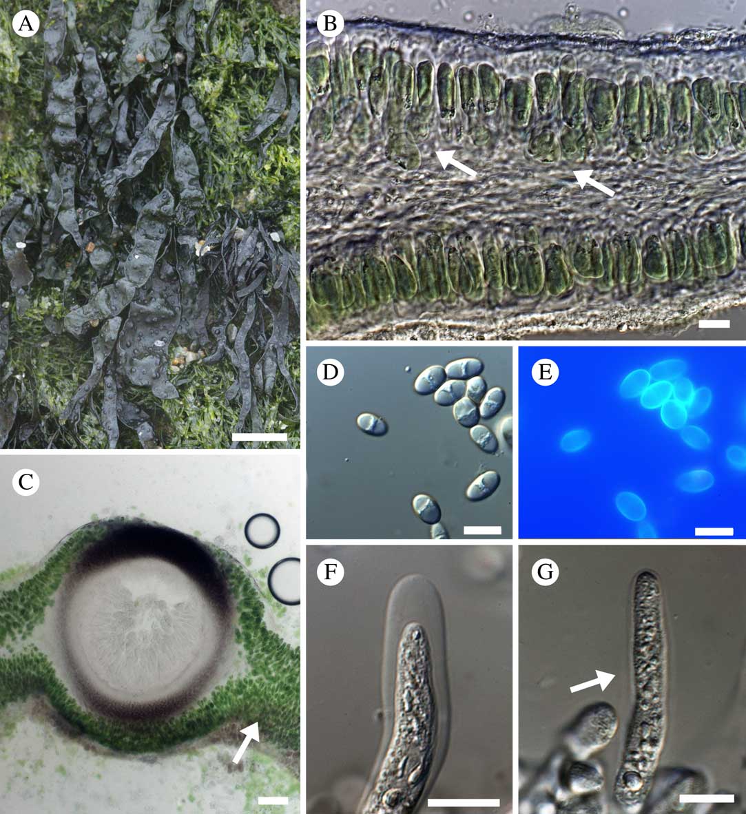
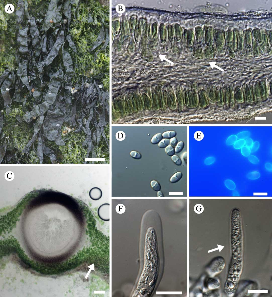
Turgidosculum ulvae (M. Reed) Kohlm. & E. Kohlm. is a fungus that forms a symbiosis with a foliose green alga and thereby transforming its morphology (Fig. 1A). It was originally described as Guignardia ulvae M. Reed, a parasite on Ulva vexata Setchell & Gardner (=Blidingia minima var. vexata (Setchell & Gardner) J. N. Norris) (Reed Reference Reed1902). The special nature of this symbiosis has led to different taxonomic interpretations and subsequent ascriptions to divergent higher taxonomic taxa (Reed Reference Reed1902; Kohlmeyer & Kohlmeyer Reference Kohlmeyer and Kohlmeyer1972; Schatz Reference Schatz1980). Recent systematic reviews dealing with the classification of lichen-forming and marine fungi have either omitted the genus (Jaklitsch et al. Reference Jaklitsch, Baral, Lücking, Lumbsch and Frey2016; Lücking et al. Reference Lücking, Hodkinson and Leavitt2016, Reference Lücking, Hodkinson and Leavitt2017) or have placed it in the Mastodiaceae (Ascomycota family incertae sedis) (Jones et al. Reference Jones, Suetrong, Sakayaroj, Bahkali, Abdel-Wahab, Boekhout and Pang2015).
Turgidusculum ulvae, habit and anatomical characters. A, thalli (dark green) surrounded by thalli of Blidingia minima (light green); B, light microscopy image of a cross-section of the thallus, arrows indicate several layers of algal cells (differential interference contrast, DIC); C, cross-section of a perithecium, arrow indicates the characteristic multi-layered algal structure near the perithecium; D, ascospores (DIC); E, ascospores dyed with Calcofluor White (fluorescence microscopy); F, young ascus with both tunicae (DIC); G, young asci only with endotunica and remains of the exotunica (arrow) (DIC). Scales: A=0·5 mm; B, D, E & G=10 µm; C=50 µm; F=20 µm.
Turgidosculum ulvae occurs along the west coast of North America from San Francisco to Vancouver Island. It has received little attention from lichenologists and its phylogenetic relationships, as well as the nature and characteristics of the fungus-alga interaction, remain unclear. In this study we propose: 1) to clarify features, such as ascus type and spore septation, that have been variously interpreted in the literature; 2) to analyze the degree of interaction between both bionts at an ultrastructural level by means of transmission electron microscopy; 3) to place T. ulvae in a phylogenetic framework in order to shed light on its systematic position and determine its closest relatives; 4) to characterize the photobiont with molecular markers.
Material and Methods
Anatomical studies
Specimens were examined under a Leica S8APO dissecting microscope. The field photograph was taken using a Canon 70D camera coupled with a Canon 100 mm f2.8 macro lens. Hand-cut sections of algal and lichen thalli, as well as ascomata and pycnidia, were observed using either a Zeiss Axioplan 2 or an Olympus BX51 microscope fitted with Nomarski differential interference contrast (DIC). Photographs were taken with a Zeiss Axiovision 4.8 image analyzer system. 10% KOH, Lugol’s iodine, without (I) or with (K/I) pretreatment with KOH, were used for tissue dissociation and examination, and for testing colour reactions in ascomatal elements. An aqueous solution (0·1%) of Calcofluor White Stain (Sigma-Aldrich) was used to enhance vegetative hyphae and ascus characters due to its high affinity for a wide range of β-glucans (Grube Reference Grube1999). Preparations were observed with a Zeiss Axioplan 2 microscope coupled with an HBO 103 W/2 lamp.
Transmission electron microscopy (TEM)
Specimens were examined using a Leica S8APO stereomicroscope fitted with a Leica EC3 image capture system. Samples for ultrastructural study were prepared following de los Ríos & Ascaso (Reference de los Ríos and Ascaso2002). In brief, lichenized algal thallus fragments were initially fixed in glutaraldehyde and then in osmium tetroxide. They were dehydrated in a graded series of ethanol before embedding in Spurr’s low-viscosity resin. Ultrathin sections (79 nm) were made using a Leica EM UC-6 ultramicrotome, post-stained with uranyl acetate followed by lead citrate, and examined with a JEOL JEM-1011 transmission electron microscope.
Taxon sampling, DNA extraction, PCR amplification, and DNA sequencing
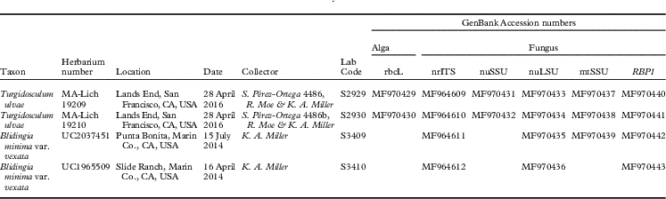
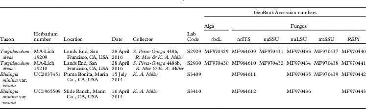
We studied four specimens collected from the Californian coast (Table 1). After the lichen thallus was cleaned of detritus with a razor blade and rinsed with sterilized water, a small fragment (c. 1 mm2) was removed. The fragment was inspected under the compound microscope to check for algal or fungal contaminants. Isolation of genomic DNA was performed using a CTAB-based method (Werth et al. Reference Werth, Reynisdóttir, Gudmundsson and Andrésson2016). We amplified partial regions of the nrITS, nrLSU, nrSSU, mtSSU and RPB1 regions of the fungal genome and a fragment of the RuBIsCO large subunit rbcL, using the primer pairs ITS1F-Kyo2 and ITS4-Kyo2 (Toju et al. Reference Toju, Tanabe, Yamamoto and Sato2012), LR0R (Rehner & Samuels Reference Rehner and Samuels1994) and LR5 (Vilgalys & Hester Reference Vilgalys and Hester1990) for nuLSU, nssu-131 and nssu-1088 (Kauff & Lutzoni Reference Kauff and Lutzoni2002) for nuSSU, mrSSU1 and mrSSU3 (Zoller et al. Reference Zoller, Scheidegger and Sperisen1999) for mtSSU, RPB1-Af (Stiller & Hall Reference Stiller and Hall1997) and RPB1-Cr (Matheny et al. Reference Matheny, Liu, Ammirati and Hall2002) for the RPB1 region A–D, and RH1 (Manhart Reference Manhart1994) and rbcL 530R (Verbruggen et al. Reference Verbruggen, Ashworth, LoDuca, Vlaeminck, Cocquyt, Sauvage, Zechman, Littler, Littler and Leliaert2009). PCR reactions were performed in a total volume of 15 µl, containing 2 µl of template DNA, 0·5 µl of each primer (10 µM), 6·5 µl of MyTaq Mix (MyTaq DNA Polymerase (Bioline) and dNTPs); distilled water was added to reach the final volume. PCR amplifications were performed in an Eppendorf Mastercycler Epgradient S thermal cycler. PCR conditions were as follows: an initial 5 min heating phase at 95°C, a touchdown of 6 cycles of 1 min at 94°C, 75 s at 56°C and 90 s at 94°C, decreasing 1°C in each cycle, and 30 more cycles of 1 min at 94°C, 75 s at 50°C and 90 s at 94 °C, and a final extension step of 7 min at 72°C. PCR products were purified using ExoSAP-IT™ PCR Product Cleanup Reagent (ThermoFisher) according to the manufacturer’s instructions and sequenced by Macrogen Inc. (Amsterdam, The Netherlands) using the same primer sets as for PCR amplification. Sequence contigs were assembled using SeqMan v.12.0 (Lasergene, DNA Star Inc., WI, USA).
Specimen data and GenBank Accession numbers for the collections used in the phylogenetic analyses of Turgidosculum ulvae and its photobiont Blidingia minima
Sequence alignment and phylogenetic analysis
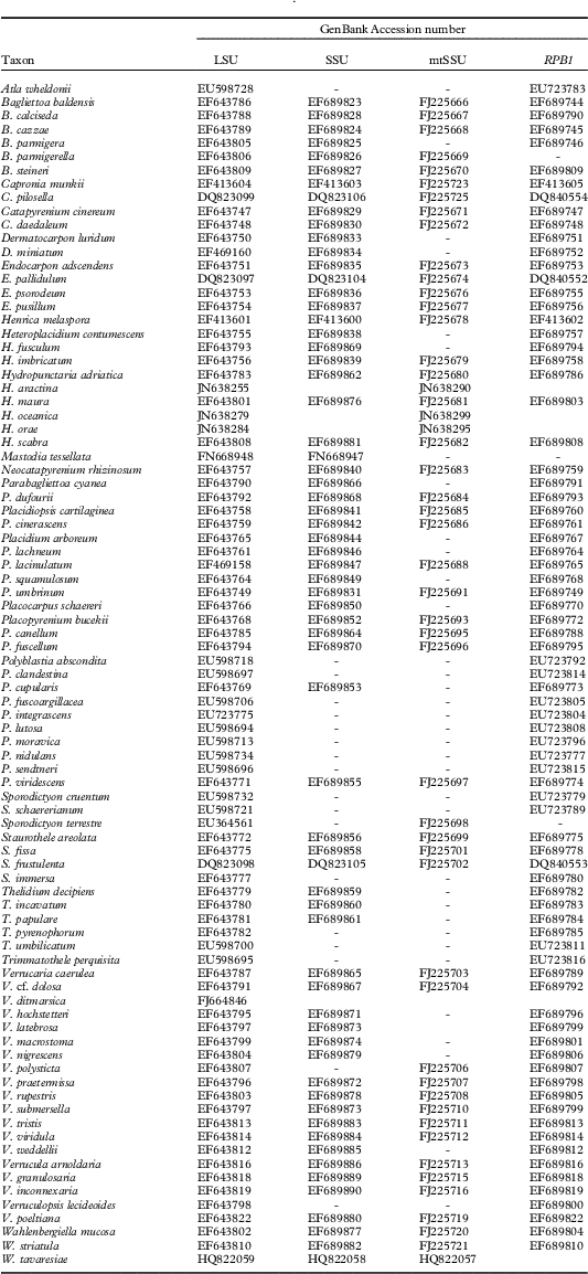
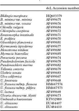
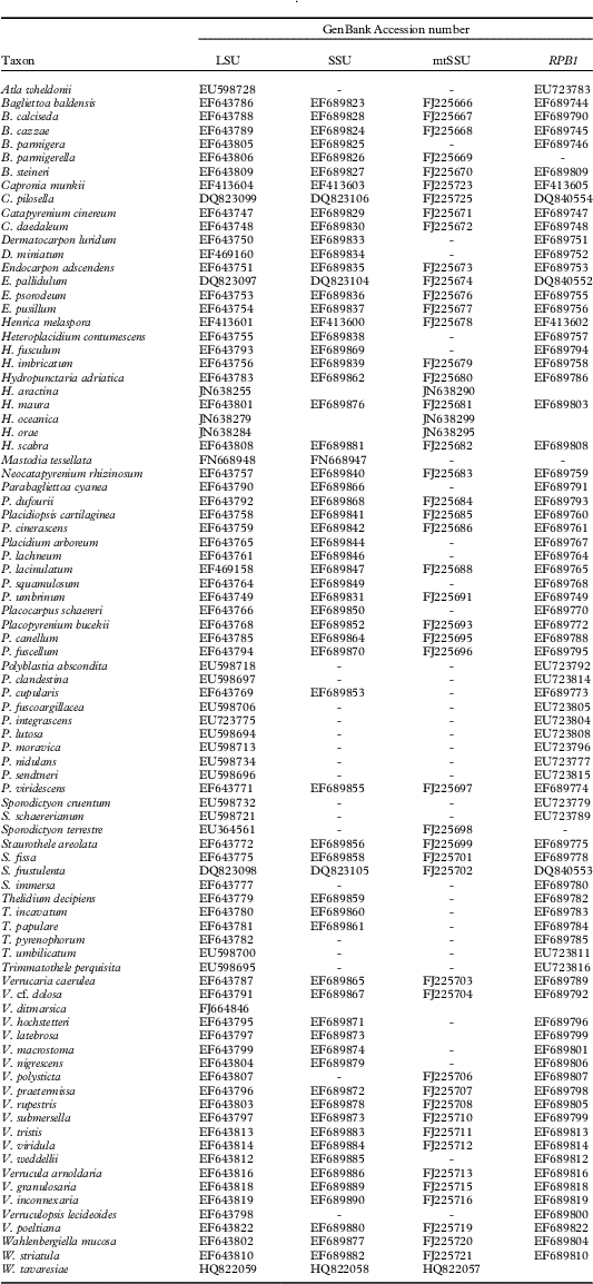
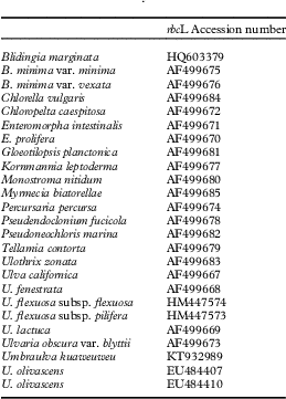
The nrLSU, nrSSU, mtSSU and RPB1 sequences obtained from four specimens of T. ulvae were aligned with representative members of the Verrucariaceae (Gueidan et al. Reference Gueidan, Savić, Thüs, Roux, Keller, Tibell, Prieto, Heiðmarsson, Breuss and Orange2009), including all marine members of the family and the closest matches of our sequences in BLAST searches (see Table 2). Two species of Capronia Sacc. were used as outgroup. rbcL sequences were aligned with representative members of the Ulvales (Table 3). Pseudoneochloris marina, Gloeotilopsis planctonica and Ulothrix zonata were used as outgroup. Alignments were carried out using MAFFT v.7.0 (Katoh et al. Reference Katoh, Misawa, Kuma and Miyata2002) as implemented in GeneiousTM v.9.0.4 (Biomatters Ltd, New Zealand) using default parameters, and inspected by eye in BioEdit v.7.0.9 (Hall Reference Hall1999). Gblocks 0.91b (Castresana Reference Castresana2000; implemented at http://molevol.cmima.csic.es/castresana/Gblocks_server.html) was used to remove ambiguously aligned regions and large gaps, applying the least stringent parameters but allowing gaps in 50% of the sequences. Optimal nucleotide substitution models for each genomic region were established by means of jModelTest v.2 (Darriba et al. Reference Darriba, Taboada, Doallo and Posada2012) using the Akaike Information Criterion (GTR+I+G for all loci).
GenBank Accession numbers for the members of the Verrucariace and outgroup used in the phylogenetic analyses of Turgidosculum ulvae
GenBank Accession numbers for the members of the Ulvales and outgroup used in the phylogenetic analyses of the photobiont of Turgidosculum ulvae
Maximum likelihood (ML) analysis was implemented in RAxML v.8.1.10 (Stamatakis et al. Reference Stamatakis, Hoover and Rougemont2008) at the CIPRES Science Gateway v.3.3 web portal (http://www.phylo.org/index.php/portal/). ML searches were implemented using the GTRGAMMA substitution model for each of the regions. Bootstrap support was calculated based on 1000 replications. Strongly supported nodes were defined as those with a bootstrap value ≥70%. Bayesian phylogenetic analyses were carried out in MrBayes v.3.2 (Ronquist et al. Reference Ronquist, Teslenko, van der Mark, Ayres, Darling, Höhna, Larget, Liu, Suchard and Huelsenbeck2012) using the nucleotide substitution model suggested by jModelTest v.2 and partitioning the dataset by loci. Settings included two parallel runs with four chains over 30 M generations for the analyses of Verrucariaceae and 1 M generations for the analyses of Ulvales and sampling was performed every 1000th and 200th step, respectively; the first 25% of saved data was discarded as ‘burn-in’. Convergence of chains was confirmed by the convergent diagnostic of the Potential Scale Reduction Factor (PSRF), which approached one. Phylogenetic trees were visualized in FigTree v.1.4.1 (http://tree.bio.ed.ac.uk/software/figtree/). Adobe Illustrator CS2® was used for artwork.
Results
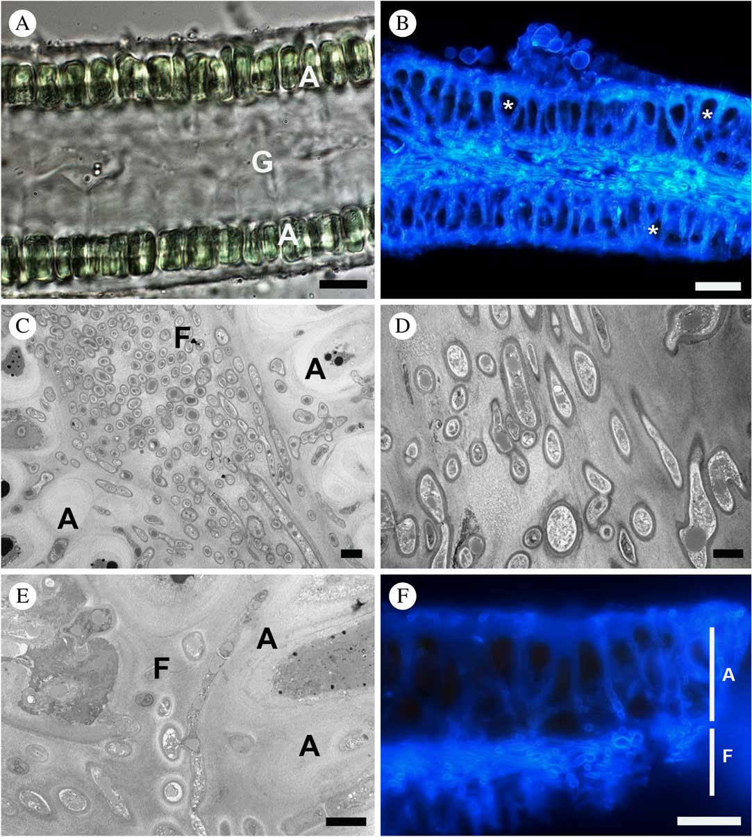
Thallus habit and morphology agreed with previous descriptions (Reed Reference Reed1902; Kohlmeyer & Kohlmeyer Reference Kohlmeyer and Kohlmeyer1972) and will not be detailed further. It should, however, be noted that lichenized Blidingia thalli (Fig. 1A, dark green) were considerably larger and much darker in colour than non-lichenized thalli (Fig. 1A, light green), indicating that lichenization clearly affects the growth of the alga. The typical double layer of cells in Blidingia tissue (Fig. 2A) was altered by the presence of the fungus (Fig. 1B), producing cell divisions and subsequent stratification (arrow in Fig. 1B). Stratification was much more pronounced near the perithecia (Fig. 1C) where the photobiont layer was up to 10 cells in depth, with almost no space between them (Fig. 1C, arrow).
Non-lichenized Blidingia minima and Turgidosculum ulvae thalli, anatomical features. A, cross-section of Blidingia thallus (differential interference contrast, DIC), G indicates the gelatinous material between both cell layers; B, thallus section of T. ulvae stained with Calcofluor White, some algal cells are denoted by an asterisk (fluorescence microscopy); C, T. ulvae medulla, consisting of a compact network of fungal hyphae (F) with algal cells (A) at both sides (TEM); D, T. ulvae fungal hyphae immersed in a fibrillar electron-dense matrix in the medulla (TEM); E, T. ulvae, detail of fungal hyphae (F) from the medulla next to the algal layer (A) (TEM); F, T. ulvae thallus, stained with Calcofluor White, showing penetration of the algal layer (A) by medullary fungal hyphae (F) (fluorescence microscopy). Scales: A & B=20 µm; C–E=2 µm; F=10 µm. In colour online.

Under DIC, ascospores appeared to be septate due to the presence of two vacuoles at either end of the ascospore with cytoplasmic material characteristically located at an equatorial position (Fig. 1D). Calcofluor White dye, with its affinity for molecules in the hyphal wall, revealed that ascospores were typically single celled under fluorescence microscopy (Fig. 1E). Asci were typically bitunicate at early stages of development (Fig. 1F). They lost the exotunica before ascospores were fully developed (Fig. 1G, arrow) so that most asci in microscopic preparations appeared to be unitunicate.
Interactions between symbionts
The fungus-free Blidingia thallus consisted of two layers of cells (Fig. 2A), with multi-layered cell walls. The area between the layers was filled with a homogenous mucilaginous substance. When the fungus was present, this space was filled by a medulla consisting of a compact network of fungal hyphae (Figs 1B & 2B). The lichenized Blidingia had a heterogenous structure with multiple layers of photobiont cells (Fig. 1B), especially near fungal perithecia.
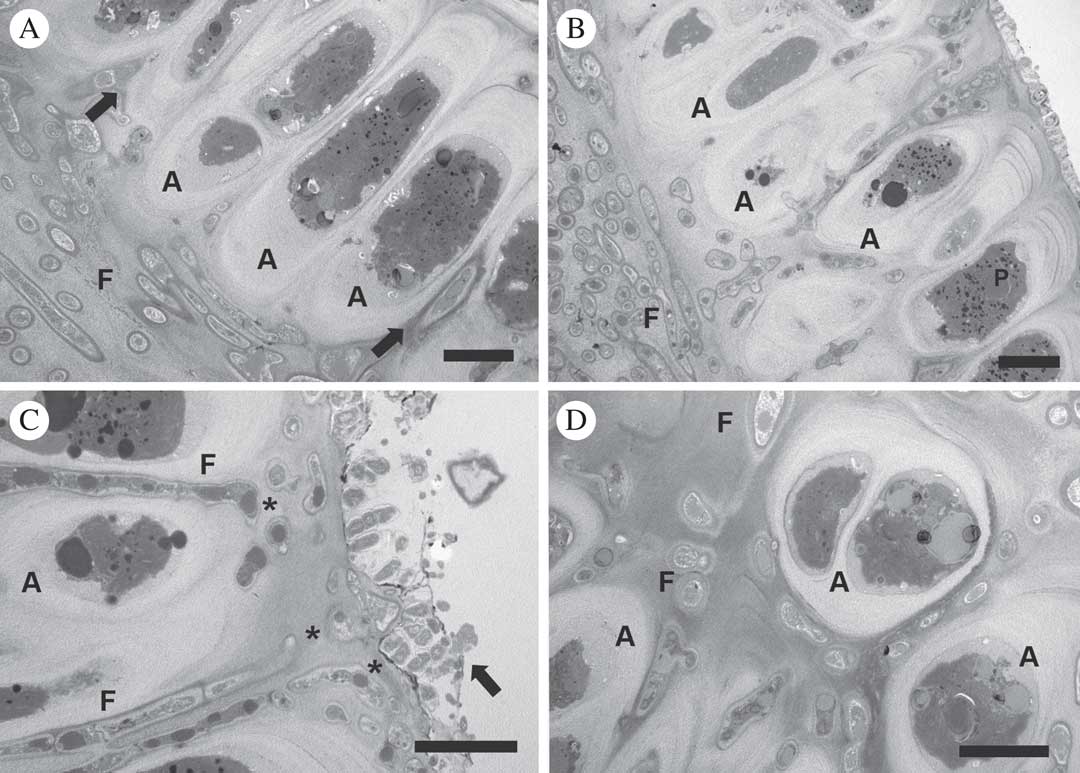
The medulla was composed of interwoven fungal hyphae (Fig. 2C), mostly oriented parallel to the thallus surface (Fig. 2D). Medullary hyphae appeared to be immersed in a fibrillar electron-dense matrix (Fig. 2D), especially near algal cells (Fig. 2E), suggesting that this matrix could correspond to remnants of the gelatinous matrix between algal cell layers.
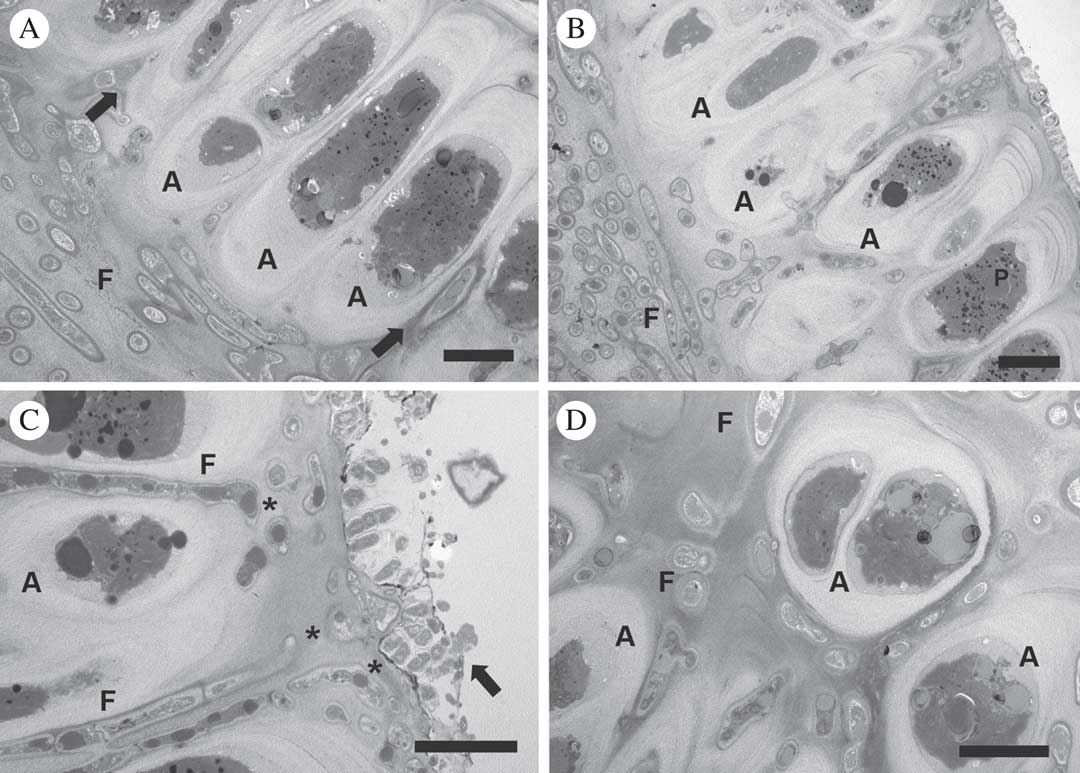
Fungal hyphae extend from the medulla, perpendicular to the surface, penetrating both layers of algal cells (Fig. 2F). Fungal cells grew between algal cells (arrow in Fig. 3A), pushing and separating them (Fig. 3B), and finally reaching the superficial cuticle of the alga (asterisks in Fig. 3C). Diverse epiphytic microbial communities were found on both surfaces of thalli (Fig. 2B, arrow in Fig. 3C). Single algal cells were surrounded by fungal hyphae and multiplied by cell division, forming isolated groups of algal cells enclosed by fungal hyphae (Fig. 3D).
Turgidosculum ulvae and Blidingia minima, relationship between both bionts at the ultrastructural level (TEM). A, penetration by medullary hyphae (F) between individual algal cells (A, arrows); B, algal layer (A) showing how the intercellular penetration by fungal hyphae (F) induces separation of the algal cells (P=pyrenoid); C, fungal hyphae (F) reaching the superficial gelatinous sheath (asterisks), arrow indicates the presence of epiphytic microbial communities on the gelatinous sheath; D, fungal cells (F) from the algal layer immersed in the electron-dense matrix and closely associated with algal cells (A). Scales: A–D=5 µm.
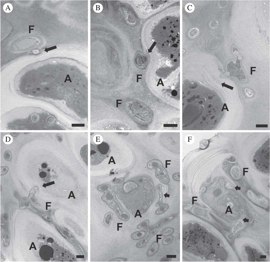
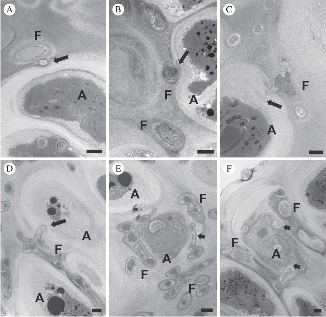
Individual algal cells were surrounded by cell walls with dense fibrillar components organized in layers and enclosed by a polymeric electron-dense matrix (Fig. 3A & B). The chloroplast was parietal with one pyrenoid (P) and numerous lipid bodies scattered in the stroma (Fig. 3B). Fungal cells in algal layers were immersed in the electron-dense matrix and closely associated with multi-layered algal cell walls (Fig. 4A). Intraparietal haustoria-like structures were common and appeared as peg-like protrusions of the fungal cell wall penetrating the layered algal cell walls (arrows in Fig. 4A & B). Electron-dense material accumulated around the hyphae at the point of interaction of haustoria with algal cell walls (arrow in Fig. 4B). Haustoria induced delamination of the algal cell wall (arrows in Fig. 4C & D). Later, haustorial hyphae expanded and grew between the separated layers of algal cell walls, increasing the surface of contact with algal cells (arrow in Fig. 4E). Often two or more haustoria occurred within a single algal cell and occasionally reached the plasmalemma (arrows in Fig. 4F), but intracellular penetrations were observed only in dying or collapsed algal cells (Fig. 4F).
Interactions between symbiont cells in Turgidosculum ulvae and Blidingia minima (TEM). A, peg-like protrusions of the fungal cell wall penetrating the algal cell wall (arrow); B, penetration by fungal peg-like protrusions through layered algal cell wall (arrow indicates accumulation of electron-dense material at the point of interaction); C, invasion by peg-like haustorial structure inducing delamination within the algal cell wall (arrow); D, deep penetration by peg-like haustorial structure within the algal cell wall (arrow); E; algal cell penetrated by different haustoria-like structures (arrow indicates the expansion of intraparietal haustoria between separated layers of algal cell wall); F, algal cell showing penetration by intraparietal haustoria (e.g. arrows) which reach but do not visibly penetrate the plasmalemma. F=fungal hyphae, A =algal cells. Scales: A–D=1 µm; E & F=5 µm.
Molecular and phylogenetic results
We successfully amplified all markers for the two most recently collected specimens. The nuSSU and mtSSU could not be amplified for specimen S3410. rbcL was amplified only for specimens S2930 and S1727. ITS sequences, which differed only in a single nucleotide, retrieved the marine species Verrucaria ditmarsica Erichsen (FJ664846, 81% cover, 89% similarity) as the closest match in a BLAST search in the GenBank database.
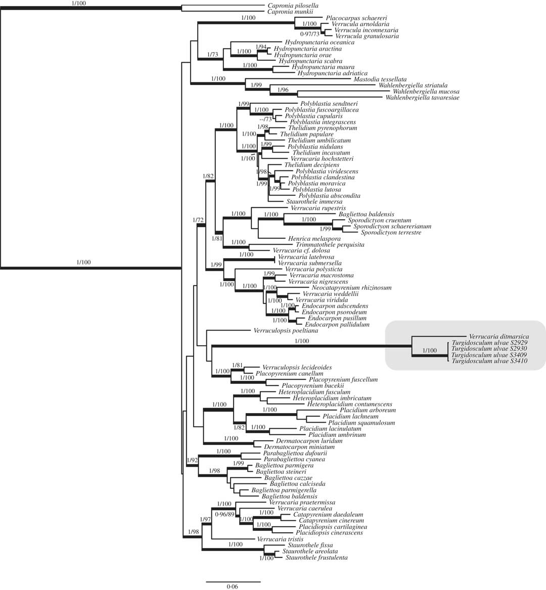
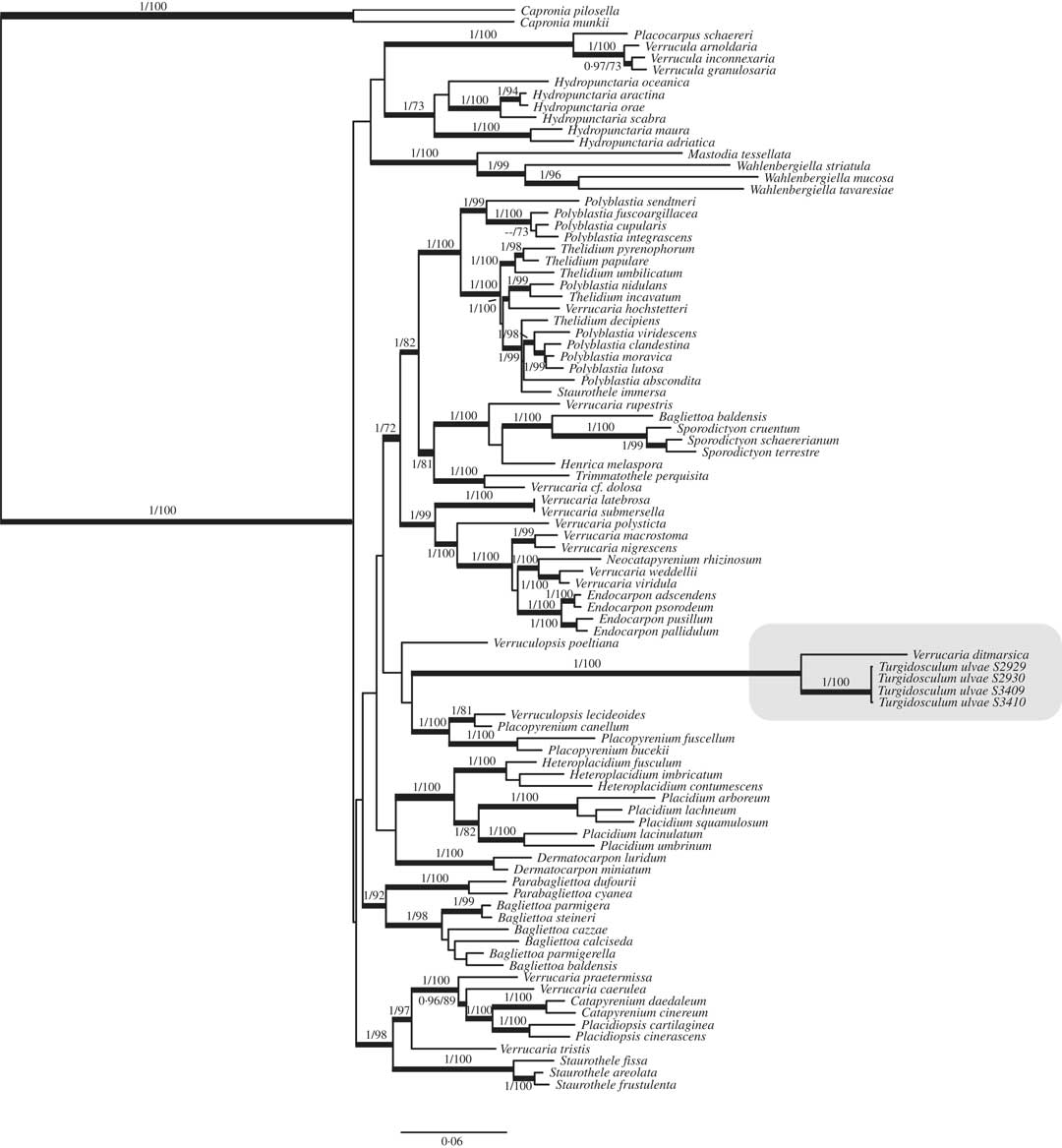
The combined data matrix for the four mycobiont markers and the rbcL matrix for the photobiont included 3253 and 507 bp, respectively, after the removal of gaps and ambiguous positions using Gblocks (693 RPB1, 526 nrLSU, 997 nrSSU, 732 mtSSU). The number of positions used in the analyses represents c. 33% of the total length of the original alignment; this is mostly due to the removal of long introns in the nuSSU marker. A single insertion of 3 nucleotides present in only c. 16% of the samples was removed from the rbcL alignment. Supported clades obtained in the most likely tree in an ML search mostly agreed with those retrieved in the 50% majority-rule tree obtained in the Bayesian analyses; an exception being the relationship between Polyblastia integrascens and P. cupularis which was supported in the ML but not the Bayesian analysis. The first topology is shown in Fig. 5 which includes supporting values for nodes retrieved in both analyses. The topology obtained agrees with previous studies (Gueidan et al. Reference Gueidan, Roux and Lutzoni2007, Reference Gueidan, Savić, Thüs, Roux, Keller, Tibell, Prieto, Heiðmarsson, Breuss and Orange2009, Reference Gueidan, Thüs and Pérez-Ortega2011; Savić et al. Reference Savić, Tibell, Gueidan and Lutzoni2008; Muggia et al. Reference Muggia, Gueidan and Grube2010), with high support for most of the generic groups but a complete lack of support for the backbone. Marine species in the Verrucariaceae were found in three different clades. The first clade included marine and freshwater species of Hydropunctaria for which relationships were not completely resolved. The second clade included species of Wahlenbergiella and Mastodia tessellata, a relationship already recovered in previous studies (Pérez-Ortega et al. Reference Pérez-Ortega, de los Ríos, Crespo and Sancho2010; Gueidan et al. Reference Gueidan, Thüs and Pérez-Ortega2011). The third clade included marine species, grouping together all specimens of T. ulvae and a single specimen of Verrucaria ditmarsica. This clade was well supported in both analyses and the long branch connecting it with its sister clade (relationship not supported) suggests that it is a distinct lineage within Verrucariaceae.
Combined nuLSU-nuSSU-RPB1-mtSSU maximum likelihood phylogenetic tree of Verrucariaceae obtained in the RAxML analysis (Bayesian analysis recovered a similar topology). Branches in bold depict both bootstrap (B) and posterior probability (PP) support; figures are given above or below branches (B/PP). Turgidosculum ulvae - Verrucaria ditmarsica clade is highlighted in dark grey.
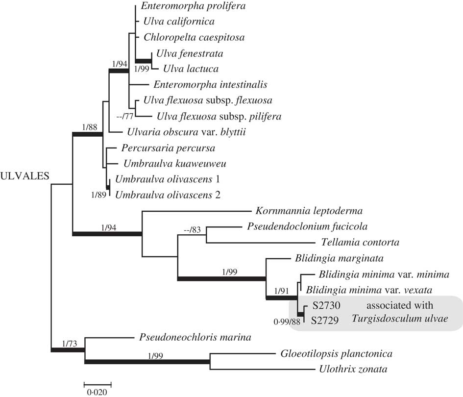
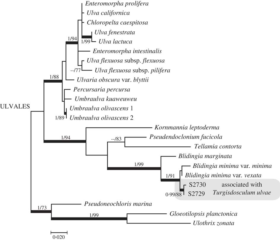
The rbcL tree (Fig. 6) showed that the photobionts of T. ulvae were in a well-supported group close to Blidingia minima var. minima and B. minima var. vexata. However, relationships among them are not well supported. Newly produced sequences differed by 1 bp between them, by 3 and 4 bp from a GenBank sequence of B. minima var. vexata (AF499676), and by 7 and 8 bp from a GenBank sequence of B. minima var. minima (AF499675). The sequences AF499676 and AF499675 differed by 4 bp.
rbcL maximum likelihood phylogenetic tree of Ulvales obtained in the RAxML analysis (Bayesian analysis recovered a similar topology). Bold branches depict both bootstrap (B) and posterior probability (PP) support; figures are given above or below branches (B/PP). Blidingia sequences obtained from Turgidosculum ulvae analyzed in this study are highlighted in grey.
Discussion
We have shown that Turgidosculum ulvae clearly belongs to the family Verrucariaceae. The systematic position of this genus had been a matter of debate. Kohlmeyer & Kohlmeyer (Reference Kohlmeyer and Kohlmeyer1972) originally placed it in the Sphaeriales, an order now split into several lineages in the Sordariomycetes (Kirk et al. Reference Kirk, Cannon, Minter and Stalpers2008) on the basis of its hamathecial and ascal characters. We have shown that the asci of T. ulvae are typically bitunicate and not unitunicate as previously thought (Kohlmeyer & Kohlmeyer Reference Kohlmeyer and Kohlmeyer1972; Schatz Reference Schatz1980), although asci quickly lose their exotunica, a character that has been interpreted in other species of lichen-forming fungi as an adaptation to their aquatic habitat (Ménard & Roux Reference Ménard and Roux1995; Pérez-Ortega et al. Reference Pérez-Ortega, de los Ríos, Crespo and Sancho2010). Schatz (Reference Schatz1980) also pointed to the presence of 2-celled ascospores in T. ulvae, but by means of fluorescence microscopy we have shown that ascospores are simple. On the other hand, on the basis of unitunicate asci and the observation of paraphyses growing downwards from the upper part of the perithecium (pseudoparaphyses), Schatz (Reference Schatz1980) placed Turgidosculum in the family Hypocreaceae (Hypocreales), emending the genus description. It has also been included in the Mastodiaceae Zahlbr. (Jones et al. Reference Jones, Suetrong, Sakayaroj, Bahkali, Abdel-Wahab, Boekhout and Pang2015), a family erected to accommodate the deviant M. tessellata (Zahlbruckner Reference Zahlbruckner1907). Pérez-Ortega et al. (Reference Pérez-Ortega, de los Ríos, Crespo and Sancho2010) have shown that M. tessellata belongs to the Verrucariaceae and is closely related to the genus Wahlenbergiella. Thus, with the phylogenetic placement of T. ulvae in Verrucariacae, it is definitively clear that Mastodiaceae represents a later synonym and is superfluous.
The closest relative to T. ulvae in BLAST searches and in the phylogenetic tree is Verrucaria ditmarsica. These species form a distinct lineage separated from the rest of Verrucariaceae by a long branch. This clade probably represents an independent colonization of rocky seashores. While Hydropunctaria occurs in the supratidal zone, both T. ulvae and V. ditmarsica live in the intertidal zone where Wahlenbergiella species also occur. Verrucaria ditmarsica was described from Germany (Erichsen Reference Erichsen1937) and has so far been recorded from the coasts of France (Roux Reference Roux2014), North America (Taylor Reference Taylor1982), the UK (Smith et al. Reference Smith, Aptroot, Coppins, Fletcher, Gilbert, James and Wolseley2009) and Spain (Paz-Bermúdez et al. Reference Paz-Bermúdez, López de Silanes and Carballal2000). The concept of this species has been very broadly interpreted and several related species are still undescribed (S. Pérez-Ortega et al. in prep.). Further research is needed to understand the breadth of this group, to discover if all species are marine and to determine if Turgidosculum is the correct generic name for the whole clade.
The alga associated with Turgidosculum ulvae belongs to Blidingia minima s. lat. Ulva vexata was described by Setchell & Gardner (Reference Setchell and Gardner1920) on the basis of its deviant thallus and cell sizes. These authors noted that differences in cell size and shape might be “a malformation due to the parasite”, but the characteristics of the cells of the “less parasitized” portions led them to describe the photobiont as a distinct species of Ulva. Later it was transferred to Blidingia (as B. minima var. vexata) by Norris (Reference Norris1971). The sequences from our two specimens differed from available sequences of B. minima and B. minima var. vexata, and relationships among them were not well supported. Thus, it is not clear if T. ulvae associates with a Blidingia species distinct from B. minima, and if that species also occurs without the fungal associate. Further molecular studies focused on the algal partner should be carried out to resolve this question.
The symbiosis between T. ulvae and Blidingia constitutes the second shift of a species in the family Verrucariaceae from a unicellular/filamentous photobiont to a species with a multicellular blade-like thallus. Recent studies have shown that Mastodia tessellata and its undescribed sister species Mastodia sp. (Garrido-Benavent et al. 2017 Reference Garrido‐Benavent, de los Ríos, Fernández‐Mendoza and Pérez‐Ortegab ) associate with Prasiola species (P. borealis Reed, P. delicata Setchell & Gardner and an undescribed Antarctic species) (Pérez-Ortega et al. Reference Pérez-Ortega, de los Ríos, Crespo and Sancho2010; Moniz et al. Reference Moniz, Guiry and Rindi2014; Garrido-Benavent et al. 2017 Reference Garrido-Benavent, Pérez-Ortega and de los Ríosa ). The family Verrucariaceae hosts the highest diversity of photobionts of all lineages of lichenized fungi (Thüs et al. Reference Thüs, Muggia, Pérez-Ortega, Favero-Longo, Joneson, O’Brien, Nelsen, Duque-Thüs, Grube and Friedl2011) including the only known symbiosis of a lichen-forming fungus, Wahlenbergiella tavaresiae (R. L. Moe) Gueidan et al., with a brown alga, Petroderma maculiforme (Wollny) Kuckuck (Moe Reference Moe1997; Sanders et al. Reference Sanders, Moe and Ascaso2004), and several reported symbioses with a member of the Xanthophyceae (Heterococcus caespitosus Vischer; Zeitler Reference Zeitler1954; Parra & Redón Reference Parra and Redón1977; Thüs & Schultz Reference Thüs and Schultz2008). Dilabifilum Tschermak-Woess (Ulvales) represents the most common photobiont associated with marine and freshwater species of Verrucariaceae (Thüs & Schultz Reference Thüs and Schultz2008; Thüs et al. Reference Thüs, Muggia, Pérez-Ortega, Favero-Longo, Joneson, O’Brien, Nelsen, Duque-Thüs, Grube and Friedl2011). Thus, it is likely that ancestors of T. ulvae had Dilabifilum as the photosynthetic partner and that the photobiont shift might have driven speciation.
Although Reed (Reference Reed1902) and subsequent authors (Kohlmeyer & Kohlmeyer Reference Kohlmeyer and Kohlmeyer1972, Reference Kohlmeyer and Kohlmeyer2013; Schatz Reference Schatz1980) provided thorough and extensive descriptions of both thallus structure and symbiont interactions, these studies based on light microscopy did not reveal the presence of haustoria-like structures. TEM images have demonstrated intimate contact between fungal and algal cell walls and the existence of intraparietal haustorium-like structures. This type of haustorium, which penetrates the cell wall but not the plasmalemma, has been attributed to morphologically complex taxa of lichen-forming ascomycetes (Honegger Reference Honegger2009). Although intraparietal haustoria have been previously reported for other marine lichens, viz. Wahlenbergiella tavaresiae (Sanders et al. Reference Sanders, Moe and Ascaso2004) and Mastodia tesellata (Perez-Ortega et al. Reference Pérez-Ortega, de los Ríos, Crespo and Sancho2010), those found in T. ulvae differ structurally. Similar peg-like protrusions of the fungal cell wall penetrating the algal cell and causing delamination of the algal cell wall were observed in the three symbioses, but thinning of the penetrated algal cell wall was not detected in T. ulvae. In Turgidosculum, after forming peg-like protrusions, haustorial cells occasionally grow between separated layers of algal cell walls, establishing striking and extensive wall-to-wall contact. However, complex haustorial systems formed by lobes which envelop the algal cells as described in Mastodia tessellata were not found in T. ulvae. Algal cells were penetrated by one or more haustoria-like structures but visible damage to the algal cells or invagination of the algal cytoplasm were not detected. Hence our TEM study has confirmed the original light microscopy observations of the lack of noticeable damage to algal cells as a result of fungal interactions (Reed Reference Reed1902). The fungal presence in the cytoplasm of dying and collapsed cells is interpreted here as saprophytic behaviour of the fungus at the senescence stage of the algal cells. However, we cannot completely discard the role of the fungus in controlling the life cycle of algal cells, promoting their death and collapse when entering into the cytoplasm.
The intercellular hyphal growth among individual cells of Blidingia and the associated interactions established between both symbionts alter the structure of its thallus. The haustorial system in Turgidosculum seems to be a stable element that could play at the mycobiont-photobiont interface, a structural role that improves mechanical stabilization and integration of tissue (Sanders et al. Reference Sanders, Moe and Ascaso2004), as well as a functional one. Haustoria, together with appressoria and intragelatinous protrusions, are the sites at which algal cells are provided with micronutrients and water, and where the fungal partner mobilizes carbon fixed by the algal cells (Honegger Reference Honegger2009).
The range and type of symbioses between fungi, algae and cyanobacteria have made it difficult to circumscribe the term ‘lichen’ (Hawksworth Reference Hawksworth1988; Grube & Hawksworth Reference Grube and Hawksworth2007). Most accepted definitions, such as that proposed by Hawksworth & Honegger (Reference Hawksworth and Honegger1994), strongly emphasize that the fungal partner acts as the exhabitant. Mastodia tessellata and T. ulvae have traditionally represented a problem to lichenologists because their foliose partners do not fit most lichen definitions. Some authors have termed these symbioses mycophycobiosis, fungal-algal permanent relationships in which fungi do not form thalli and occur endophytically. Well-known examples are Mycophycias ascophylli (A. D. Cotton) Kohlm. & Volkm.-Kohlm. on Ascophyllum nodosum (L.) Le Jolis, and Pelvetia canaliculata (Linnaeus) Decaisne & Thuret, and Blodgettia bornetii E. P. Wright on Cladophora caespitosa (C. Agardh) Kützing and C. fuliginosa Kützing (Kohlmeyer & Kohlmeyer Reference Kohlmeyer and Kohlmeyer2013). Hawksworth (Reference Hawksworth1988) stated that whereas M. tessellata differs from the concept of mycophycobiosis, T. ulvae is closer to the symbiosis that is produced by Mycosphaerella ascophylli (=Mycophycias ascophylli).
It seems clear that a more inclusive definition of lichens should include morphogenetic effects of the symbiosis (Lücking et al. Reference Lücking, Hodkinson and Leavitt2016). In the cases of both Mastodia and Turgidosculum, there exists a clear effect of the mycobiont on the algal thallus. In Turgidosculum, in addition to the anatomical transformation (Fig. 3B & C) in which the mycobiont hyphae separate algal cells and promote cell division, there are changes in the overall appearance clearly visible to the naked eye (Fig. 1A). Lichenized thalli are considerably larger in length and width than typical Blidingia specimens. Furthermore, those thalli show a different coloration, having a blackish green tone, probably related to the melanization of fungal hyphae. It has been reported that non-lichenized thalli of Blidingia are preferred to lichenized thalli by intertidal invertebrates (Cubit Reference Cubit1975), and that melanin synthesis affects the food quality and preferences in their diet (Scheu & Simmerling Reference Scheu and Simmerling2004).
In conclusion, we have shown that Turgidosculum ulvae represents an independent photobiont shift to a foliose alga in the Verrucariaceae, a family that has independently colonized the rocky seashores of the world several times. In this environment, species of all kingdoms have developed key innovations to cope with extreme conditions. Species in the Verrucariaceae have acquired unusual and unique photobiont partners ranging from a brown alga to two different foliose green algae. Further research is needed on the taxonomy of marine Verrucariaceae and the identity of the Turgidosculum photobiont.
It is our pleasure to dedicate this study to our colleague Professor Ana Crespo on her 70th birthday. We appreciate her contribution to the development and advancement of lichenology and her positive influence on our careers.
We are grateful to Richard Moe for his support during fieldwork in California and his helpful comments on the manuscript. We also thank William Sanders and an anonymous reviewer for providing comments that improved the manuscript. SPO was supported by the grant RYC-2014-16784 from the Spanish Ministry of Economy, Industry and Competitiveness. TEM study was supported by the grant CTM2015-64728-C2-2-R to ADLR. We thank Cristina Patiño and Javier Bueno (Centro Nacional de Biotecnología, UAM, Madrid) for technical support in the TEM.