Introduction
Salinity has a major influence on the distribution of marine fishes in the oceanic environment (Holliday, Reference Holliday, Hoar and Randall1969). Adults, juveniles and most larvae are well adapted to changing environments (Holliday & Blaxter, Reference Holliday and Blaxter1960), while egg and embryonic stages tend to be more sensitive to ambient conditions. Salinity optima and tolerance limits for fertilization and early embryonic development are apparent for marine fish species, even for those inhabiting variable environments such as plaice, Pleuronectes platessa (Holliday & Blaxter, Reference Holliday and Blaxter1960). Based on catches of young larvae the spawning area for European eel, Anguilla anguilla is delimited to the southern Sargasso Sea, however spawning adults or eggs have still not been encountered (Tsukamoto et al., Reference Tsukamoto, Chow, Otake, Kurogi, Mochioka, Miller, Aoyama, Kimura, Watanabe, Yoshinaga, Shinoda, Kuroki, Oya, Watanabe, Hata, Ijiri, Kazeto, Nomura and Tanaka2011). Captive reproduction of eel is challenged by an endocrinological inhibition of maturation during the silvering stage, which in nature precedes spawning migration to the Sargasso Sea (Dufour et al., Reference Dufour, Burzawa-Gerard, Belle, Sbaihi, Vidal, Aida, Tsukamoto and Yamauchi2003). Eel broodstock are therefore matured via hormone therapy (Dufour et al., Reference Dufour, Burzawa-Gerard, Belle, Sbaihi, Vidal, Aida, Tsukamoto and Yamauchi2003) and recent advancements in reproduction methods of European eel have advanced to a state in which large quantities of viable gametes, eggs and yolk-sac larvae can be regularly obtained and cultured using standardized methods (Tomkiewicz, Reference Tomkiewicz2012, Butts et al., Reference Butts, Sørensen, Politis and Tomkiewicz2014; Sørensen et al., Reference Sørensen, Skov, Lauesen, Tomkiewicz, Bossier and De Schryver2014). This situation now allows us to conduct studies on basic eco-physiological factors that are not known in the natural environment for European eel, such as salt and salinity effects on eggs/embryos, that are important for gamete activation and fertilization.
Egg activation is a key process in early embryonic development of fish, triggered by the contact between eggs, sperm and an aqueous medium (i.e. seawater or freshwater). The process of egg activation, although not fully understood (Webb & Miller, Reference Webb and Miller2013), follows a general mechanism initiated by the release of intracellular stored Ca2+ between the chorion (egg envelope) and the yolk membrane (Coward et al., Reference Coward, Bromage, Hibbitt and Parrington2002; Alavi et al., Reference Alavi, Linhart, Coward, Rodina, Alavi, Cosson, Coward and Rafiee2008). Ca2+ release mediates the cortical alveoli, embedded in the plasma membrane around the yolk to exocytose cortical proteins (Laale, Reference Laale1980; Cerdà et al., Reference Cerdà, Fabra, Raldúa, Babin, Cerdà and Lubzens2007). Cortical proteins are then broken into smaller units by proteolysis and form the osmotic gradient that facilitate uptake of seawater from the environment across the chorion (Lønning & Davenport, Reference Lønning and Davenport1980; Govoni & Forward, Reference Govoni, Forward, Finn and Kapoor2008). This process forms and expands the perivitelline space (PVS) and is facilitated by the flexible structure of the chorion. The release of intracellular stored Ca2+ is relatively high during this period and, besides triggering the cortical reaction, it is assumed to counteract polyspermy, affect early cell cleavage patterns and embryonic development (Coward et al., Reference Coward, Bromage, Hibbitt and Parrington2002; Claw & Swanson, Reference Claw and Swanson2012). The size of the PVS determines final egg size and differs between species (Davenport et al., Reference Davenport, Lønning and Kjørsvik1981). The PVS serves as an important ‘cushioning’ incubation medium for the embryo and provides an essential buffer and sink for nitrogenous wastes during embryogenesis (Finn, Reference Finn2007).
Activated eggs of European eel and Japanese eel (A. japonica) have, as seen for other primitive species, a remarkably large PVS (Govoni & Forward, Reference Govoni, Forward, Finn and Kapoor2008) and a prominent oil droplet that is part of the egg yolk (Heinsbroek et al., Reference Heinsbroek, Støttrup, Jacobsen, Corraze, Kraiem, Holst, Tomkiewicz and Kaushik2013). The natural egg size of the European eel remains unknown, but the diameter of Japanese eel eggs in nature ranges from 1490–1710 μm in contrast with the somewhat smaller eggs from captivity-bred broodstock, i.e. 1300–1600 μm (Tsukamoto et al., Reference Tsukamoto, Chow, Otake, Kurogi, Mochioka, Miller, Aoyama, Kimura, Watanabe, Yoshinaga, Shinoda, Kuroki, Oya, Watanabe, Hata, Ijiri, Kazeto, Nomura and Tanaka2011). In early reproduction experiments of European eel, unfertilized egg sizes ranged from 930–1400 μm (Fontaine et al., Reference Fontaine, Bertrand, Lopez and Callamand1964), 1000–1100 μm (Villani & Lumare, Reference Villani and Lumare1975; Boëtius & Boëtius, Reference Boëtius and Boëtius1980) and up to 1200–1600 μm (Kokhnenko et al., Reference Kokhnenko, Bezdenezhnykh and Gorovaya1977). Pedersen (Reference Pedersen2003) obtained fertilized eggs of ~1000 μm and recorded unfertilized eggs from a spontaneous spawning female to be 1400 μm. Additionally, Palstra et al. (Reference Palstra, Cohen, Niemantsverdriet, van Ginneken and Van den Thillart2005) obtained a few developing embryos and presents an illustration with an egg of unspecified size (Palstra et al., Reference Palstra, Cohen, Niemantsverdriet, van Ginneken and Van den Thillart2005), although this appears large, presumably ~1500 μm. Such variation among eggs together with the apparent larger size of wild-caught Japanese eel eggs, points to a suboptimal activation process in the assisted reproduction procedures. Insights into factors that affect egg swelling are therefore seen as crucial for successful in vitro production of eel offspring (Yoshinaga et al., Reference Yoshinaga, Miller, Yokouchi, Otake, Kimura, Aoyama, Watanabe, Shinoda, Oya, Miyazaki, Zenimoto, Sudo, Takahashi, Ahn, Manabe, Hagihara, Morioka, Itakura, Machida, Ban, Shiozaki, Ai and Tsukamoto2011).
Besides the protective PVS, marine pelagic fish eggs depend on a hydrostatic lift to facilitate embryonic development in their environment (Govoni & Forward, Reference Govoni, Forward, Finn and Kapoor2008) and lack of sufficient buoyancy is a known defect of eel eggs produced in captivity (Seoka et al., Reference Seoka, Yamada, Iwata, Yanagisawa, Nakagawa and Kumai2003). Egg buoyancy is determined during oocyte maturation in the ovary and pelagic species exhibit a particularly high proteolysis of yolk proteins into free amino acids (FAA) (Thorsen & Fyhn, Reference Thorsen and Fyhn1996; Cerdà et al., Reference Cerdà, Fabra, Raldúa, Babin, Cerdà and Lubzens2007). FAA increase the osmotic potential and causes an aquaporin-mediated water uptake of maternal isosmotic water during the hydration process. In turn, this action causes the oocyte to swell (Fabra et al., Reference Fabra, Raldúa, Power, Deen and Cerdà2005; Cerdà et al., Reference Cerdà, Fabra, Raldúa, Babin, Cerdà and Lubzens2007; Govoni & Forward, Reference Govoni, Forward, Finn and Kapoor2008). The difference between egg osmolality and that of the seawater environment promotes final egg buoyancy for which 80% is due to maternal fluids absorbed by the oocyte and the remaining ~20% is facilitated by oil droplets and yolk lipids (Cerdà et al., Reference Cerdà, Fabra, Raldúa, Babin, Cerdà and Lubzens2007; Govoni & Forward, Reference Govoni, Forward, Finn and Kapoor2008).
Preliminary studies on the European eel revealed that use of natural seawater was a prerequisite for obtaining viable eggs (Tomkiewicz & Jarlbæk, Reference Tomkiewicz and Jarlbæk2008). Use of natural seawater in experiments is however problematic, while collection of water is influenced by the natural variability. A selection of commercial available marine salt types, varying in composition (Atkinson & Bingman, Reference Atkinson and Bingman1997), facilitate a test whether artificial seawater could be a more stable medium for in vitro fertilization and experiments. Investigation of the applicability of artificial seawater would therefore be of value for understanding the eco-physiological response of oocytes in different media and could lead to an improvement of in vitro fertilization conditions.
In this study, activation and fertilization processes were explored using a range of salinities, seawater and salt types in order to reveal their effects on: (1) magnitude and size of the PVS; (2) resulting egg size; (3) fertilization success and early cleavage patterns; and (4) neutral buoyancy of the eggs.
Materials and methods
Broodstock and gamete management
Female silver eels were obtained from a freshwater lake (Lake Vandet, Denmark). Eels were transported to a research facility of the Technical University of Denmark (DTU) and stocked in 300-l tanks in a recirculation system at a density ≤ 30 kg per m3. Broodstock were acclimatized to artificial seawater adjusted by Tropic Marin® Sea Salt (Dr Biener Aquarientechnik, Germany) to ~35 psu and maintained at ~20°C under dimmed light conditions at ~20 lux, imitating natural daylight, dusk/dawn; local diurnal periodicity. Prior to experiments, fish were anaesthetized (ethyl p-aminobenzoate, 20 mg l−1; Sigma–Aldrich Chemie, Steinheim, Germany) and tagged with a passive integrated transponder (PIT tag). No feed was provided during experiments, as maturing eels cease feeding (Pankhurst & Sorensen, Reference Pankhurst and Sorensen1984).
To induce vitellogenesis, females received weekly injections of salmon pituitary extract (SPE; 18.75 mg kg−1 body weight; Argent Chemical Laboratories, Washington, USA). Females were weighed weekly and, at 10% increase, biopsies were routinely made for evaluation of oocyte development. At approximately oocyte stage 3 (Palstra et al., Reference Palstra, Cohen, Niemantsverdriet, van Ginneken and Van den Thillart2005), females received another injection of SPE as a priming dose to stimulate final maturation. Females were injected, ~24 h after receiving the SPE priming, with an injection of maturation-inducing steroid (MIS; 17α,20β-dihydroxy-4-pregnen-3-one; Sigma-Aldrich Denmark A/S,) (Ohta et al., Reference Ohta, Kagawa, Tanaka, Okuzawa and Hirose1996). In the timespan of 12–14 h after receiving MIS, eggs were stripped into dry and sterilized trays by applying slight pressure to the abdomen of the fish and stripped eggs were weighed (± 1 g).
Male broodstock were reared at Stensgård Eel Farm in Jutland, Denmark. Males (mean ± standard deviation (SD) standard length and body weight were 37.6 ± 2.1 cm and 110.2 ± 13.6 g, respectively) were transported to the DTU facility (husbandry as above) and received weekly injections of human chorionic gonadotropin (Sigma-Aldrich, Denmark A/S) at 1.5 IU g−1 fish. Milt was collected after 7–8 weeks of hormonal treatment (~12 h after administration of hormone) by applying slight pressure along the abdominal region and for each fertilization event a pool of milt from 3–4 males was utilized. Prior to milt collection, the urogenital pore was wiped dry using deionized water and the initial ejaculate was omitted to avoid contamination. Milt was collected into sterile beakers.
Sperm motility was characterized, within 30 s of activation, using a Nikon Eclipse 55i microscope (Nikon Corporation, Tokyo, Japan), equipped with a Nikon ×400 magnification objective (×40 magnification CFI Plan Flour) and motilities <50% were discarded. Sperm concentration was measured using spermatocrit (Haematokrit 210, Andreas Hettich GmbH & Co. KG, Tuttlingen, Germany), obtained from spinning milt for 10 min at 6000 g (Sørensen et al., Reference Sørensen, Gallego, Pérez, Butts, Tomkiewicz and Asturiano2013). After motility analyses, milt was diluted in artificial seminal plasma, P1 medium, described by Asturiano et al. (Reference Asturiano, Pérez, Garzón, Marco-Jiménez, Peñaranda, Vicente and Jover2014) (Butts et al., Reference Butts, Sørensen, Politis and Tomkiewicz2014) at a ratio of 1:99. Diluted milt was kept in sterile culture flasks at 20˚C, prior to fertilization (within 2 h post-stripping).
Experiment 1: Activation salinity
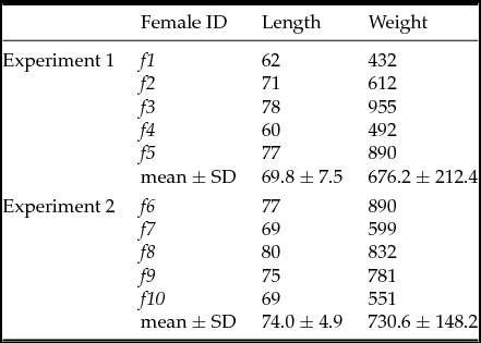
Eggs from each female (n = 5; Table 1), extruded as described above, were fertilized with sperm from an independent pool of 3–4 males, using a ratio of 1 ml of diluted milt per 2 g of newly stripped eggs. Activation seawater from the North Sea, kept in a 2000-l aerated tank, was adjusted to targeted salinities (±0.1 psu) using an electronic conductivity meter (WTW Multi 3410 + TetraCon325, Wissenschaftlich-Technische Werkstätten GmbH, Weilheim, Germany); taking into consideration the salinity of P1 milt diluent being 10.3 psu. The seawater was adjusted to the desired salinities using Tropic Marin Sea Salt. pH was measured using pH-indicator strips (pH 6.5–10.0, Merck Millipore, KGaA, Darmstadt, Germany) and adjusted to pH 8.2 using HCl.
Length (cm) and weight (g) for wild-caught female European eels Anguilla anguilla, used in Experiments 1 and 2

SD, standard deviation.
Following mixing of gametes, samples of 3.5 g eggs (~7000 eggs) were distributed into triplicate weigh boats (100 ml, 7 × 7 cm) for each of the treatments: 30, 35, 40, 45 and 50 psu. Each replicate was immediately activated, after mixing of gametes, using 11.7 ml of activation medium from pre-filled disposable 20 ml syringes. Loading and gamete activation were performed randomly across treatments. After activation, gametes were gently poured (10 min post-activation) into 250 ml glass beakers containing 200 ml of the corresponding salinity seawater, supplemented with penicillin G at 45 ppm and streptomycin sulphate at 65 ppm (Sigma-Aldrich, St. Louis, Missouri, USA). Egg/embryos were incubated in light below 50 lux at 20 ± 0.2°C. After 3.5–5 h of incubation, eggs were randomly sampled from each replicate beaker by gently mixing eggs to obtain a homogenous sample (see section: Egg evaluation). Digital images of sampled egg were then taken (see section: Egg evaluation).
Experiment 2: Activation salt type
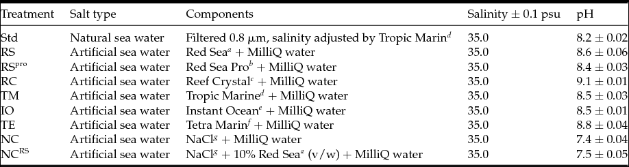
Following stripping, samples of 3.5 g of eggs from each of females (n = 5; Table 1) were distributed randomly into dry weigh boats (100 ml, 7 cm × 7 cm). There were triplicate samples for each activation treatment, which consisted of commercial salts types and natural seawater (Table 2). pH was measured using an electronic pH meter (model 827, Metrohm Inc. Riverview, Florida, USA), measuring three replicate 35 psu seawater mixtures 1 h after mixing (± 0.01 pH). Treatments were mixed with MilliQ water prior to use and adjusted to 35 ± 0.1 psu, taking into consideration the salinity of P1 milt diluent being 10.3 psu. Activation salinity was confirmed using an electronic conductivity meter. Each replicate was activated as described in Experiment 1 and sampling for imaging was performed similar to Experiment 1.
Specification of salt type treatments used in Experiment 2 for activation and incubation of eggs of European eel, Anguilla anguilla

Sea salt brands: a Red Sea, Red Sea International, Eilat, Israel; b Red Sea Coral Pro salt, Red Sea International, Eilat, Israel;
c Reef Crystal, Aquarium system, Sarrebourg, France; d Tropic Marin® Sea Salt, Dr. Biener GmbH, Wartenberg, Germany;
e Instant Ocean, Aquarium system, Sarrebourg, France; f Tetra Marine, TetraEurope, Paris, France;
g NaCl, Sigma-Aldrich, 0.2 μm filtered.
Egg evaluation
Egg sizes
Four digital images, from each replicate (4× ~20 eggs), formed the basis for analysis of egg sizes as well as egg categories (see section: Egg categorization and fertilization per cent). Egg sizes were measured using NIS Elements image software (v.3.32, Nikon Corporation, Tokyo, Japan). Except for the egg categorization (see Fig. 1), all egg measurements were based solely on fertilized eggs. Image software was used to calculate egg diameter by marking five reference points around each egg membrane circumference thereby deriving a diameter equivalent to a circle with sphericity of 1. The chorion and the yolk (vitelline membrane) circumference hence formed egg chorion diameter and yolk diameter, respectively. Sizes were measured within 3.5–5 h post-fertilization and obtained in association to egg categories described in section: Egg categorization and fertilization per cent. In Experiments 1 and 2, ≥15 eggs were measured per replicate.
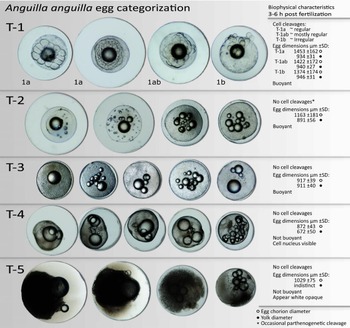
Characteristics of egg types used to categorize eggs from European eel, Anguilla anguilla, after in vitro activation and fertilization. Five main types of eggs (T1–T5) are displayed based on morphology, size and buoyancy at 35 psu, 3–6 h post fertilization and incubation at 20 ± 0.5°C. Category T-1 includes fertilized eggs and is subdivided into three subcategories based on cell cleavage pattern: (T-1a) regular and symmetric; (T-1ab) mostly regular with minor irregularities; and (T-1b) irregular.

To quantify the rate of eggs having egg chorion diameters >1600 μm a dataset was formed based on egg measurements from treatments of salt and salt type from both Experiment 1 and Experiment 2. Treatments of salt and salt type providing significantly smaller egg sizes were not included in this dataset.
Egg categorization and fertilization per cent
Four digital images, from each replicate (4× ~20 eggs) were acquired at 3.5–5 h post-fertilization and a categorization scheme was established (Fig. 1) that distinguished fertilized, unfertilized and dead eggs, outlining specific characteristics within each of five egg types. Fertilized eggs were categorized as type 1 (T-1), having >4 visible cells (pathogenic first cleavage occurs) and represent the proportion of fertilized eggs. To evaluate the regularity of cell cleavage patterns, fertilized eggs were subdivided into three subcategories according to early cell cleavage symmetry: (T-1a) regular cleaved cells showing cleavage symmetry and well defined cell margins with full adhesion between cells; (T-1ab) cells with an overall even appearance and margin but minor cleavage irregularity; and (T-1b) irregular cleavage planes with uneven cell sizes and low or no adhesion between cells (Fig. 1). Unfertilized eggs were divided into four categories: (T-2) unfertilized but activated, showing a PVS, buoyant with mean diameter (± SD) of 1163 μm ± 181 (N = 234) and variable oil droplets; (T-3) eggs failing to activate and form a PVS, buoyant with a mean diameter of 917 μm ± 39 (N = 219) and variable oil droplets; (T-4) unripe, non-ovulated eggs with visible nucleus, non-buoyant with mean diameter of 872 μm ± 43 (N = 378) and variable oil droplets; and (T-5) white or opaque eggs, resembling dead eggs, non-buoyant with mean diameter of 1029 μm ± 75 (N = 481).
Egg size difference between T-1a, T-1ab and T-1b
To test for overall size effects between each subcategory of fertilized eggs (T-1a, T-1ab and T-1b) data obtained from Experiments 1 and 2 were used. In specific, the 30, 35, 40 and 45 psu treatments were used from Experiment 1, while the Std, RS (Red Sea Salt), RSpro, RC (Reef Crystal) and TM (Tropic Marin) treatments were used from Experiment 2.
Egg activation with and without milt
Following stripping of female f5 (Table 1), 3.5 g of eggs were distributed randomly into three replicate dry weigh boats per treatment (100 ml, 7 × 7 cm) and two activation treatments were tested, differing in presence and absence of milt. Eggs were activated and incubated as described for Experiments 1 and 2 using 35 psu seawater. Digital images were obtained and analyzed as described in section: Egg evaluation, above.
Measurement of egg neutral buoyancy
Eggs from two females were used for buoyancy measurements; one used in Experiment 1 and one in Experiment 2. Neutral buoyancy of eggs, following treatments and incubation, were conducted at 7 and 30 h post-fertilization. A glass tank (thermoglass preventing temperature gradients) 100 × 10 × 10 cm was filled from the bottom using methods modified from Coombs (Reference Coombs1981). A peristaltic pump (Economy Console fitted Easy-Load® 3 pumphead, Masterflex, Cole-Parmer, Vernon Hills, Illinois, USA) provided steady addition of high saline water (58 psu, 2.5–2.8 ml × min−1) into a closed stirred mixing beaker containing low saline water (9 psu). A gradual increasing salinity gradient formed from the bottom of the glass tank within ~6 h. Water was premixed using demineralised water and Tropic Marine sea salt. Four density glass floats covering the range of 5.5–38 psu (Martin Instruments Co. Welwyn Garden City, Hertfordshire, UK) were inserted in the salinity gradient and positions used to establish the logarithmic function connection positions and salinity within the gradient. A correlation coefficient lower than r < 0.98 was chosen as the lower limit for a usable gradient. All salinity-density conversions were temperature compensated by measurements in the gradient (± 0.1˚C) (Chapman, Reference Chapman2006).
From each replicate, 5–10 eggs (~20 eggs per treatment) were gently inserted into the gradient using a disposable inoculation needle (1.0 mm), avoiding addition of water to the salinity gradient, which can disturb the gradient. Position of the eggs and reference floats were read on a metric scale (± 1 mm) 30 min after loading. Positioning was aided by a self-levelling laser on a tripod in darkness (SuperCross 2, Laserliner®, Frauenfeld, Switzerland) giving a thin-lit cross-section in the gradient to ensure an unbiased reading of position in the gradient. A new salinity gradient was established between each time point. In the reading at 7 h post-fertilization (HPF) in Experiment 1, eggs were left for 210 min to estimate their sinking rate. Sinking rate was measured as egg density change in psu min−1.
Statistical analyses
Data were analyzed using SAS (v.9.1; SAS Institute Inc., Cary, NC, USA) and SigmaPlot (v.11; Systat Software Inc., Hounslow, UK). Residuals were tested for normality (Shapiro-Wilk test) and homogeneity of variance (plot of residuals versus predicted values). When necessary, data were transformed to meet the assumptions of normality and homoscedasticity. Treatment means were contrasted using a Tukey's test. All data are presented as mean ± standard deviation (SD). Significance was set at α of 0.05.
Relationships between female body size (independent variable, i.e. length and weight), egg chorion diameter, yolk diameter and overall fertilization percentage (depended variables) were examined using linear regression.
Effects of salinity and salt type on fertilization success and egg traits were analyzed using a series mixed-model analysis of variance (ANOVA) (PROC MIXED), in which salinity and salt type were considered fixed factors and the female and female × salinity and/or salt type interaction were considered random factors. Analyses a posteriori were not performed on random effects. Instead, variance components (VC) were constructed using the restricted maximum likelihood estimation method in SAS PROC Mixed and expressed as a percentage. To test for significant variability among VC that were > 0, likelihood ratio statistics were generated (PROC MIXED). To further explore within female effects, salinity and salt type were analyzed using a series of one-way ANOVA models.
T-tests were used to compare egg and yolk size between the treatments activated with and without milt, while egg buoyancy data were analyzed using one-way ANOVA models. Relationships between salinity difference (activation salinity subtracted the measured buoyancy) (dependent variable) and the activation salinity (independent variable) were analyzed using linear regression.
Results
Experiment 1: Activation with different salinities
Egg size
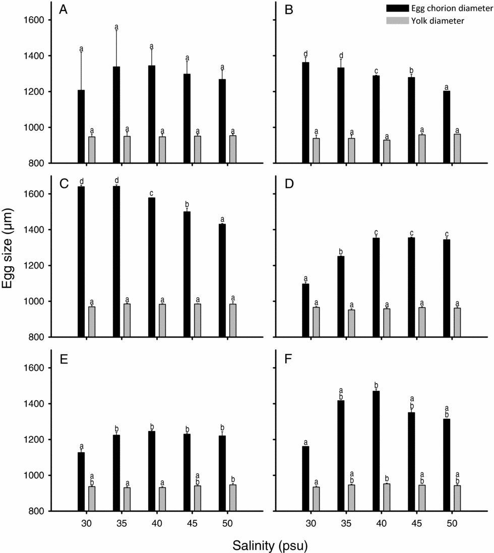
No relationship was detected between female body size and egg characteristics (R2 < 0.11). Average egg chorion diameter for the five females ranged from 1207 ± 212 to 1338 ± 203 μm across the salinity gradient (Fig. 2 A). For the mixed-model ANOVA including all females, salinity had no significant effect on egg chorion diameter (Fig. 2 A). For the model's random effects, the female × salinity interaction was significant and explained the majority of the models variance (VC = 99.7%), while the female VC was not significant (VC = 0.28%). For within female effects, salinity had a significant effect on egg chorion diameter for all females (Fig. 2 B–F). Egg chorion diameter was largest for female f1 and f2 at 30 and 35 psu (Fig. 2 B, C), f3 at 40–50 psu (Fig. 2 D), f4 at 35–50 psu (Fig. 2 E) and f5 at 35–50 psu (Fig. 2 F).
Egg size of European eel, Anguilla anguilla, in relation to activation salinity (psu). Measurements include chorion and yolk diameter (μm ± standard deviation (SD)) of fertilized eggs 3.5–5 h post in vitro fertilization and incubated at 20 ± 0.5°C. (A) Average egg sizes all females. (B–F) egg sizes for female f1–f5. a–dLetters indicate significant differences (α = 0.05) among salinities within groups.

Yolk diameter ranged from 947 ± 16 to 953 ± 9 μm (Fig. 2 A). The mixed-model ANOVA showed that salinity had no effect on yolk diameter (Fig. 2 A), while for the models random effects, both the female (VC = 74.2%) and female × salinity VC were significant (VC = 10.8%). For within female effects, salinity had no effect on yolk diameter for f1 (Fig. 2 B), f2 (Fig. 2 C) and f3 (Fig. 2 D), but significant effects were detected for both f4 (Fig. 2 E) and f5 (Fig. 2 F).
Fertilization rate and zygote development
No relationship was detected between female body size and fertilization percent (R2 < 0.05).
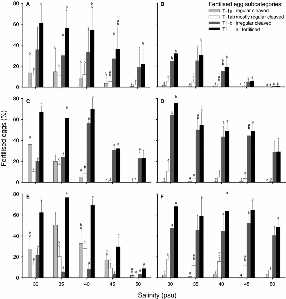
Overall fertilization success ranged from 22.1 ± 18.6 to 60.9 ± 16.9% across the salinity gradient (Fig. 3 A). When considering all fertilized eggs (egg category = T-1), salinity had a significant effect on fertilization per cent, such that the 30–40 psu treatments had the highest fertilization rates (Fig. 3 A). For the model's random effects, both the female (VC = 58.4%) and female × salinity interaction were significant (VC = 23.1%). When considering within female effects, salinity had a significant effect on fertilization success for females f1, f2, f3, f4, (Fig. 3 B–E) although not for f5 (Fig. 3 F). The highest fertilization rate was achieved for f1 (Fig. 3 B), f2 (Fig. 3 C) and f4 (Fig. 3 E) at 30–40 psu and f3 at 30–35 psu (Fig. 3 D). Typically, the lowest fertilization percentage was detected at 45–50 psu (Fig. 3).
Fertilization success and cleavage pattern of in vitro fertilized eggs of European eel, Anguilla anguilla, in relation to activation and incubation salinity. Fertilization percentage and distribution for subcategories T-1a, T1ab and T-1b were assessed 3.5–5 h post fertilization and following incubation at 20 ± 0.5°C. Bars show percentage + standard deviation (SD). (A) Average egg sizes all females. (B–F) are egg sizes for female f1–f5. a–cLetters indicate significant differences (α = 0.05) among salinities within groups.

The overall rate of regular cleaved fertilized eggs (egg subcategory = T-1a), ranged from 0.65 ± 0.98 to 14.71 ± 21.57% (Fig. 3 A). The mixed-model ANOVA showed that salinity had no effect on the proportion of regular cleaved fertilized eggs (Fig. 3 A). The female VC was significant and explained 58.7% of the models variance. The female × salinity VC was also significant and explained 35.0% of the variance. For within female effects, f2 (Fig. 3 C) and f4 (Fig. 3 E) were significant, where the percentage of regular cleaved fertilized eggs declined across the salinity gradient.
For the fertilized eggs that showed mostly regular cell cleavages (egg subcategory = T-1ab), salinity had an impact (Fig. 3 A), where the percentage of fertilized eggs declined at the 50 psu treatment. For the models random effects, both the female VC (VC = 52.2%) and female × salinity interaction were significant (VC = 17.0%). For within female effects, salinity had a significant effect on the distribution of T-1ab fertilized eggs for all females (Fig. 3) except for f3 (Fig. 3 D).
Salinity had no effect on the proportions of fertilized eggs with irregular cell cleavages, (egg subcategory = T-1b) (Fig. 3 A). On the contrary, both the female (VC = 72.9%) and female × salinity (VC = 19.4%) random VCs were significant. When comparing the rate of irregular cleaved eggs with overall fertilization per cent, the proportion of irregular cleaved eggs increased with increasing salinity (Fig. 3 A). For within female effects, salinity had a significant effect on the distribution of T-1b fertilized eggs for f1, f2, f3, f4 (Fig. 3 B–E), but not for f5 (Fig. 3 F).
Egg chorion diameter did not differ between eggs activated with and without the presence of milt, however yolk diameter was larger in the activated unfertilized eggs (Fig. 4 A). Eggs activated without the presence of sperm also formed a PVS (Fig. 4 B), i.e. T-2 eggs in the categorization scheme (Fig. 1) and occasionally showed a disk-like structure (Fig. 4 B egg 1) and first cell cleavage (Fig. 4 B; egg 2 and 3).
Eggs of European eel, Anguilla anguilla, activated with and without the presence of sperm. (A) Box plot of egg chorion and yolk diameter at 3.5 h post fertilization. (B) Images of unfertilized eggs 1.5–4 h post fertilization with signs of cell cleavage. Characteristics of egg 1: large perivitelline space (PVS) and a single disk; egg 2 and egg 3: negligible PVS and parthenogenetic cleavage.

Egg neutral buoyancy
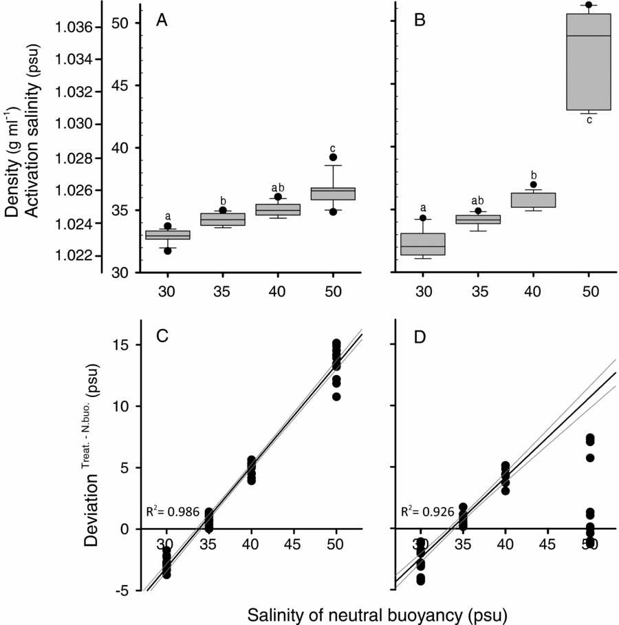
Activation salinity had a significant effect on egg buoyancy at 7 HPF (Fig. 5 A), such that fertilized eggs in the 50 psu treatments had the highest buoyancy, while eggs from the 30 and 35 psu treatments showed lower and similar buoyancy. At 30 HPF, salinity continued to significantly affect buoyancy, showing a similar trend (Fig. 5 B). Sinking speed of eggs recorded over a period of 210 min, from 7–10.5 HPF, in the salinity gradient column from the 35 psu treatment were on average 0.04 ± 0.0009 mm × min−1 corresponding to 0.0025 ± 0.001 psu × min−1 (N = 13).
Buoyancy characteristics of eggs from European eel, Anguilla anguilla, activated and fertilized at different salinities, and incubated at 20 ± 0.5˚C. (A, B) Neutral buoyancy of eggs (N ≥ 15) from each treatment after: (A) 7 HPF, (B) 30 HPF. a–cLetters indicate significant differences (α = 0.05) among groups. (C, D) Deviation between activation salinity and salinity at neutral buoyancy in relation to activation salinity at (C) 7 HPF (linear regression ± 95% CL, R2 = 0.99, y = 0.825X – 27.87); and (D) 30 HPF (linear regression excluding 50 psu ± 95% CL, R2 = 0.93, y = 0.655X – 22.073).

The deviation between measured buoyancies and salinity in activation and incubation, in relation to treatments, is shown in Fig. 5 C for 7 HPF and Fig. 5 D for 30 HPF. The deviation was significantly related to the activation salinity (R2 = 0.99, y = 0.825X – 27.87). At 30 HPF, data violated statistical assumptions as buoyancy of the 50 psu treatment increased more than the remaining treatments; therefore, the 50 psu treatment was omitted and the model was re-run. After re-running the model, a significant relationship between the salinity deviation (activation salinity subtracted the measured buoyancy) was also detected at 30 HPF (R2 = 0.93, y = 0.655X – 22.073).
Experiment 2: Activation of eggs with different salt types
Egg size
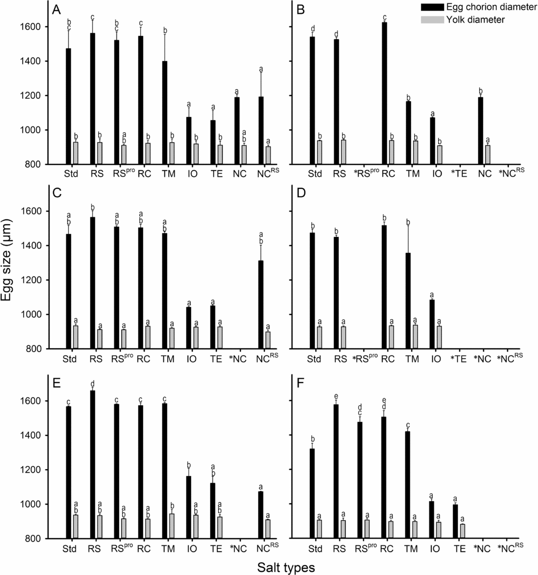
Average egg chorion diameter ranged from 1055 ± 59 to 1562 ± 75 μm across the nine salt types (Fig. 6 A). The mixed-model ANOVA showed that salt type had a significant effect on chorion diameter, such that the RS, RC, RSpro and Std treatments had the largest egg chorion diameter (Fig. 6 A). The female VC was non-significant (VC = 8.5%), while the female × salt type interaction was significant and explained the majority of the models variance (VC = 83.5%). For within female effects, salt type significantly influenced egg chorion diameter for all females (Fig. 6). For instance, the RC salt type evoked the largest chorion diameter for f6 (Fig. 6 B), while the RS salt type caused the largest egg chorion diameter for females f9 (Fig. 7 E). For f7, f8, f10 (Fig. 6 C, D, F), the largest egg chorion diameter was observed across a wide-range of salt types. For all females, the IO (Instant Ocean), TE, NCRS and NC salt types typically resulted in small egg chorion diameters compared with the other treatments (Fig. 6).
Egg size of European eel, Anguilla anguilla, in relation to salt type treatment adjusted to 35.0 ± 0.1 psu. Measurements include chorion and yolk diameter (μm ± standard deviation (SD)) of fertilized eggs 3.5–5 h post in vitro fertilization and incubated at 20 ± 0.5˚C. Salt type treatment: Std, natural seawater; RS, Red Sea salt; RSpro, Red Sea Pro; RC, Reef Crystal; TM, Tropic Marine; IO, Instant Ocean; TE, Tetra Marine; NC, NaCl; NCRS, NaCl + 10% Red Sea salt. (A) Average of all females. (B–F) are for female f6–f10. a–dLetters indicate significant differences (α = 0.05) among groups. *Treatments without fertilized eggs or not tested for a particular female.

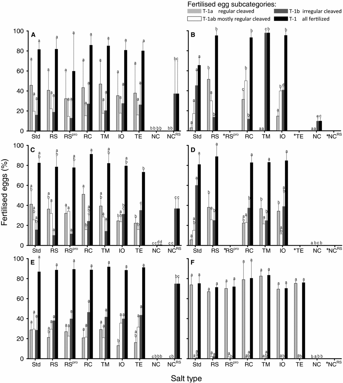
Fertilization success and cleavage pattern of in vitro fertilized eggs of European eel, Anguilla anguilla, in relation to salt type treatment adjusted to 35.0 ± 0.1 psu. Fertilization percentage and distribution of subcategories T-1a, T1ab and T-1b were assessed 3.5–5 h post fertilization and incubation at 20 ± 0.5˚C. (A) Average of all females. (B–F) are for female f6–f10. Letters indicate significant differences (α = 0.05) among groups. Bars show percentage + SD. Salt type treatment: Std, Natural seawater; RS, Red Sea salt; RSpro, Red Sea Pro; RC, Reef Crystal; TM, Tropic Marine; IO, Instant Ocean; TE, Tetra Marine; NC, NaCl; NCRS, NaCl + 10% Red Sea salt. *Treatments without fertilized eggs or not tested for a particular female.

Salt type had a significant effect on yolk diameter (Fig. 6 A). The female VC was significant and explained 46.1% of the variance, while the female × salt type VC was also significant and explained 31.8% of the variance. For within female effects, salt type had no effect on yolk diameter for female f8 (Fig. 6 D) and f10 (Fig. 6 F), while salt type had a significant effect for the other females.
Fertilization rate and zygote development
Considering all fertilized eggs, (egg category = T-1), fertilization percentage for the five females ranged from 0.3 ± 0.7 to 85.7 ± 9.0% across the nine salt types (Fig. 7 A). The mixed-model ANOVA showed a significant salt type effect in the fertilization per cent. Within this regard, RS, RC, RSpro, Std, TM, IO and TE had higher fertilization success than the NC and NCRS treatments (Fig. 7 A). For the models random effects, the female VC was significant (VC = 27.4%). The female × salt type interaction was also significant and explained 33.7% of the models variance. Salt type had a significant effect on fertilization for all females, such that the NC treatment always had the lowest fertilization while the RS, RC, TM and IO treatments had the highest fertilization (Fig. 7 B–F).
The proportion of fertilized eggs with regular cell cleavage (egg subcategory = T-1a), differed significantly depending on salt type (Fig. 7 A), with low proportions in NC and NCRS treatments compared with other salt types. The female VC was significant (VC = 62.1%) and similarly the female × salt type interaction, explaining 30.5% of the model's variance. For each female, the effect of salt type on the proportion of eggs with regular cell cleavage was also significant (Fig. 7 B–F).
With respect to the fertilized eggs that showed mostly regular cell cleavage, (egg subcategory = T-1ab), salt type had a significant impact on its distribution (Fig. 7 A). Furthermore, for the model's random effects the female VC was significant (VC = 41.7%) and the female × salt type interaction was also significant (VC = 39.0%). Salt type had a significant effect on the percentage of mostly regular cleaved eggs for the females f5, f7, f8, f9 (P ≤ 0.002; Fig. 7), while the effect of salt type was non-significant for f10 (Fig. 7 F). The NC and NCRS salt type treatment resulted in the lowest percentage of T-1ab for the five females, while for the remaining salt types showed no general pattern (Fig. 7 B–F).
Salt type had a significant effect on the proportion of eggs with irregular cell cleavages (egg subcategory = T-1b) (Fig. 7 A), where the NC differed from the NCRS treatment (Fig. 7 A). The female VC was significant (VC = 42.9%) and the female × salt type VC was significant and explained 49.5% of the model. Salt type effects were significant for all females with respect to proportion of T-1b eggs (Fig. 7 B–F).
Egg neutral buoyancy
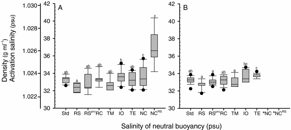
Mean neutral buoyancy of eggs from the different salt type treatments ranged from 32.4 ± 0.5 to 37.1 ±1.9 psu at 7 HPF (Fig. 8 A) and 32.8 ± 0.5 to 33.8 ± 0.2 psu at 30 HPF (Fig. 8 B). At 7 HPF, activation and incubation salt type had a significant effect on egg buoyancy, with no significant difference between Std, RS, RSpro, RC TM and TE salt types, which all had the lowest egg buoyancy. The NC and NCRS salt types had the highest buoyancy (Fig. 8 A). Additionally, RS was also significantly lighter than IO. At 30 HPF, the buoyancy of eggs was also significantly affected by activation and incubation salt type (Fig. 8 B) with NC and NCRS treatments no longer buoyant and RS, RSpro and TM having lower neutral buoyancy than IO and TE.
Neutral buoyancy of eggs from European eel, Anguilla anguilla, in vitro activated and fertilized, in relation to salt type treatment adjusted to 35.0 ± 0.1 psu and incubated at 20 ± 0.5°C. Box plot including outliers shows density and salinity at neutral buoyancy of (A) eggs (N ≥ 15) at 7 HPF; and (B) eggs (N ≥ 15) at 30 HPF. a–cLetters indicate significant differences (α = 0.05) among salt type treatments. *Treatments with non-buoyant eggs.

Size difference among fertilized egg types
Selection of treatments from Experiment 1 (30–45 psu) and Experiment 2 (Std, RS, RSpro, RC and TM) formed a dataset of 896 eggs with regular cell cleavages (T-1a); 753 eggs having mostly regular cell cleavages (T-1ab) and 1535 eggs with irregular cell cleavages (T-1b) (Table 3). The fertilized egg types significantly differed with respect to egg diameter, yolk diameter, egg volume, yolk volume and the size of the perivitelline space. For all egg metrics, the three fertilized egg types differed significantly from each other (Table 3).
Average metrics of in vitro fertilized eggs from European eel, Anguilla anguilla according to egg categories T-1a, T-1ab and T-1b; defined by cleavage symmetry at 3.5–5 h after fertilization

a–c Within each egg measure sub-letters indicate significant differences (α = 0.05).
Discussion
Egg sizes of marine fish show both inter- and intra-species variation (Kennedy et al., Reference Kennedy, Geffen and Nash2007). Maternal effects on egg diameter have been studied in many fish species and are generally found significant, depending on diet, broodstock condition and timing in relation to the reproductive season (Laale, Reference Laale1980; Kjørsvik et al., Reference Kjørsvik, Mangor-Jensen, Holmefjord, Blaxter and Southward1990; Morley et al., Reference Morley, Batty, Geffen and Tytler1999). Similarly, paternal effects are important, impacting processes related to fertilization (Evans & Geffen, Reference Evans and Geffen1998; Kroll et al., Reference Kroll, Peck, Butts and Trippel2013). In our study, wild-caught females were crossed with farmed males and we found a clear female effect that accounted for a large proportion of the models variance, e.g. yolk size had a female VC of 74.2%. However, no relationship was found between female size and egg chorion diameter, yolk diameter, or overall fertilization percentage.
Currently, no information is available regarding the size of European eel eggs in nature and we were limited to compare our observations with wild-captured eggs from Japanese eel. These wild-captured eggs are reported larger than eggs from their captivity-bred counterparts (Tsukamoto et al., Reference Tsukamoto, Chow, Otake, Kurogi, Mochioka, Miller, Aoyama, Kimura, Watanabe, Yoshinaga, Shinoda, Kuroki, Oya, Watanabe, Hata, Ijiri, Kazeto, Nomura and Tanaka2011; Yoshinaga et al., Reference Yoshinaga, Miller, Yokouchi, Otake, Kimura, Aoyama, Watanabe, Shinoda, Oya, Miyazaki, Zenimoto, Sudo, Takahashi, Ahn, Manabe, Hagihara, Morioka, Itakura, Machida, Ban, Shiozaki, Ai and Tsukamoto2011). In captive breeding of Japanese eel, egg sizes were in the range of 1300–1600 μm, while eggs captured in nature ranged from 1490–1710 μm with a mean of 1610 μm ± 70 μm (Tsukamoto et al., Reference Tsukamoto, Chow, Otake, Kurogi, Mochioka, Miller, Aoyama, Kimura, Watanabe, Yoshinaga, Shinoda, Kuroki, Oya, Watanabe, Hata, Ijiri, Kazeto, Nomura and Tanaka2011; Yoshinaga et al., Reference Yoshinaga, Miller, Yokouchi, Otake, Kimura, Aoyama, Watanabe, Shinoda, Oya, Miyazaki, Zenimoto, Sudo, Takahashi, Ahn, Manabe, Hagihara, Morioka, Itakura, Machida, Ban, Shiozaki, Ai and Tsukamoto2011). Overall, we found 13% of 3442 fertilized eggs to be in that range (1600–1785 μm), thus comparable with wild-caught Japanese eel. Therefore, our study pointed to effects of salinity and salt composition on egg size. Within females, we found that activation and incubation salinities had an effect on egg size and typically the largest eggs were obtained within the 30–40 psu interval. The variation among females regarding salinity tolerance and optima did however preclude a clear trend. Salinity-induced swelling was described in the long rough dab, Hippoglossoides platessoides where PVS is accounting for ≤85% of the egg volume (Lønning & Davenport, Reference Lønning and Davenport1980), which is close to the average of 74% PVS in regular cleaved eggs that we reported. The mechanisms during activation of a fish egg are still unknown (Webb & Miller, Reference Webb and Miller2013) and, specifically, research on the initial swelling following activation has been urged in relation to improving captive rearing in eels (Yoshinaga et al., Reference Yoshinaga, Miller, Yokouchi, Otake, Kimura, Aoyama, Watanabe, Shinoda, Oya, Miyazaki, Zenimoto, Sudo, Takahashi, Ahn, Manabe, Hagihara, Morioka, Itakura, Machida, Ban, Shiozaki, Ai and Tsukamoto2011).
As seen in our study, regardless of sperm being present, eel eggs swell upon activation by contact with seawater. Activation is known to be triggered by release of intracellular stored Ca2+ in between the yolk and chorion membrane (Finn, Reference Finn2007). This stimulates the cortical alveoli in the yolk membrane to exocytose glycoprotein into the perivitelline space, leaving the broken cortical alveoli to fuse with the plasma membrane and form the new protective and impermeable vitelline membrane surrounding the yolk (Hart & Yu, Reference Hart and Yu1980). The glycoproteins are cleaved into smaller colloid proteins (Gallo & Costantini, Reference Gallo and Costantini2012) and form a colloid osmotic gradient between the exterior and the PVS across the chorion (Shephard, Reference Shephard1989). This osmotic gradient has a charge potential, called the perivitelline potential (PVP) and the more negative the PVP, the stronger the force of water uptake from the environment (Peterson & Martin-Robichaud, Reference Peterson and Martin-Robichaud1986). This force is possibly imposed by diffusion through the chorion by cations such as sodium, Na+ rather than chloride, Cl− (Peterson & Martin-Robichaud, Reference Peterson and Martin-Robichaud1986), however the mechanism of water influx through the chorion in fish is not fully known (Yoshinaga et al., Reference Yoshinaga, Miller, Yokouchi, Otake, Kimura, Aoyama, Watanabe, Shinoda, Oya, Miyazaki, Zenimoto, Sudo, Takahashi, Ahn, Manabe, Hagihara, Morioka, Itakura, Machida, Ban, Shiozaki, Ai and Tsukamoto2011; Webb & Miller, Reference Webb and Miller2013). In freshwater fish, the ionic effects on swelling shows that polyvalent ions like Al3+ and Zn2+ inhibit PVS formation with increasing concentration (Eddy, Reference Eddy1983; Li et al., Reference Li, Jenssen and Fyhn1989; Shephard, Reference Shephard1989). Marine fish are fundamentally different as they face a hypertonic environment but are likewise sensible to ion composition, as shown in herring by cadmium Cd2+ affecting the osmotic gradient plus water uptake and egg size, especially at lower salinities (Alderdice et al., Reference Alderdice, Rao and Rosenthal1979a,Reference Alderdice, Rosenthal and Velsenb). The increase in egg diameter we observed could hardly be due to the same effects, as increasing salinity would increase inhibiting ions and thus decrease water uptake. A more likely explanation seems to be related to a selective influx of Na2+, as suggested by Peterson & Martin-Robichaud (Reference Peterson and Martin-Robichaud1986). This process would enforce a more negative PVP and create a stronger force of water uptake. Alone or playing in concert, osmotic shock exerted by the activation salinity may also contribute by triggering activation; similar to the mechanical touch of the chorion found to onset the activation process in Medeka, Oryzias latipes (Webb & Miller, Reference Webb and Miller2013).
The diameter of egg yolk showed no consistent size change in relation to salinity supporting the impermeable nature of the vitelline membrane; precluding further yolk size increases due to osmotic pressure as described by Hart & Yu (Reference Hart and Yu1980). This finding is in line with other studies, such as those on lumpsucker, Cyclopterus lumpus (Kjørsvik et al., Reference Kjørsvik, Davenport and Lönning1984) or long rough dab (Lønning & Davenport, Reference Lønning and Davenport1980) where even strong hyperosmotic pressure had a low impact on yolk osmolality and size. The long rough dab, Hippoglossoides platessoides limandoides, which has an expandable chorion, shows egg properties similar to the eel with a large expansion of the PVS and no size change in yolk plasma diameter (Lønning & Davenport, Reference Lønning and Davenport1980). Studies have shown the cortical reaction and formation of vitelline membrane, is faster once initiated at the egg animal pole, i.e. when sperm penetrates (Iwamatsu & Ito, Reference Iwamatsu and Ito1986) as compared with when activated merely by contact with seawater. Consequently, a delayed formation of this membrane is expected once activated without sperm cells. This may validate why we found a slightly larger yolk diameter once activated without sperm cells, as the slower activation process may cause extended membrane exposure to hypertonic medium and increase the yolk membrane diameter by osmotic pressure.
In two out of five females, the proportion of regular cleaved eggs (T-1a) showed significant peaks around 30 and 35 psu. Despite the fact that the proportion of regular cleaved eggs was low for the remaining three females, a general decrease in higher quality blastulas, T-1a as well as T-1ab, was observed with increasing salinity. Coincidentally at high salinity, the irregular cleaved eggs, T-1b, accounted for a high proportion of overall fertilized eggs, along with an increase in unactivated and dead eggs, T3 and T-5 respectively. Early cell cleavage symmetry, evenness and mutual adhesion at the 8–128-cell stage are known to correlate with larval survival in several species (Shields et al., Reference Shields, Brown and Bromage1997; Kjørsvik et al., Reference Kjørsvik, Hoehne-Reitan and Reitan2003). Activation and incubation salinity are likely to affect the success of in vitro fertilization in eel and the optimum is presumably close to the salinity of the proposed spawning area, i.e. ~36.5 psu (Munk et al., Reference Munk, Hansen, Maes, Nielsen, Castonguay, Riemann, Sparholt, Als, Aarestrup, Andersen and Bachler2010). The mechanisms of salinity effects on the zygote are scarcely known, but they are overall related to external and internal supplies of Ca2+. The initial wave of calcium upon sperm entry is released mainly from internal stores, while external supplies found in the seawater environment act to renew these stores and facilitate complex mechanisms; e.g. inositol triphosphate-mediated calcium release (IP3) (Stricker Reference Stricker1999). The different levels of action of this calcium wave may be an underlying explanation for the variation in fertilization quality patterns that we observed at different salinities. A study on Japanese eel embryos illustrated the importance of salinity changes, as eggs activated at 33 psu, followed by incubation treatments from 24–45 psu, showed a discrete effect on hatching but reduced larval survival above 36 psu and increased deformities below 30 psu (Okamoto et al., Reference Okamoto, Kurokawa, Gen, Murashita, Nomura, Kim, Matsubara, Ohta and Tanaka2009).
Egg buoyancy characteristics are fundamental to European eel egg development both in nature and hatcheries as after spawning the PVS fills with seawater, equaling the surrounding salinity (Davenport et al., Reference Davenport, Lønning and Kjørsvik1981). Neutral buoyancy is therefore determined by a balance between the seawater salinity and the yolk osmolality and lipids sealed by the vitelline membrane. Neutral buoyancy of eggs were correlated with the activation salinity. By using a range of salinities for activation and incubation it was possible to deduce a regression intercept at 33.8 psu, which indicates neutral buoyancy regardless of activation salinity. A similar pattern was confirmed at 30 HPF, although 50 psu had damaging effects. The measurement of neutral buoyancy in a salinity gradient can, however, lead potentially to overestimation of egg density because pelagic eggs with time tend to equilibrate across the chorion membrane with the surrounding water (Coombs et al., Reference Coombs, Boyra, Rueda, Uriarte, Santos, Conway and Halliday2004; Govoni & Forward, Reference Govoni, Forward, Finn and Kapoor2008). As defined by Stokes’ Law, sinking speed is related to egg size (Coombs et al., Reference Coombs, Boyra, Rueda, Uriarte, Santos, Conway and Halliday2004) and we observed a rapid sinking speed until the point of neutral buoyancy. Sardine eggs were shown to continue sinking from point of neutral buoyancy in a salinity gradient, challenging estimation of neutral buoyancy (Coombs et al., Reference Coombs, Boyra, Rueda, Uriarte, Santos, Conway and Halliday2004). In our study, however, we found eel eggs remained at the point of neutral buoyancy and sank only marginally from 7–10.5 HPF. This result means that from the time of the fully distended chorion and probably during the period of chorion hardening, the exchange of water across the chorion is very limited and validates our neutral buoyancies measures.
Commercial synthetic salt products are often branded as being similar in composition to seawater and thus potentially useful for standardizing water conditions for fish culture. Our results showed that several salt types were able to activate eggs at the same level as natural seawater (Std), but do so more consistently, thereby reducing variability. Similarly, some artificial sea salt brands as RS; Red sea salt pro, (RSpro;) Reef Crystal, (RC) and Tropic Marin, (TM) provided egg sizes as large as, or larger than, natural seawater (Std). In contrast, IO and Tetra Marine (TE), consistently resulted in eggs with a small PVS, which also pertained to pure NaCl, (NC) and NaCl +10% RS, (NCRS). In summary RS and RC seemed consistent in forming large eggs. Hardly any eggs could be fertilized in NC, however the addition of only 10% RS proved, in several cases, to enable fertilization and improve PVS formation and thereby added important input to the discussion of external calcium during zygote formation. NC lacks ions needed for activation and fertilization such as K+ and Ca2+, but indeed also affects eel sperm functionality that further needs K+, Na+ and HCO3 − (Ohta et al., Reference Ohta, Kagawa, Tanaka and Unuma2001; Gallego et al., Reference Gallego, Martínez-Pastor, Mazzeo, Peñaranda, Herráez, Asturiano and Pérez2014).
The mineral compositions for the RS, RC, TM and IO brands are published (Atkinson & Bingman, Reference Atkinson and Bingman1997; Hovanec & Coshland, Reference Hovanec and Coshland2004; Arnold et al., Reference Arnold, Cotsifas, Winter, Klinck, Smith and Playle2007) however variation between studies and composition of minor elements between reported brands is considerable (Atkinson & Bingman, Reference Atkinson and Bingman1997; Hovanec & Coshland, Reference Hovanec and Coshland2004; Arnold et al., Reference Arnold, Cotsifas, Winter, Klinck, Smith and Playle2007). Minor differences in elements make up more than 20 components in the salt types, including several of the metals believed to affect egg activation. A reported difference between brands relates to the alkalinity and pH (Atkinson & Bingman, Reference Atkinson and Bingman1997). IO and RC differed in their ability to induce a large PVS, resembled each other in composition (Atkinson & Bingman, Reference Atkinson and Bingman1997; Hovanec & Coshland, Reference Hovanec and Coshland2004), but showed a difference in pH of 0.6. Variation in pH of 0.6 is, however, similar to magnitude of pH difference between the salt types leading to consistently large PVS, why pH within the range observed cannot determine PVS formation. Variability in alkalinity, not determined in this study, could have an impact on the sperm via HCO3 −, which has been seen to increase motility in Japanese eel sperm (Ohta et al., Reference Ohta, Kagawa, Tanaka and Unuma2001).
Artificial salt types contain many different ions and several are known to block the cortical reaction e.g. Al3+ in Atlantic salmon (Finn, Reference Finn2007) or inhibit it, such as Zn2+, Mg2+ and SO4 2− (Eddy, Reference Eddy1983). A study on blue mussel Mytilus edulis, found that salt type IO contained a higher amount of Cu ions from industrial processing (Arnold et al., Reference Arnold, Cotsifas, Winter, Klinck, Smith and Playle2007). Until recently, aquaporin was only described as acting in fish tissues and in the fish oocyte during the maturation process (Cerdà & Finn, Reference Cerdà and Finn2010). Amaroli (Reference Amaroli2013) showed for sea urchins Paracentrotus lividus, that water uptake during activation was highly dependent on aquaporin channels in the chorion (Amaroli, Reference Amaroli2013) and that PVS formation and fertilization was highly inhibited by ions such as Cu, Hg and Ni. Assuming aquaporins are present in fish egg chorion, then variability in the concentrations of metal ions in marine salt types could affect PVS formation.
Egg neutral buoyancy at 7 HPF did not differ among salt types, reflecting the remarkable deviation in egg chorion diameter observed in the eggs from IO and TE. However, at the embryo formation stage (30 HPF), the small eggs obtained using IO and TE became heavier and this could indicate the first signs of insufficient PVS size. This influences metabolism and wastes from the embryo, and causes suboptimal incubation conditions (Eddy, Reference Eddy1983). This scenario resembles previous reported observations in which artificial seawater invoked small eggs that were unable to hatch (Tomkiewicz & Jarlbæk, Reference Tomkiewicz and Jarlbæk2008). The neutral buoyancy found from the salts that provided large eggs was around 33.8 psu, a value that is similar to buoyancy estimates for Japanese eel eggs (Tsukamoto, Reference Tsukamoto2009).
In summary, salinity and sea salt type significantly influenced egg activation, fertilization, buoyancy and early embryonic development, although with substantial variation in optimal conditions and tolerance between females. Fertilization success was highest in the salinity range 30–40 psu. Salt type significantly affected egg chorion diameter; RS and RC lead to consistently large eggs while IO, TE, NC and NCRS generated eggs with limited PVS. Yolk diameters showed low variability across treatments, supporting the impermeability of the vitelline membrane during early embryonic development. Activation and incubation salinity affected neutral buoyancy; only within a salinity range of 30–40 psu could egg neutral buoyancy be sustained during incubation to embryo formation. Salt types were found to induce only small egg chorion diameters and failed to sustain buoyancy. In conclusion, salt concentration and elemental composition strongly affected processes during egg activation and embryonic development. The effects would often play in concert and these mechanisms are not fully understood. Successful activation and development of the eggs of European eel are crucial for progress in the artificial propagation of the species; further studies on these quality aspects of the activation media are encouraged.
Acknowledgements
Funding was provided by the project ‘Reproduction of European Eel: Toward a Self-sustained Aquaculture’ (PRO-EEL) supported financially by the European Commission's 7th Framework Programme under Theme 2 ‘Food, Agriculture and Fisheries and Biotechnology’, Grant Agreement no. 245257. We appreciate the assistance of Christian Graver, Peter Lauesen and Maria Krüger-Johnsen in managing broodstocks and performing experiments. All fish were handled in accordance with the European Union regulations concerning the protection of experimental animals (Dir. 86/609/EEC).
Conflicts of interest
There are no conflicts of interest.
Ethical standards
The authors assert that all procedures contributing to this work comply with the ethical standards of the relevant national and institutional committees on human experimentation and with the Helsinki Declaration of 1975, as revised in 2008. Furthermore we assert that all procedures contributing to this work comply with the ethical standards of the relevant national and institutional guides on the care and use of laboratory animals.