Introduction
Platinum is used as a catalytic electrode in fuel cells. Fuel-cell performance is often governed by the oxygen-reduction reaction (ORR) at an air electrode made of platinum.[Reference Stephens, Bondarenko, Grønbjerg, Rossemeisl and Chorkendorff1] Platinum is a rare metal and is found disproportionately in the earth's crust. Hence, the platinum price is high and fluctuates, which prevents fuel cells from being used widely. Many efforts have been made to improve the catalytic activity of platinum in the ORR. One strategy is nanostructuring of platinum; that is, a reduction in the typical length scale of platinum is advantageous in terms of an effective surface area. Platinum nanoparticles[Reference Wikander, Ekström, Palmqvist and Lindbergh2, Reference Li, Zhao, Cheng, Fortunelli, Chen, Yu, Zhang, Gu, Merinov, Lin, Zhu, Yu, Jia, Guo, Zhang, Goddard, Huang and Duan3] and hollow platinum nanoparticles are good candidates for efficient ORR catalytic electrodes.[Reference Wang, Xu, Tsai, Li, Liu, Zhao, Liu, Yuan, Abild-Pedersen, Prinz, Nørskov and Cui4, Reference Zhang, Ma, Zhu, Si, Cai, Wang and Adzic5]
On the other hand, the synthesis of nanoporous metals by dealloying or selective dissolution of one element from a binary alloy is another strategy for nanostructuring. Such a three-dimensional nanoporous structure is obtained in various metals.[Reference Forty6, Reference Pugh, Dursun and Corcoran7] Nanoporous copper (NP-Cu) can be also produced by dealloying ductile Cu–Mn alloys,[Reference Keir and Pryor8–Reference Hayes, Hodge, Biener, Hamza and Sieradzki10] and similarly, porous Raney copper is synthesized by dealloying Cu–Al alloys.[Reference Kong, Ma, Zhang, Sun, Chen and Liu11]
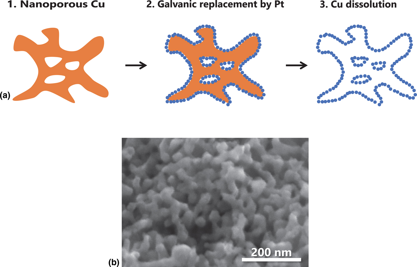
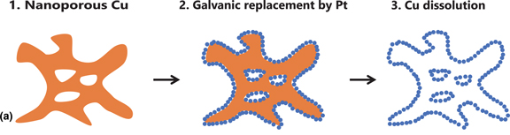
Platinum can be deposited in the form of a dense film or fine particles on copper by electroless plating or galvanic replacement.[Reference Zhang, Ma, Zhu, Si, Cai, Wang and Adzic5, Reference Feng, Liu, Wang and Jin12] Thus, NP-Cu can be converted to nanoporous platinum with a skeletal structure replicated from NP-Cu and deposits of fine platinum particles. The supporting NP-Cu can be removed readily by HNO3 which dissolves copper selectively out through the porous particulate platinum layer, and leaves a bimodal nanoporous platinum (BNP-Pt) [Fig. 1(a)]. Here we show the synthesis of BNP-Pt for ORR by electroless plating of platinum on the sacrificial NP-Cu and subsequent dissolution of NP-Cu. We focus on the contraction of the crystal lattice of platinum in BNP-Pt as a source of high ORR catalytic activity per surface area.
(a) Schematic illustration of the synthesis of bimodal nanoporous platinum using nanoporous copper as a sacrificial support. (b) Scanning electron microscopy image of nanoporous copper fabricated by dealloying of Cu–Mn alloy.

Material and methods
Initial Cu0.3Mn0.7 alloy button-shaped ingots were fabricated by the arc melting of pure copper and manganese in an argon atmosphere. After heating at 1123 K for 24 h in an Ar atmosphere, the alloy ingot was water-quenched,[Reference Okamoto13] followed by cold rolling to 0.2-mm thickness.
Dealloying to fabricate NP-Cu was conducted by free corrosion in 1 mol L−1 HCl for 24 h, where manganese was dissolved selectively and copper formed a nanoporous structure spontaneously [Fig. 1(b)].[Reference Keir and Pryor8–Reference Hayes, Hodge, Biener, Hamza and Sieradzki10] After washing in pure water, the NP-Cu working electrode was pulverized manually in an agate bowl to a powder.
NP-Cu (0.05 g) was mixed with 1 g of 5 wt% Nafion ethanolic solution and 5 g of water. After homogenization in an ultrasonic bath, this suspension was fixed on a glassy carbon (GC) rotation disk electrode (RDE) with 5-mm diameter. The displacement deposition of platinum on the fixed NP-Cu was conducted by immersing the GC RDE in 1 mmol L−1 H2PtCl4 aqueous solution at 273 K for 1 h at 600 rpm.[Reference Coleman and Co14] Thus, the GC electrode includes NP-Cu with a fine platinum deposition. The GC electrode that includes BNP-Pt was immersed in 0.1 mol L−1 HNO3 for 24 h to dissolve the copper.
We conducted cyclic voltammetry (CV) at room temperature by using the GC electrode loading BNP-Pt as a working electrode. To evaluate the electrochemical surface area (ECSA) of the BNP-Pt samples, CV was conducted in degassed 0.5 mol L−1 H2SO4 with a scan rate of 10 mV s−1. A reversible hydrogen electrode (RHE) and Pt wire were used as a reference and counter electrode, respectively. For electrochemical cleaning, CV was repeated until a steady current-potential cyclic curve was attained. The charge that is associated with hydrogen adsorption (Q H) was calculated by using the cathodic peak between 0.1 and 0.35 V (versus RHE), which was then converted to the ECSA (A) of the sample;[Reference Trasatti and Petrii15]
 $$A_{{\rm sample}}/A_{{\rm bulk}} = Q_{{\rm H,sample}}/Q_{{\rm H,bulk}}$$
where the subscripts “sample” and “bulk” represent the BNP-Pt and bulk platinum, respectively.
The working GC-RDE-loaded BNP-Pt was rotated for ORR activity measurements during the CV in an electrolyte of O2-saturated 0.1 mol L−1 H2SO4 with a scan rate of 10 mV s−1. The kinetically-controlled current i K, which is an index of the ORR activity of the BNP-Pt, was calculated according to:
 $$1/i = 1/i_{\rm K} + 1/B\omega ^{1/2}$$
where i is a measured current; B is a constant and ω is the RDE rotation speed. i K was derived from the intersection at ω −1/2 = 0 by the Koutechy–Levich plot of i −1 (at 0.65 V versus RHE) against ω −1/2.[Reference Garsany, Baturina, Swider-Lyons and Kocha16–Reference Zheng, He, Chen, Wang, Xue and Feng19] The specific activity per ECSA was calculated by dividing i K by A sample (ECSA).
Substances on the GC RDE were collected for SEM, scanning transmission electron microscopy (STEM) and high-resolution transmission electron microscopy (HR-TEM). Energy-dispersive X-ray spectroscopy (EDXS) was used for elemental analyses in the microscopic observations. X-ray diffraction (XRD) patterns of samples (and conventional Pt black) were collected under a parallel-beam configuration at 298 K. The concentration of platinum and other elements in samples was determined by inductively-coupled plasma atomic emission spectroscopy (ICP-AES) after dissolving the samples in 20 mL of nitrohydrochloric acid.
Results and discussion
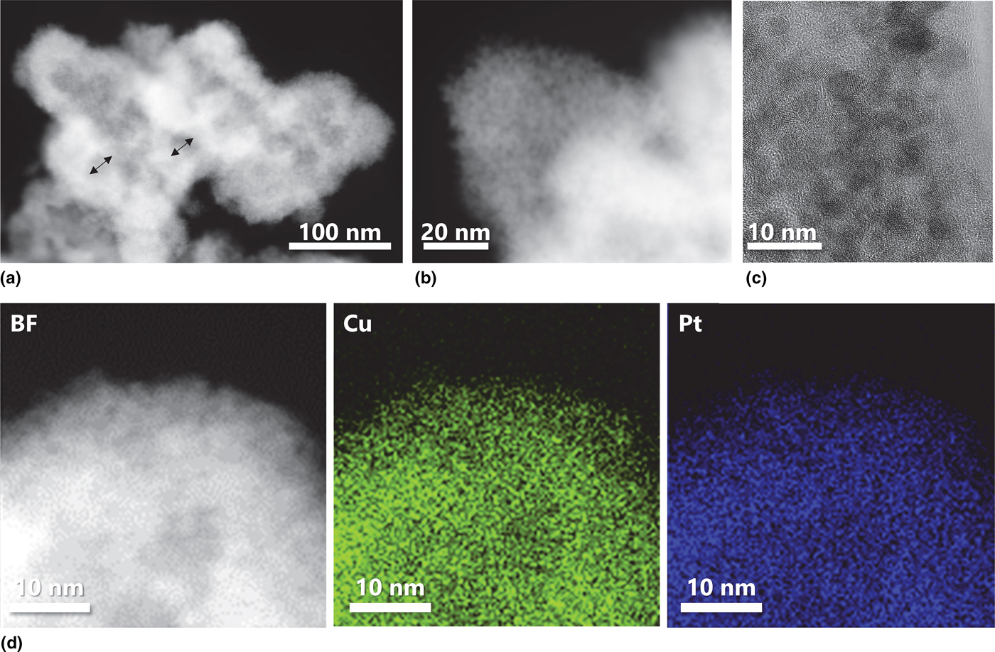
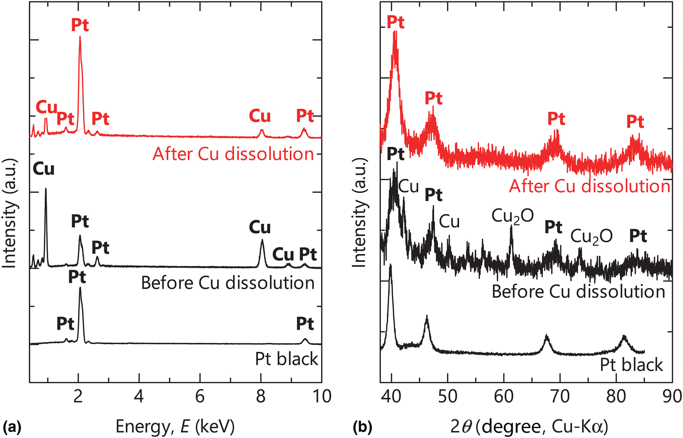
STEM images (Fig. 2) indicate the bimodal structure of the sample. The nanoporous hollow structure with an average pore diameter of 23 nm [Fig. 2(a)], which is replicated from NP-Cu [Fig. 1(b)], was composed of finer Pt particles with 3–5 nm diameters [Figs. 2(b) and 2(c)]. STEM-EDXS reveals that Cu remains even after dissolution by HNO3, although Cu and Pt exhibited a homogeneous distribution without surface segregation [Fig. 2(d)]. The remaining Cu in the sample after dissolution by HNO3 was confirmed by SEM-EDXS [Fig. 3(a)], where the atomic ratio of Cu/Pt was 11.5 before dissolution and 0.54 after dissolution. On the other hand, manganese was not detected by EDXS and ICP-AES in BNP-Pt before and after the dissolution of copper. Thus, the contribution of manganese is eliminated in the following discussion on the catalytic properties of BNP-Pt samples.
Scanning transmission electron microscopy (a, b, d) and high-resolution transmission electron microscopy (c) images of bimodal nanoporous platinum. Panels (a), (b), and left panel in (d) are high-angle annular dark-field images, whereas the center and right panels in (d) show the elemental mapping by energy-dispersive X-ray spectroscopy. Arrows in panel (a) show pore size (=20 nm) replicated from skeletal nanoporous copper, while panel (b) and (c) show the finer porous structure of platinum.

(a) Energy-dispersive X-ray spectroscopy results and (b) X-ray diffraction patterns of bimodal nanoporous platinum before and after copper dissolution. The pattern for Pt black is shown for comparison in (b).

XRD patterns of BNP-Pt before and after copper dissolution are shown in Fig. 3(b). Before copper dissolution, the broad peaks for platinum are accompanied by peaks for metallic copper and copper oxide (Cu2O). Minor Cu2O appears to form during the preparation of NP-Cu by dealloying. Metallic copper and Cu2O were not detected after dissolution by HNO3. Core NP-Cu and accompanying minor Cu2O dissolved into HNO3, whereas Pt remained undissolved. An inspection of the XRD patterns showed that the peaks of Pt in BNP-Pt shifted to higher diffraction angles compared with referenced Pt black.
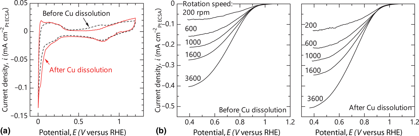
CV curves for BNP-Pt before and after copper dissolution [Fig. 4(a)] are typical for platinum[Reference Coleman and Co14–Reference Zheng, He, Chen, Wang, Xue and Feng19] with peaks for hydrogen and oxygen adsorption/desorption. Copper dissolution by HNO3 caused no significant change in the redox process during CV scanning.
(a) Cyclic voltammetry curves for bimodal nanoporous platinum before and after copper dissolution. The scan rate was 10 mV s−1 and the electrolyte was 0.1 mol L−1 H2SO4. (b) Polarization curves for rotation disk electrodes that load bimodal nanoporous platinum before and after copper dissolution. The scan rate was 10 mV s−1 and the electrolyte was 0.1 mol L−1 H2SO4.

The ORR activity of the BNP-Pt samples was examined by RDE [Fig. 4(b)]. In the polarization curves, the current density at a high potential of 1.1 V was independent of the electrode rotation rate, which corresponds to the kinetic region of the ORR. As the potential decreased from 1.1 V, transitional regions from a kinetic- to a diffusion-controlled reaction rate were observed, with a lower current density at a higher electrode rotation rate. Below 0.4 V, the current density was almost constant and a diffusion-controlled reaction occurred.
The specific activities of BNP-Pt per ECSA of Pt before and after copper dissolution were 1.48 and 4.18 mA cm−2, respectively, according to the RDE results. On the other hand, activity per mass of BNP-Pt before and after copper dissolution was 0.14 and 0.35 A mgPt−1, respectively. These values are significantly higher than those of the conventional Pt/C[Reference Hu, Kuttiyiel, Sasaki, Su, Yang, Park, Zhang, Chen and Adzic17] and comparable with the values reported recently for other nanoporous Pt-based alloys.[Reference Stephens, Bondarenko, Grønbjerg, Rossemeisl and Chorkendorff1, Reference Cui, Mei, Han, Chen, Zhang, Quan, Gu, Zhang, Fang, Qian, Jiang and Han20–Reference Godínez-Salomón, Mendoza-Cruz, Arellano-Jimenez, Jose-Yacaman and Rhodes25] Furthermore, the copper dissolution increased the specific activity of the BNP-Pt. Above all, a higher activity per ECSA (and not per mass or geometric area) for BNP-Pt after copper dissolution means that the intrinsic catalytic nature of the platinum is enhanced by the disappearance of the supporting skeletal NP-Cu, although the overpotential itself is not so different between the two BNP-Pt samples.
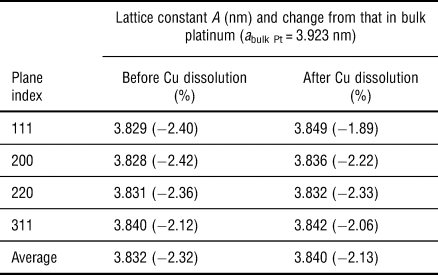
Lattice straining affects the overall catalytic performance in platinum.[Reference Strasser, Koh, Anniyev, Greeley, More, Yu, Liu, Kaya, Nordlund, Ogasawara, Toney and Nilsson21, Reference Rhen and McKeown26, Reference Gasteiger, Kocha, Sompalli and Wagner27] Strasser et al.[Reference Strasser, Koh, Anniyev, Greeley, More, Yu, Liu, Kaya, Nordlund, Ogasawara, Toney and Nilsson21] discuss the effect of lattice strain on the activity of core-shell platinum catalysts for ORR, where the Pt–Cu nanoparticles are dealloyed partially to form nanoparticles with a platinum shell and a Pt–Cu core. A moderate reduction in lattice constant, namely, lattice contraction of the platinum is important to enhance the ORR activity of the platinum, although an excessive compressive lattice strain (approximately below −2.5%) deteriorates the ORR activity. Table I shows the lattice constants of BNP-Pt calculated from the XRD peaks [Fig. 3(a)]. The lattice constants of the BNP-Pt before and after copper dissolution were lower than those of the bulk platinum, which suggests that misfit strain and/or alloying occurs during the displacement electrodeposition of platinum on NP-Cu. The reduction in lattice constants is milder in the BNP-Pt after copper dissolution than in the BNP-Pt before copper dissolution. This moderate lattice contraction in BNP-Pt after copper dissolution is responsible for the improved ORR activity per ECSA. A partial dissolution of supporting NP-Cu from BNP-Pt leads to a relaxation of the misfit strain that is generated between the platinum shell and the copper core.
Lattice constants of platinum in bimodal nanoporous platinum.

The SEM-EDXS showed that the residual copper in the BNP-Pt is not negligible even after immersion in HNO3. According to Vegard's law, lattice constant a is supposed to be 3.815 nm for Pt0.65Cu0.35 (BNP-Pt after copper dissolution), which is smaller than the present value (Table I). That is, the lattice contraction is milder than expected from the atomic ratio of Pt/Cu and Vegard's law. After copper dissolution, other copper-containing phases in the BNP-Pt were not detected by XRD [Fig. 3(a)]. The STEM-EDXS also showed no significant distribution and separation of copper and platinum. These results indicate that the nanoporous and replicated hollow structure intrinsically cause a peculiar lattice straining due to the large positive and negative curvatures.[Reference Hakamada, Nakano, Furukawa, Takahashi and Mabuchi28, Reference Fujita, Guan, McKeena, Lang, Hirata, Zhang, Tokunaga, Arai, Yamamoto, Tanaka, Ishikawa, Asao, Yamamoto, Erlebacher and Chen29] The nanoporous nickel that is fabricated by dealloying of Ni–Mn alloys shows larger lattice constants than those in bulk nickel.[Reference Hakamada, Takahashi, Furukawa and Mabuchi30] Similarly, the nanoporous and replicated structure may relax the severe lattice compression in BNP-Pt expected by Vegard's law.
Further discussion on the ORR performance of BNP-Pt requires determination of active site density, turnover frequency, crystallographic structure, and chemical (oxidation) state of BNP-Pt. The Brunauer–Emmett–Teller measurement, extended X-ray absorption fine structure analysis, X-ray photoelectron spectroscopy are useful tools; nevertheless, the specific activity per ECSA is a first parameter representative for the performance of ORR. Also, the role of copper in the straining and subsequent catalysis on ORR is not fully understood at this stage. There is much room for investigation.
Conclusions
BNP-Pt catalysts for ORR were fabricated by using NP-Cu as a sacrificial metallic support. The high ORR specific activity of BNP-Pt after copper dissolution is attributed to the moderate lattice contraction in platinum, which is induced by alloying with copper and/or a misfit strain at the Pt/Cu interfaces, but which is relaxed by the replicated nanoporous hollow structure.
Acknowledgments
M. H. acknowledges financial support by Kansai Research Foundation for Technology Promotion. TEM and STEM observations were conducted at the Kyoto University Nano Technology Hub in the “Nanotechnology Platform Project” sponsored by the Ministry of Education, Culture, Sports, Science and Technology (MEXT), Japan.