The palaeontology and stratigraphy of northern Central Kazakhstan has been the subject of research for more than seventy years. Nevertheless, diverse Late Ordovician trilobite faunas, known mainly from publications by Weber (Reference Weber1948) and Koroleva (Reference Koroleva1982 and references therein), remain inadequately documented, and the taxonomic composition and ages of these faunas often require considerable revision. This study focuses on the Late Ordovician (Katian) trilobite associations recovered in the vicinity of Lake Atansor, in the eastern part of the Stepnyak region.
The occurrence of Late Ordovician trilobites on the northern coast of Lake Atansor (Figs 1, 2B) was first documented by Shlygin in 1930. His collection was subsequently studied by Weber (Reference Weber1948), who briefly described and illustrated several taxa, including Ceraurus? sp., Pliomerina (Encrinurella) sulcifrons Weber, 1948 [= Pliomerina sulcifrons (Weber, Reference Weber1948)], Remopleurides cf. pisiformis Weber, 1948, Sphaerexochus hisingeri Warburg, Reference Warburg1925 [= Sphaerexochus conusoides Koroleva, Reference Koroleva1959] and Toernquistia shlygini Weber, 1948. All these specimens were sampled from the unit of limestone up to 100 m thick exposed on the cliff along the northern shore of Lake Atansor, but the precise position of the locality within the unit remains unknown. In 1951, the locality was revisited by Koroleva, who gave a brief geological outline of the area (Koroleva Reference Koroleva1954) and later described several more trilobite taxa from the Sandbian Mailisor Formation (Koroleva Reference Koroleva1965). She also provided a detailed sketch map, which is helpful in identifying the precise geographical position of the sampled trilobite localities. The Atansor area is situated within the Stepnyak tectonofacies unit of Şengör & Natal'in (Reference Şengör, Natal'in, Yin and Harrison1996), which may represent a Late Ordovician Andean- or Japan-type continental volcanic arc on the margin of the Kalmykkol–Kokchetav Microcontinent (Krőner et al. Reference Kröner, Hegner, Lehmann, Heinhorst, Wingate, Liu and Ermelov2008; Degtyarev Reference Degtyarev2012).
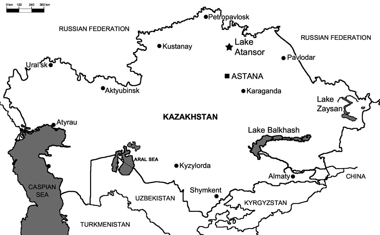
Map showing the location of Lake Atansor in Northern Kazakhstan (star symbol).

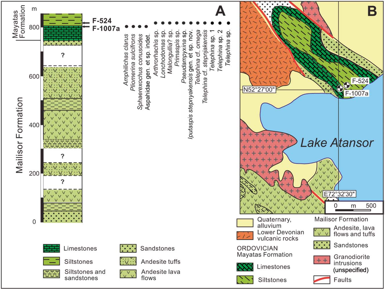
Simplified stratigraphic column and geological map, showing the location of samples F-524 and F-1007a in the Mayatas Formation, Northern Kazakhstan.

1. Localities and stratigraphy
The upper Ordovician deposits of the eastern and central part of the Stepnyak unit are subdivided into the Lidievka, Mailisor and Mayatas formations (Nikitin Reference Nikitin1972, Reference Nikitin1991). The Lidievka Formation comprises volcanic sandstones, siltstones with units of andesitic volcanic rocks and tuffs. The Mailisor Formation is characterised by the predominance of volcanoclastic deposits, andesite tuffs and lava flows; whereas the Mayatas Formation comprises siliciclastic rocks with lens-like bodies of limestone up to 300–400 m thick, associated with microbial build-ups in the lower part. The limestones contain a diverse trilobite fauna partly described by Weber (Reference Weber1948) and Koroleva (Reference Koroleva1965), but it remains inadequately known. Nikitin (Reference Nikitin1972) provided extensive lists of the upper Ordovician trilobites, brachiopods and graptolites from the Mailisor and Mayatas formations, but they are outdated and mostly based on preliminary identifications, and there is inadequate monographic coverage of these fauna.
The collection used in the present study was sampled by Popov and Tsai in 1974 from two localities (samples F-524 and F-1007a) on the northern shore of Lake Atansor, from the uppermost part of the limestone unit and overlying unit of siliceous graptolitic shales, assigned to the lower part of the Mayatas Formation (Fig. 2A, B). The limestone unit, up to 100 m thick, includes bacterial build-ups (so-called ‘mud-mounds’); however, to date these have not been the subject of detailed sedimentological study. The early Katian age of the limestone unit is supported by the occurrence of the tabulate coral Amsassia chaetetoides Sokolov, identified by Kovalevskii (Nazarov & Popov Reference Nazarov and Popov1980), because elsewhere in Kazakhstan it occurs in rocks of known early Katian age, dated by graptolites (Nikitin Reference Nikitin1972).
Sample F-1007a was taken from the uppermost 0.2–0.3 m of dark-grey bioclastic limestone, probably deposited on the flanks of a mud-mound. An oligotaxic trilobite assemblage recovered from that sample includes Amphilichas clarus (Koroleva, Reference Koroleva1959), Pliomerina sulcifrons (Weber, Reference Weber1948), Sphaerexochus conusoides Koroleva, 1959 and an indeterminate asaphid. The associated fauna includes a diverse linguliform microbrachiopod association described by Nazarov & Popov (Reference Nazarov and Popov1980) and Popov (Reference Popov2000), and an ostracod fauna described by Melnikova (Reference Melnikova1986), comprising Kazakhstanian endemics (Laccochilina indigena Melnikova, Reference Melnikova1980, L. multispinosa Melnikova, Reference Melnikova1986, Junctusina bicalcarata Melnikova, Reference Melnikova1986, Quadritia aperta Melnikova, Reference Melnikova1986, Steusloffina amphla Melnikova, Reference Melnikova1986 and Ockerella lateralis Melnikova, Reference Melnikova1986). Preservation of the trilobites in Sample F-1007a is variable, with some preserved as internal moulds, whilst others partially retain original cuticle. However, all specimens exhibit some damage and are incomplete to varying degrees.
Sample F-524 was collected from about 3 m above the base of the siliceous argillite unit overlying the limestones. The assemblage contains Arthrorhachis sp., Lonchodomas sp., Pseudampyxina sp., Malongullia? sp., Primaspis sp., Telephina cf. omega Koroleva, Reference Koroleva1982, Telephina cf. stepnjakensis Koroleva, Reference Koroleva1982, a possible additional Telephina species, and a new trinucleid, Iputaspis stepnyakensis gen. et sp. nov. The fauna is dominated by blind trilobites (a trinucleid, an agnostid and three raphiophorids) and three species of large-eyed Telephina, suggesting that they occupied the disphotic zone in deep water offshore (BA5-6). A single cranidium of the odontopleurid Primaspis is also present. Preservation of the trilobites in Sample F-524 is generally poor, with specimens preserved as internal or external moulds, which in some cases have undergone some surface weathering.
2. Significance of the fauna
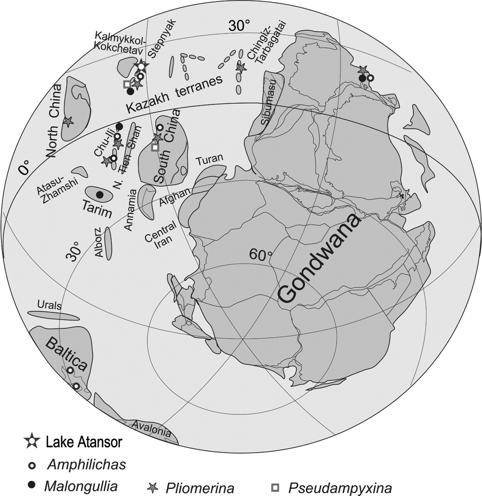
The present day Kazakhstanian orogen incorporates microplates and volcanic arc systems, which formed a significant archipelago during the Late Ordovician, extending far along subequatorial latitudes west of the Australasian sector of Gondwana (Popov et al. Reference Popov, Bassett, Zhemchuzhnikov, Holmer and Klishevich2009). The faunas described here originate from a cluster of terranes in north-central Kazakhstan (Fig. 3), which correspond to the Kalmykkol–Kokchetav unit of Şengör & Natal'in (Reference Şengör, Natal'in, Yin and Harrison1996) or to the Shatsk and Kokchetav microplates of Dobretsov et al. (Reference Dobretsov, Buslov, Zhimulev, Travin and Zayachkovskii2006). Koroleva (Reference Koroleva1982) summarised Late Ordovician trilobite distributions in the Selety, Ishim and Stepnyak regions, but these records are incomplete and their taxonomic affiliations require revision.
Palaeogeographical reconstruction of Gondwana, satellite microcontinents and island arcs for the early Katian (455 Ma), showing the geographical distribution of selected biogeographically informative trilobite genera; mainly after Torsvik & Cocks (Reference Torsvik and Cocks2009), with emendations. Large star shows inferred position of the Atansor locality.

The fauna from sample F-1007a is derived from a carbonate build-up, and such faunas became widespread throughout Kazakhstanian island arcs and microcontinents in the Sandbian–Katian, and represent the illaenid–cheirurid biofacies (Fortey Reference Fortey1975a). The Atansor fauna includes some taxa commonly associated with the illaenid–cheirurid biofacies, namely Amphilichas and Sphaerexochus, but lacks the nominate genera and is of much lower generic diversity than was typical in some other parts of Kazakhstan. The presence of Amplilichas and Pliomerina suggests affinity with Australian faunas (Webby Reference Webby1971, Reference Webby1974; Edgecombe et al. Reference Edgecombe, Banks and Banks2004; Edgecombe & Webby Reference Edgecombe and Webby2007).
The fauna from sample F-524 represents a very different environment, the disphotic, deeper offshore setting of the raphiophorid biofacies. The Atansor fauna completely lacks remopleuridids, which are a characteristic component of this biofacies in both the southern cluster of Kazakhstanian terranes (in Chu–Ili) and the volcanic island arc systems of the northeast (Chingiz–Tarbagatai and Boshchekul terranes). There are at least two species of Telephina in the fauna but, although this genus is also known from the Australian sector of Gondwana, South China (e.g., Zhou et al. Reference Zhou, Li and Qu1982; Tripp et al. Reference Tripp, Zhou and Pan1989) and Thailand (Fortey Reference Fortey1997), it is a globally widespread epipelagic genus of limited biogeographic value. The occurrence of the unusual three-segmented raphiophorid Pseudampyxina at Lake Atansor represents the first record of the genus outside South China (Tripp et al. Reference Tripp, Zhou and Pan1989). Malongullia is also found in Tarim and Australia. However, it is likely that the Atansor species described here forms a natural group with the South Chinese species, closely allied to Malongullia but lacking eye tubercles, and that they are closely related to species such as M. biloba (Chugaeva, Reference Chugaeva1958) and M. gracilis (Lu, Reference Lu1975) in which the eye tubercles are relatively small.
The faunas from Atansor have a limited number of taxa in common with other parts of Kazakhstan, indicating that there was no significant faunal exchange with other Kazakhstanian terrane clusters. A number of genera in common with Australia and South China suggests that there may have been some exchange with the Australasian part of east Gondwana and the South China microcontinent, suggesting a peri-Gondwanan location for the Stepnyak terrane; whilst palaeomagnetic data obtained by Bazhenov et al. (Reference Bazhenov, Levashova, Degtyarev, Van der Voo, Abrajevitch and McCausland2012) suggest its position in subequatorial latitudes (although polarity is uncertain). The occurrence in this fauna of a single specimen of Primaspis, a genus previously only known from Bohemia, is unusual, but some cold-water elements can be expected in deep-water raphiophorid assemblages. This may also be an indicator of strong thermal stratification of the water column. A rthrorhachis is also recorded from European peri-Gondwana; however, its cosmopolitan distribution also includes Avalonia, Baltica and Northern China (see Budil et al. Reference Budil, Fatka, Kolář and David2011), as well as Kazakhstan, so it is impossible to isolate a biogeographic signal from it.
3. Systematic palaeontology
All studied and illustrated specimens are housed in the Natural Sciences Department of the National Museum of Wales under Accession Numbers NMW 2008.34G and 2015.31G.
Order Agnostida Salter, 1864
Superfamily Agnostoiidea M‘Coy, Reference M'Coy1849
Family Metagnostidae Jaekel, Reference Jaekel1909
Genus Arthrorhachis Hawle & Corda, Reference Hawle and Corda1847
Type species. Battus tardis Barrande, 1846 from the Králův Dvůr Formation (upper Katian) of Libomysl, Czech Republic (by monotypy).
Arthrorhachis sp.
(Fig. 7j)
Material. One external mould of a pygidium (NMW 2015.31G.1). Sample F-524.
Description. Pygidium moderately convex, sub-circular in outline, maximum width (tr.) around 1.2 times length. Axis (excluding articulating half ring) around 50 % of pygidium length (sag.), with maximum width occupying around 40 % of pygidium width, tapering gently backwards. Axis divided into three lobes, of which posterior lobe (M3) is the longest (sag.), accounting for around 40 % of axial length. A median ridge is present on M1 and M2, but preservation is too poor to assess whether this extends onto posterior lobe. Pleural fields gently convex, wider postaxially (sag.) than laterally (tr.), sloping down posteriorly and laterally to border furrow. Border gently convex, widest posterolaterally, narrower posteriorly and anterolaterally. No evidence of posterolateral border spines. Anterior border with prominent, down-sloping articulating facets, separated from pleural fields by moderately impressed furrows. Specimen is too effaced to preserve details of external ornament.
Remarks. Fortey (Reference Fortey1980) restricted the genus Trinodus M'Coy, Reference M'Coy1846 to the single, poorly preserved specimen of the type species, T. agnostiformis, and placed all other species assigned to Trinodus in Arthrorhachis Hawle & Corda, Reference Hawle and Corda1847, whose type species (A. tarda) is much better preserved and documented. Budil et al. (Reference Budil, Fatka, Kolář and David2011) revised A. tarda based on all available material from its type area in the Prague Basin, including several previously unpublished specimens. They also assessed, and dismissed as too poorly preserved, additional specimens assigned to T. agnostiformis by Owen & Parkes (Reference Owen and Parkes2000), and rejected their suggestion that Arthrorachis should be regarded as a junior subjective synonym of Trinodus. We concur with Budil et al. (Reference Budil, Fatka, Kolář and David2011) and feel that Fortey's (Reference Fortey1980) position on the two genera should be maintained, pending the discovery of better-preserved material of T. agnostiformis.
The new pygidium from Kazakhstan has an axis that makes up 50 % of total pygidial length, and has a long posterior lobe. It appears to belong in the elspethi species group of Arthrorhachis as defined by Nielsen (Reference Nielsen1997).
Koroleva assigned a pygidium from the Sandbian of Terek River in north-central Kazakhstan to Trinodus cf. tardus (Koroleva Reference Koroleva1982, pl. 1, fig. 6); however, it has a relatively long axis (around 50 % pygidial length), with a posterior lobe that roughly matches in length (sag.) the total length of the other two lobes, and so does not appear to belong within the tardus species group. The new pygidium figured here may represent the same species, but it is too poorly preserved to make a detailed comparison. Koroleva's pygidium has a pair of spines on the posterior border, which appear to be absent from the new pygidium, and its posterolateral border is relatively wider; but these features are difficult to assess accurately from a poorly preserved external mould.
Apollonov's (Reference Apollonov1974) pygidia of A. tarda, from the Zharyk Beds (upper Katian) of the Boshchekul region of Kazakhstan, have short axes consistent with assignment to that species group, and Budil et al. (Reference Budil, Fatka, Kolář and David2011) regarded them as conspecific with specimens from the Prague Basin type area. The pygidium assigned by Koroleva (Reference Koroleva1982, pl. 1, fig. 14) to T. indigenus has an axis that is less than half the total pygidial length, placing it within the tardus species group, and distinguishing it from the pygidium figured here. The remaining specimens assigned to Trinodus by Koroleva (Reference Koroleva1982) were regarded as different genera by Nielsen (Reference Nielsen1996), an opinion which we share. Balashova's (Reference Balashova1960, Reference Balashova1961) material of T. agnostiformis was identified as a mixed assemblage of a Pseudorhaptagnostus (Machairagnostus) pygidium and a geragnostid cephalon; Koroleva's (Reference Koroleva1982) cephala assigned to the same species likely also represent Geragnostus. Trinodus kirgizicus (Weber), Trinodus sp. 1, Trinodus sp. 2 and Trinodus sp. 4 Koroleva (Reference Koroleva1982) all belong to Geragnostus (Novoagnostus) Nielsen; Trinodus sp. 3 was placed within Koroleva's (Reference Koroleva1982) genus Dividuagnostus; Trinodus sp. 5 and T. sp. 6 were left unassigned, but were thought to probably belong to Geragnostus.
Order Asaphida Salter, 1864
Superfamily Asaphidae Burmeister, 1843
Family Asaphidae Burmeister, 1843
Asaphidae gen. et sp. indet.
Material. Eight pygidia (NMW 2015.31G.2–9), three hypostomata (NMW 2015.31G.10–12) and one librigena (NMW 2015.31G.13). Sample F-1007.
Remarks. Three incomplete hypostomata from sample F-1007 resemble those of Farasaphus Ghobadi Pour & Popov in Ghobadi Pour et al., Reference Ghobadi Pour, Popov and Vinogradova2009 (late Darriwilian of the Betpak–Dala Desert, central Kazakhstan), in having a very small posterior lobe, separated from the median body by suboval maculae (Fig. 9e). The anterior of all three specimens is damaged and incomplete, but the median body appears to have a subquadrate outline comparable to Farasaphus. Eight pygidia from sample F-1007 have similar sub-semicircular outlines and proportions to Farasaphus, with maximum length around 60–70 % of maximum width (Fig. 9i, k). The axis has a similar subconical outline, and reaches around 80 % of pygidial length. However, the Lake Atansor pygidia differ from those of Farasaphus in having much more faintly expressed axial ring furrows and pleural furrows, which are only detectable in exfoliated specimens. On specimens with an intact cuticle, only very shallow axial furrows and border furrow are visible. A large fragmentary librigena may be conspecific with the hypostomata and pygidia described here. The librigena has a narrow, gently convex border, separated from the librigenal field by a wide, shallow furrow. The librigenal field narrows (tr.) backwards, but the genal spine is not preserved.
Superfamily Trinucleoidea Hawle & Corda, 1847
Family Raphiophoridae Angelin, 1854
Subfamily Endymioniinae Raymond, Reference Raymond1920
Genus Malongullia Webby, Moors & McLean, Reference Webby, Moors and McLean1970
[=Ampyxinops Zhang, Reference Zhang1980]
Type species. Malongullia oepiki Webby, Moors & McLean Reference Webby, Moors and McLean1970, from the Malongulli Formation (Katian), New South Wales, Australia.
Other assigned species. M. bilobus (Chugaeva, Reference Chugaeva1958), M. balaschovae (Koroleva, Reference Koroleva1959) and M. gracilis (Lu, Reference Lu1975)
Remarks. Fortey (Reference Fortey1975b) tentatively placed subfamily Ampyxinellinae Koroleva within the Raphiophorinae, one of two subfamilies he recognised within Raphiophoridae. He characterised the other subfamily, Endymioniinae, as having seven thoracic segments, lacking a frontal spine, and having a prominent glabellar tubercle far back on the glabella. Zhou et al. (Reference Zhou, Webby and Yuan1995) considered that a variation in the number of thoracic segments and the presence/absence of a glabellar tubercle were insufficient to justify subfamilial separation, and proposed a revised concept of the Endymioniinae, including Ampyxinellinae Koroleva within it, a view supported by Vaccari et al. (Reference Vaccari, Waisfeld, Chatterton and Edgecombe2006).
Zhang (Reference Zhang1980) viewed the Raphiophoridae and Ampyxinellidae as separate families, the former possessing a long glabellar spine and lacking glabellar lobes, the latter possessing prominent lateral glabellar lobes and eye tubercles but lacking a glabellar spine. He defined Ampyxinellidae as raphiophorids with prominent lateral glabellar lobes and five, six or seven thoracic segments. He characterised the Subfamily Ampyxinellinae as having eye tubercles and lacking a frontal glabellar spine; and the Subfamily Ampyxinae as having a short slender frontal glabellar spine but no eye tubercles.
Koroleva (Reference Koroleva1959) assigned Ampyxina biloba Chugaeva to her new genus Ampyxinella. Zhang (Reference Zhang1980) made Ampyxinella biloba (Chugaeva) the type species of Ampyxinops, this new genus being distinguished from Ampyxinella by having eye tubercles situated just in front of the lateral glabellar lobes and almost attached to the lateral glabella margin, and a prominent narrow posterior border furrow; and by lacking a posterior caecal furrow and v-shaped ridges and furrows on the glabella. Ampyxinella biloba, A. balaschovae (Koroleva, Reference Koroleva1959) and A. gracilis (Lu, Reference Lu1975) were all reassigned to Ampyxinops by Zhang (Reference Zhang1980) on this basis. Ampyxinella rugosa, A. nana, A. venulosa and A. costata were all retained within Ampyxinella, on the basis that they have eye tubercles situated far apart from the lateral glabellar lobes and from the lateral glabella margin, they have a prominent posterior caecal furrow, and a faint posterior border furrow. Zhang (Reference Zhang1980) erected two new Ampyxinella species, A. rotundata Zhang, 1980 and A. ovata Zhang, 1980.
Zhou et al. (Reference Zhou, Webby and Yuan1995) regarded Ampyxinops as a junior subjective synonym of Malongullia Webby et al., Reference Webby, Moors and McLean1970, an opinion which we share. Webby (Reference Webby1974) noted the similarity between Malongullia and Edmundsonia from the Lower Edinburg Formation of Virginia and Tennessee, although the latter apparently lacks eye tubercles and a median glabellar tubercle. Kobayashi & Hamada (Reference Kobayashi and Hamada1971) and Apollonov (Reference Apollonov1974) regarded Malongullia as a junior synonym of Ampyxinella, an opinion rejected by Zhang (Reference Zhang1980), who distinguished Malongullia from Ampyxinella on the basis that it possesses a median tubercle at the highest point of the glabella and a transverse glabellar furrow between the lateral glabellar lobes. Zhou et al. (Reference Zhou, Webby and Yuan1995) concurred, stating that Ampyxinella differs in lacking a transglabellar furrow and a distinct posterior border furrow, and in possessing lateral occipital lobes and caecal furrows.
Malongullia? sp.
(Fig. 4a)
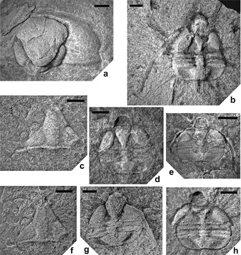
(a) Malongullia? sp., NMW 2015.31G.14, incomplete cranidium, internal mould, dorsal view. (b, d, e, g, h) Pseudampyxina sp.: (b) NMW 2015.31G.19, articulated incomplete exoskeleton, internal mould, dorsal view; (d) NMW 2015.31G.22, articulated incomplete exoskeleton, internal mould, dorsal view; (e) NMW 2015.31G.18.1, articulated incomplete exoskeleton, internal mould, dorsal view; (g) NMW 2015.31G.24.1, articulated incomplete exoskeleton, latex cast of external mould; (h) NMW 2015.31G.16.1, incomplete articulated exoskeleton, internal mould. (c, f) Lonchodomas sp., NMW 2015.31G.28: (c) NMW 2015.31G.28a, incomplete cranidium, internal mould, dorsal view; (f) NMW 2015.31G.28b, incomplete cranidium, latex cast of external mould, dorsal view. All from sample F-524, base of the Mayatas Formation, Upper Ordovician, Katian. Scale bars:=1 mm (b, d, g, h); 2 mm (a, e); 3 mm (c, f).

Material. Two incomplete cranidia, one very poorly preserved (NMW 2015.31G.14–15). Sample F-524.
Description. Cranidium with semicircular outline. Glabella flask-shaped, convex, around 90 % of total cranidium length (sag.). Posterior lobe of glabella around 10 % total glabella length, transversely elongate-subelliptical; separated from large frontal lobe by deep, backwardly-curved transglabellar furrow. Large bacculae elongate subelliptical, around one half of total glabella length; separated from glabella by deep furrows. Preglabellar field 10 % of cranidial length (sag.); gently convex and sloping down fairly steeply to anterior border. Anterior border around half length of preglabellar field; flat, angled gently upwards. Eye tubercles absent. Fixigena on one side of better preserved cranidium has two caecae, the upper one starting at anterior end of baccula and extending back towards genal angle, converging with lower caeca, which begins one-quarter baccula length back from anterior end and extends at a very gentle angle towards genal angle. Occipital ring narrow (sag.), convex, separated from glabella by deep, narrow furrow. Posterior border gently convex, up to 10 % of cranidium length, separated from fixigenae by narrow, relatively shallow but distinct furrow.
Remarks. Both cranidia from Atansor apparently lack eye tubercles and glabellar tubercles, although the surface of one specimen is too eroded to confidently distinguish these features. There is a well-developed preglabellar field and anterior border. The better-preserved specimen is similar to M. gracilis (Lu, Reference Lu1975) from the Middle Ordovician Miaopo Formation of central China. However, M. gracilis differs in having very small eye tubercles, albeit these are virtually obscured by caecae. Lu (Reference Lu1975) noted the similarity of M. gracilis to M. bilobus (Chugaeva, Reference Chugaeva1958) from the Uzunbulak Formation of Kuyandy–Sai in the Chu–Ili Range, Kazakhstan, distinguishing the two species by the relatively shorter (sag.) bacculae and preglabellar field of the former. The Atansor specimens are very similar to M. bilobus in cranidial outline and proportions, in possessing a deep transglabellar furrow, and in the outline of the glabella and relative size of the bacculae. However, M. bilobus differs in having obvious eye tubercles next to the glabellar margin and a longer (sag.) preglabellar field.
Zhou et al. (Reference Zhou, Webby and Yuan1995) noted that several species assigned to Ampyxinella from South China differ from typical members of the genus in possessing a transglabellar furrow, a well-defined posterior border furrow, large prominent bacculae and diminished or obsolete eye tubercles. They suggested that these species should possibly be transferred to another genus, but that further material is required to make generic assignments. The South Chinese species are similar to the Atansor specimens in cranidial and glabellar outline, in possessing large bacculae, and in apparently lacking eye tubercles. Ampyxinella jingshanensis Sun, Reference Sun1984 differs in having a very short preglabellar field; A. jiangningensis Liu in Qui et al., Reference Qui, Lu, Zhu, Bi, Lin, Zhou, Zhang, Qian, Ju, Han and Wei1983 and A. rectilimbata Zhang in Qui et al., Reference Qui, Lu, Zhu, Bi, Lin, Zhou, Zhang, Qian, Ju, Han and Wei1983 both differ in having a prominent tubercle at the posterior end of the anterior glabellar lobe. It is likely that the Atansor species described here form a natural group with the South Chinese species, closely allied to Malongullia but lacking eye tubercles, and that they are closely related to species such as M. bilobus and M. gracilis, in which the eye tubercles are relatively small. However, generic assignment must await the recovery of more specimens, as these species are represented by scant cranidial material.
Subfamily Raphiophorinae Angelin, Reference Angelin1854
Genus Pseudampyxina Ju in Qui et al., Reference Qui, Lu, Zhu, Bi, Lin, Zhou, Zhang, Qian, Ju, Han and Wei1983
Type species. Pseudampyxina trisegmentata Ju in Qiu et al., Reference Qui, Lu, Zhu, Bi, Lin, Zhou, Zhang, Qian, Ju, Han and Wei1983, from the Huangnehkan Formation (early Ashgill), Quzian, Zhejiang, China.
Remarks. Zhang (Reference Zhang1980) proposed the Taklamakaniinae as a subfamily of the Raphiophoridae to accommodate genera with three thoracic segments, namely Nanshanaspis Zhang & Fan, Reference Zhang, Fan and Yin1960, Taklamakania Zhang, Reference Zhang1980 and Pseudampyxina. However, Zhou et al. (Reference Zhou, Webby and Yuan1995) concluded that these genera independently evolved three thoracic segments through heterochrony from different raphiophorine ancestors. We concur with that view and, hence, retain Pseudampyxina within the Subfamily Raphiophorinae. Pseudampyxina differs from Taklamakania in possessing long, slender genal spines, a smooth genal region, fewer axial rings on the pygidium, and in lacking a frontal glabellar spine. Nanshanaspis also lacks a frontal spine, but differs in that its pygidium has eight pleural furrows, whilst Pseudampyxina only has three to four axial rings and two pleural ribs. In addition, Nanshanaspis has smaller fixigenae than Pseudampyxina and lacks bacculae. Caganaspis was erected by Kolobova (Reference Kolobova1985) for a single species, C. unica, from the lower Sandbian of the West Balkhash region. However, Caganaspis agrees in overall outline, cranidial and glabellar features with Nanshanaspis, and also possesses the three thoracic segments and eight axial rings typical of that genus and corresponding pleural furrows that are only clearly impressed around the border of the pygidium. Hence, Caganaspis unica is here reassigned to Nanshanaspis. Nanshanaspis unica (Kolobova, Reference Kolobova1985) differs from Nanshanaspis murrayi Stait & Laurie in Burrett et al., Reference Burrett, Stait and Laurie1983, in possessing a relatively narrower glabella and wider fixigenae, and pleural furrows that are much more restricted in expression to the most distal borders of the pygidium.
Pseudampyxina sp.
Material. Twelve dorsal shields (including three external moulds) (NMW 2015.31G.16.1, NMW 2015.31G.18.1–3, NMW 2015.31G.19, NMW 2015.31G.20.1, NMW 2015.31G.21–22, NMW 2015.31G.24.1–2, NMW 2015.31G.26.1–2); seven cranidia (including two external moulds) (NMW 2015.31G.16.2, NMW 2015.31G.17.2, NMW 2015.31G.18.4, NMW 2015.31G.23, NMW 2015.31G.25, NMW 2015.31G.26.3–4); three cranidia with attached thorax (including one external mould) (NMW 2015.31G.17.1, NMW 2015.31G.20.2–3); one external mould of a pygidium (NMW 2015.31G.27); and one partial pygidium (NMW 2015.31G.18.5). Sample F-524.
Description. Dorsal shield with subcircular outline overall, although anterior of glabella extends beyond anterior margin of fixigenae by around a quarter of total dorsal shield length. Cranidium 54–76 % as long as wide, glabella and fixigenae convex. Glabella maximum width (tr.) 50–70 % of its length (sag.); rounded anteriorly, narrowing strongly backwards from around mid-length; with strong transverse convexity. Fixigenae obliquely oval in outline; of similar maximum width (tr.) to glabella; moderately convex; extending to 54–72 % of cranidium length (sag.). No frontal glabellar spine present. Specimens comprise poorly preserved internal moulds, which do not retain any evidence of surface sculpture on cranidium. Librigenae narrow (tr.); lateral border appears to be continuous with genal spines, which are gently curved and backwards directed, tapering gradually and extending back beyond pygidium.
Thorax with three segments, rounded abaxially. Axis convex; pleurae flatter with prominent posterior bands and weakly expressed anterior bands.
Pygidium sub-semicircular, short and wide with median embayment due to upwards flexure of posterior border; length 30–40 % of maximum width (tr.). Axis moderately convex; poorly preserved as a single mass, in which it is impossible to distinguish number of axial rings. Maximum axis width (tr.) anteriorly, 30–50 % of maximum pygidium width, tapering posteriorly. Poor preservation of the specimens precludes any evidence for surface sculpture on the pygidium.
Discussion. Poor preservation prevents detailed comparison of the Atansor Pseudampyxina specimens with those of P. trisegmentata Ju described in Qui et al. (Reference Qui, Lu, Zhu, Bi, Lin, Zhou, Zhang, Qian, Ju, Han and Wei1983) and Tripp et al. (Reference Tripp, Zhou and Pan1989). The specimens are of similar overall shape, cranidial and pygidial proportions; however, on the pygidia described here it is impossible to distinguish individual axial rings and therefore, to establish if they are comparable to P. trisegmentata. Preservation of the Atansor material as internal and external moulds, which have undergone some erosion, precludes any possibility of detecting the distinct granular surface sculpture illustrated by Tripp et al. (Reference Tripp, Zhou and Pan1989) on both cranidia and pygidia of P. trisegmentata. The preservational quality of the specimens is too poor to determine whether they represent P. trisegmentata or a different Pseudampyxina species. Tripp et al. (Reference Tripp, Zhou and Pan1989) state that Pseudampyxina is endemic to China, so the specimens described here represent the first record of the genus outside China. The type (and only named) species is known from the Huangnehkan Formation (early Ashgill), Quzian, Zhejiang Province (Ju in Qui et al. Reference Qui, Lu, Zhu, Bi, Lin, Zhou, Zhang, Qian, Ju, Han and Wei1983), and the Tangton Formation, Jiangsu Province (Tripp et al. Reference Tripp, Zhou and Pan1989), both part of the South China terrane.
Genus Lonchodomas Angelin, Reference Angelin1854
Type species. Ampyx rostratus Sars, Reference Sars1835, from the Sandbian Vollen Formation (formerly Ampyx Limestone) of Bygdøy, Oslo, Norway.
Lonchodomas sp.
Material. Internal and external moulds of a damaged, incomplete cranidium (NMW 2015.31G.28). Sample F-524.
Description. Cranidium subtriangular in outline, width (tr.) estimated at around one-and-a-quarter to one-and-a-third times sagittal length, excluding anterior spine. Glabella diamond-shaped in outline, with maximum width a little anterior of mid-length (sag.) – convex (tr.) and carinate, with anterior produced into a narrow spine, only the base of which is preserved, so its total length is unknown. Occipital furrow shallow, separating off short (sag., exsag.), gently convex axial ring and posterior border, which slope gently forwards towards furrow. Posterior border and axial ring are continuous. A pair of small depressions in the occipital furrow, positioned a short distance inside the axial furrows, may mark the position of a basal pair of muscle scars, but the irregular, eroded surface precludes the identification of scars on the glabella.
Remarks. The single damaged cranidium is too incomplete to allow specific assignment. Lonchodomas tecturmasi Weber (see Chugaeva Reference Chugaeva1958, pl. 2, figs 3–5) from the Sandbian of south Kazakhstan, and L. latus Koroleva, Reference Koroleva1965 from the Sandbian of north-central Kazakhstan, both have a relatively wider (tr.) and shorter (sag.) cranidium. Lonchodomas karakanensis Weber (see Chugaeva Reference Chugaeva1958, pl. 2, figs 1–2) from the Darriwilian Karakan and Uzunbulak Formations in south Kazakhstan has a similarly proportioned cranidium overall, but the glabella is relatively wider than that of the Atansor specimen.
Family Trinucleidae Hawle & Corda, Reference Hawle and Corda1847
Subfamily Trinucleinae Hawle & Corda, Reference Hawle and Corda1847
Iputaspis gen. nov.
Type species. Iputaspis stepnyakensis sp. nov.; Upper Ordovician; Katian, Mayatas Formation, Stepnyak region, north-central Kazakhstan.
Derivation of name. After the unusual distribution of I pits on the fringe; from puteus, Latin, meaning pit.
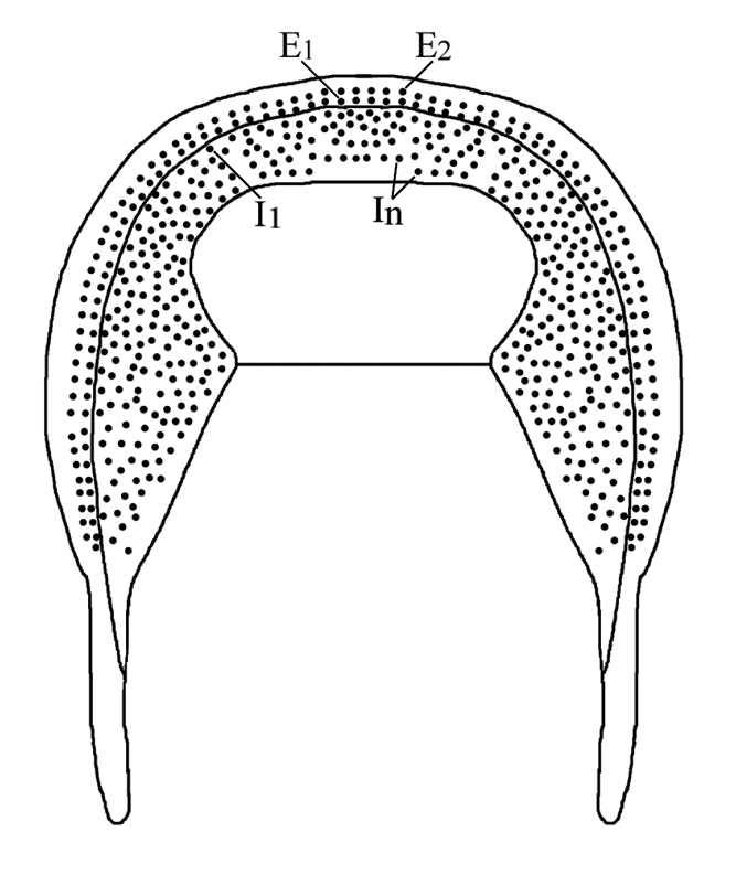
Diagnosis. Fringe with E1-2, I n-1 complete; no F pits present. E1 and E2 pits generally radially aligned with pit pairs set in sulci, but with occasional aberrant pits unpaired and out of alignment. In-1 pits radially aligned with those of In anteriorly, but posteriorly In-1 irregularly distributed and sometimes very close to In ; In pits not cut off by In-1 pits; I1 apparently complete; 2 to 3 I arcs between I1 and In-1 frontally, up to approximately 10 I arcs postero-laterally, randomly distributed. Glabella anterior greatly expanded and gently convex. Occipital ring lacks occipital spine. No lateral eye tubercles or eye ridges present. Pygidium short and wide, with width approximately three-and-a-third times length; axis with five axial rings.
Remarks. The generally effaced, poor preservation of the specimens made it difficult to place this trinucleid within a subfamily. The glabella expands anteriorly, but there is no clear evidence for differentiation of a typical Trinucleine pseudo-frontal lobe, although one specimen has a very shallow furrow across the glabella just posterior of the expanded portion. The presence of E pits excludes the Hanchungolithinae, and the absence of F pits rules out Marrolithinae and Cryptolithinae. The Reedolithinae generally possess lateral eye tubercles, eye ridges and occipital spines, and none has been observed in these specimens. Overall, subfamily Trinucleinae is the most plausible identification, as there are no fringe or other cranidial features that exclude this designation.
Iputaspis stepnyakensis sp. nov.
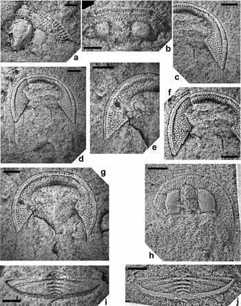
Iputaspis stepnyakensis gen. et. sp. nov. (a) NMW 2015.31G.35, paratype, incomplete cranidium, internal mould, dorsal view. (b) NMW 2015.31G.33, paratype, incomplete cranidium, internal mould, dorsal view. (c, d, f) NMW 2015.31G.29, holotype: (c) lower lamella of fringe, internal mould, ventral views showing close-up of left side of fringe; (d) entire fringe with genal spines; (f) close-up of middle and right side of fringe. (e, g) NMW 2015.31G.36, paratype: (e) lower lamella of fringe, internal mould, ventral views showing close-up of right side of fringe; (g) entire fringe with genal spines. (h) NMW 2015.31G.34, paratype, incomplete cranidium, latex cast of external mould, dorsal view. (i–j) NMW 2015.31G.30, paratype: (i) pygidium, external mould; (j) latex cast of external mould, dorsal view. All from sample F-524, base of the Mayatas Formation, Upper Ordovician, Katian. Scale bars=1 mm (a); 2 mm (b–j).

Iputaspis stepnyakensis gen. et sp. nov. Schematic drawing showing the arrangement of the pits on the fringe. Not to scale.

Derivation of name. After Stepnyak terrane in north-central Kazakhstan.
Holotype. Internal mould of lower lamella of fringe (NMW 2015.31G.29).
Paratypes. One external mould of a pygidium (NMW 2015.31G.30) and ten partial cranidia of varying degrees of completeness (NMW 2015.31G.31, NMW 2015.31G.32.1–32.4, NMW 2015.31G.33–37).
Locality. Northern shore of Lake Atansor, north-central Kazakhstan. Mayatas Formation, about 3 m above the base of the siliceous argillite unit overlying the limestones, sample F-524.
Diagnosis. As genus.
Description. Fringe with E1 and E2 complete, set close to each other and radially aligned, set a constant distance apart all around the fringe, set in shallow sulci; I arcs present and very irregularly distributed; In apparently complete, deflected forward around anterior end of glabella; on one specimen pits in In-1 arc appear to be radially aligned with those of In but this is inconsistently expressed and breaks down posteriorly where pits of In-1 are irregularly distributed and sometimes very close to In pits; In pits not cut off by In-1 pits; I1 apparently complete; 2 to 3 I arcs between I1 and In-1 frontally, up to approximately 10 I arcs postero-laterally, randomly distributed, not in regular radial or concentric alignment; no F pits present. E pits of a similar or slightly larger diameter to I pits, which are of a uniform size on all parts of fringe. E1 and E2 pits generally radially aligned with pit pairs set in sulci, but with occasional aberrant pits that are unpaired and out of alignment. Glabella effaced and poorly preserved on the few specimens that include it (e.g. Fig. 5a); anterior greatly expanded and gently convex, but no clear evidence for divided off pseudofrontal lobe. Glabellar furrows difficult to distinguish; one specimen with relatively shallow furrow running along lateral edge of posterior half of glabella, deepening at base of anteriorly expanded glabella, perhaps representing a 2p furrow (Fig. 5a). Occipital ring apparently lacks occipital spine. No lateral eye tubercles or eye ridges present. Genal spines long but bluntly terminated.
Thorax unknown. Pygidium maximum width (tr.) approximately three-and-a-third times length, anterior margin transverse, posterolateral margins evenly rounded (Fig. 5i, j). Axis gently convex, maximum width (tr.) around one quarter pygidial width, tapering gently towards posterior margin, where it merges with steeply sloping border. Axis with five axial rings plus articulating half ring, demarcated by wide furrows, which are shallow medially but much deeper distally. Four pairs of pleural furrows present; first pair clearly expressed, angled gently backwards, curve backwards slightly distally and reach border. Three other pairs shallower, angled more strongly backwards and successively shorter (tr.). Border steeply declined; relatively narrow anteriorly, widening posteriorly. Border poorly preserved in posterior view, but there appears to be a shallow medial notch.
Remarks. The highly irregular distribution of I pits in this trinucleid is unusual. Parkesolithus dictyotus Webby, Reference Webby1974, from New South Wales, Australia, has a similar number of pit arcs and the outer I arcs (I1-2 anteriorly, plus more I arcs postero-laterally) are irregularly, ‘haphazardly arranged’. However, P. dictyotus has In to In-3 regularly and radially arranged, whilst Iputaspis gen. nov. has all of its I arcs irregularly arranged, with the exception of one specimen where In and In-1 are radially arranged frontally, but this completely breaks down postero-laterally. Ghobadi Pour et al. (Reference Ghobadi Pour, Popov, McCobb, Percival, Gutiérrez-Marco, Rábano and García-Bellido2011) described the trinucleine genus Kimakaspis, from the Karagach Formation of Kazakhstan, in which the I pits are irregularly arranged laterally. However, Kimakaspis differs from Iputaspis in possessing regular, radially arranged I pits frontally, arranged in sulci with E1-2.
Order Proetida Fortey & Owens, Reference Fortey and Owens1975
Family Telephinidae Marek, Reference Marek1952
Genus Telephina Marek, Reference Marek1952
Type species. Telephus fractus Barrande, 1852, from the Králův Dvůr Formation (upper Katian) of Králův Dvůr, Bohemia.
Telephina cf. stepnjakensis Koroleva
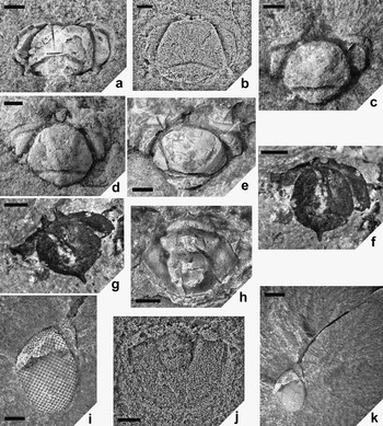
(a–b) Telephina cf. stepnjakensis Koroleva, 1982: (a) NMW 2015.31G.38, cranidium, internal mould, dorsal view; (b) NMW 2015.31G.39, cranidium, latex cast of external mould, dorsal view. (c–d) Telephina cf. omega Koroleva, 1982: (c) NMW 2015.31G.40, cranidium, internal mould, dorsal view; (d) NMW 2015.31G.41, cranidium, internal mould, dorsal view. (e) Telephina sp. 1: NMW 2015.31G.42, damaged cranidium, internal mould, dorsal view. (f–g) Telephina sp. 2, NMW 2015.31G.43.1: (f) cranidium, external mould, ventral view; (g) cranidium, external mould, oblique ventral view. (h) Telephina sp.: NMW 2015.31G.43.2, pygidium, internal mould, dorsal view. (i, k) Telephina sp., NMW 2015.31G.44: (i) close-up of damaged eye, showing square lenses, oblique dorsolateral view; (k) librigena, internal mould, oblique dorsolateral view. (j) Arthrorhachis sp.: NMW 2015.31G.1, pygidium, latex cast of external mould, dorsal view. All from sample F-524, base of the Mayatas Formation, Upper Ordovician, Katian. Scale bars=1 mm.

cf. 1982 Telephina stepnjakensis Koroleva, p. 48, pl. IV, fig. 2
Material. Two cranidia, one internal mould and one external mould (NMW 2015.31G.38–39). Sample F-524.
Description. Cranidium wide, length (sag., excluding occipital spine) 0.50–0.68 times maximum width, which occurs at anterior of palpebral lobes. Glabella moderately convex (tr.; sag.), gently tapering forwards in posterior half, more rapidly narrowing in anterior half, bluntly truncated in front, producing a sub-pentagonal outline. Glabella length (sag.) around 0.7–0.8 times cranidium length. Anterior of glabella slopes steeply down to preglabellar furrow, anterior curve of glabella slightly overhanging it, so furrow is not visible in dorsal view. Anterior border narrow (sag.), bearing a pair of spines directed ventrally and slightly forwards, so they are visible in dorsal view, their tips converging inwards towards each other; spines set close together. Palpebral area convex, relatively narrow, a maximum of 0.26–0.28 times cranidium width (tr.); sloping down steeply laterally and anterolaterally to palpebral furrow. Palpebral lobe sub-crescentic in outline, length about 0.8 times cranidium length; flat to gently convex; wider anteriorly than posteriorly. Occipital furrow wide (sag.), relatively deep, shallowing and narrowing abaxially in one specimen; furrow curved forward medially; does not reach axial furrows. Occipital ring longest (sag.) medially, one quarter total cranidial length, tapering rapidly abaxially; bearing a short, slender, posteriorly-directed spine. Posterior border with a pair of stout spines, directed outwards and backwards (Fig. 7a). Preservation of surface of specimens too poor to provide evidence of original ornament.
Remarks. The cranidia described here have similar outlines and proportions to those of T. stepnjakensis Koroleva, from the Sandbian of the Stepnyak terrane, although the occipital furrow is more strongly curved forward medially than in Koroleva's type specimen. Koroleva's specimen does not preserve the anterior border; if the Lake Atansor specimens are conspecific, they show that T. stepnjakensis had a pair of close-set, converging anterior spines. In addition, the Atansor cranidium that preserves the occipital ring shows a small medial occipital spine, a feature not evident on the incompletely preserved occipital ring of Koroleva's specimen. Telephina cf. stepnjakensis differs from T. omega Koroleva in possessing: sub-crescentic rather than sub-triangular palpebral lobes; a glabella with a bluntly truncated rather than evenly rounded anterior; an occipital furrow that is strongly deflected forwards medially, rather than more or less straight; anterior spines that are smaller, closer together, converging and more strongly ventrally directed, rather than larger, further apart and directed more straight ahead with only slight ventral incline.
Telephina cf. omega Koroleva
cf. 1982. Telephina omega Koroleva, p. 47, pl. 4, fig. 1
? 1982. Telephina aff. impunctatus (sic) (Ulrich) Koroleva, p. 50, pl. 4, fig. 3
? 1982. Telephina sp. 2 (T. cf. omega) Koroleva, p. 51, pl. 4, figs 5, 6
Material. Two cranidia (NMW 2015.31G.40–41), one damaged cranidium (NMW 2015.31G.42). Sample F-524.
Description. Cranidium wide, length (sag.) 0.62–0.64 times maximum width, which occurs at anterior of palpebral lobes. Glabella moderately convex (tr.; sag.), maximum width (tr.) immediately in front of occipital furrow, 0.67–0.68 times glabella length, tapering forwards, evenly rounded anteriorly; glabella length (sag) 0.65 times cranidium length. Anterior of glabella slopes straight down to preglabellar furrow, without overhanging it. Surfaces of two undamaged, poorly preserved cranidia show a pair of glabellar depressions, starting about a quarter of glabella length forward from the occipital furrow, and running diagonally forwards and out towards the axial furrow. Anterior border narrow, bearing a pair of spines directed forwards and slightly ventrally, so they are clearly visible in dorsal view; border width (tr.) between lateral extremities of spines around 0.4 times width of occipital ring. Palpebral lobe subtriangular in outline, length about 0.6–0.7 times cranidium length; gently convex; widest anteriorly, narrowing posteriorly from backwards bend in lobe. Occipital furrow straight medially, subtly bowed back abaxially; wide (sag.), relatively deep, shallows and narrows slightly medially where posterior glabella margin is deflected back a little; furrow also shallows abaxially and does not reach axial furrows. Occipital ring longest (sag.) medially, 0.16–0.18 times total cranidium length, tapering rapidly abaxially; no evidence for occipital spine. Posterior border not visible on available specimens. Preservation of surface of specimens too poor to provide evidence of original ornament.
Remarks. The two better-preserved Atansor cranidia (Fig. 7c, d) are very similar in outline and proportions to those of T. omega Koroleva, from an unspecified Sandbian locality in north Kazakhstan; have palpebral lobes with similar subtriangular outline and proportions; and have a comparable pair of forwardly-directed spines on the anterior border. Koroleva's (Reference Koroleva1982) description mentions the stump of an occipital spine, which was not observed on either of the Atansor specimens. However, the poor preservation of the specimens means that the original presence of a spine cannot be ruled out.
A third, damaged cranidium (Fig. 7e) has similar overall outline and proportions. Although the glabella is relatively shorter (sag.) and wider (tr.), fracturing of the specimen may have distorted its original proportions. A strong diagonal fracture, splitting off one corner of the glabella, and a corresponding groove on the opposite side, strongly resembles in position the pair of glabellar depressions on the other two cranidia. The three cranidia also have in common the relative size and outline of palpebral lobes; the apparently straight occipital furrow; the narrowness (tr.) of the anterior border; and the orientation of the anterior spines.
Telephina aff. impunctata (Ulrich) and T. sp. 2 of Koroleva (Reference Koroleva1982) are similar to T. omega and may be synonymous with it, although Koroleva's photographic plates are not clear enough to be confident about this. Koroleva's (Reference Koroleva1982) T. sp. 1 and 3 and 4 are too poorly preserved to assess their taxonomic identity.
Telephina sp. 1
Material. Damaged external mould of cranidium plus partial counterpart (NMW 2015.31G.43.1). Sample F-524.
Description. Cranidium length (sag.) about 0.64 times maximum cranidium width, which occurs at anterior of palpebral lobes. Glabella moderately convex (tr., sag.); glabella and occipital ring together have subcircular outline, widening rapidly forwards to reach maximum width (tr.) at around one-third glabella length, then tapering rapidly forwards to a semicircular front. Anterior border has a pair of subparallel spines directed strongly ventrally and slightly forwards. Tips of spines appear asymmetrically bifurcated, with a narrow slit separating the distal two-thirds of the spine from the median third (Fig. 7g). However, this may simply represent a longitudinal section through the tubular structure of the spine. Anterior border narrow (tr.), with width between extremities of spines around 30 % of width of occipital ring. Occipital furrow moderately deep and wide (sag.), curving gently forwards medially. Occipital ring longest (sag.) medially, tapering rapidly abaxially; with long, slender occipital spine. Palpebral lobes subtriangular in outline, gently convex, widest anteriorly, tapering posteriorly.
Remarks. The outline and proportions of this cranidium are most similar to T. omega Koroleva, but it differs in possessing anterior spines that are directed more strongly ventrally. This specimen also preserves a long, slender occipital spine, which is not seen in T. omega, although Koroleva (Reference Koroleva1982) mentions the stump of a spine in her description. The Atansor specimen may represent a new species of Telephina, but poor preservation renders it inadequate as a holotype.
Telephina sp.
Material. Internal and external moulds of pygidium (NMW 2015.31G.43.2), three partial librigenae, two including eye (NMW 2015.31G.44–46). Sample F-524.
Description. Pygidium subtriangular in outline, maximum width about 1.25 times length (sag.). Axis strongly convex, with one clearly separated ring and a second fused with the terminal piece; axis length around 0.9 of pygidium length. Maximum axial width at anterior, 0.6 of pygidium width, tapering backwards to bluntly rounded posterior, which slopes down almost vertically to short (sag.) postaxial field. Pleural areas narrow, convex; sub-horizontal adaxially, steeply downsloping laterally to narrow, convex border, which is separated from pleural areas by very shallow border furrow. Shallow, weakly impressed pair of pleural furrows at anterior of pleural area; a second pair of furrows is indicated by very shallow depressions at around half pleural area length. Axial furrows moderately deep and well impressed.
Articulating half-ring convex, just over half length (sag.) of first axial ring; clearly defined by wide, deep articulating furrow, which is deepest medially, shallowing slightly laterally. First and second axial rings separated by wide ring furrow, which is moderately deep medially, becoming shallower and less clearly defined laterally. Second axial ring poorly defined, fused with terminal piece, shorter than first axial ring. Terminal piece short (sag.) with a posterior pair of widely spaced circular depressions. Axial surface poorly preserved, but anterior of both axial rings with remnants of closely set pair of tubercles on midline; antero-lateral margins of first axial ring developed into a raised rim.
The three librigenae are all incomplete, one comprising just the eye; a second the eye plus a deep groove marking the path of the genal spine (Fig. 7i, k); and a third preserving a partial outline of the genal field, border and spine but lacking the eye. Genal spine long (over twice maximum eye length), slender, gradually tapering and gently curved. Damage and incomplete preservation makes it difficult to be confident about the original orientation, but it appears to be approximately laterally directed and gently bowed back, similar to the genal spine of T. bicuspis (Angelin, Reference Angelin1854) from the middle Ordovician of Sweden and Norway (Ahlberg Reference Ahlberg1995). One Telephina librigena (NMW 2015.31G.44) has a fractured, but otherwise well-preserved eye surface, showing square lenses in diagonal rows (Fig. 7i, k).
Remarks. The pygidium was found very close to the cranidium of T. sp. 1, but they do not belong to the same individual as both specimens are of similar maximum width and in micropygous Telephina the pygidium is significantly smaller than the cranidium. More than one species of Telephina is represented by cranidia in sample F-524, so it is impossible to make a confident specific identification for the pygidium.
For the same reason, it is impossible to confidently identify the librigenae to specific level. The librigena that shows good preservation of the eye clearly shows square lenses in diagonal rows, an unusual feature for trilobite eyes that has been noted before in other Telephina species (Fortey Reference Fortey1997; Han Reference Han2001; Bruton & Høyberget Reference Bruton and Høyberget2006). Fortey (Reference Fortey1997) discussed the possible function of square lenses, which are also found in the ‘reflecting superposition eyes’ of modern macruran crustaceans. In crustaceans, the inside walls of the lens are mirrored to reflect light coming from all angles down to a common source, but it is not known how this lens shape functioned in trilobite eyes. Han (Reference Han2001) claimed that each square ‘interlens’ in the Telephina eye was produced from one side of four close-lying eight-sided lenses, but there is no evidence for this preserved in the Atansor specimens. Fractured areas of the two Atansor librigenae that include part of the eye were examined under SEM to search for evidence of the structure of the lenses. However, the coarse-grained sediment precluded preservation of any of the internal structures of the eye.
Order Lichida Moore, Reference Moore1959
Superfamily Lichacea Hawle & Corda, Reference Hawle and Corda1847
Family Lichidae Hawle & Corda, Reference Hawle and Corda1847
Subfamily Lichinae Hawle & Corda, Reference Hawle and Corda1847
Tribe Tetralichini Phleger, Reference Phleger1936
Genus Amphilichas Raymond, Reference Raymond1905
[= Acrolichas Foerste, Reference Foerste1919]
Type species. Platymetopus lineatus Angelin, Reference Angelin1854, from the Boda Limestone Formation (late Katian–early Hirnantian), Dalarne, Sweden (by original designation).
Remarks. We concur with previous authors (Tripp Reference Tripp1957; Thomas & Holloway Reference Thomas and Holloway1988; Carlucci et al. Reference Carlucci, Westrop and Amati2010) in regarding Acrolichas as a junior subjective synonym of Amphilichas.
Amphilichas clarus (Koroleva, Reference Koroleva1959)
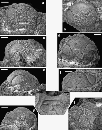
Amphilichas clarus (Koroleva Reference Koroleva1959). (a–c) NMW 2008.34G.155, cranidium: (a) dorsal view; (b) dorso-anterior view; (c) lateral view. (d–g) NMW 2015.31G.47, cranidium: (d) dorsal view; (e) lateral view; (f) anterior view; (g) posterior view. (h) NMW 2015.31G.54.1, incomplete hypostome, dorsal view. (i) NMW 2008.34G.156, incomplete pygidium, dorsal view. All from sample F-1007, base of the Mayatas Formation, Upper Ordovician, Katian. Scale bars=2 mm (a–h); 3 mm (i).

1959 Acrolichas clarus Koroleva, p. 89, pl. 1, figs 15, 16
2011 Acrolichas clarus Koroleva; Ghobadi Pour et al., p. 174, figs 2.5, 10
Holotype. Accession No. 1101 (for whole collection) in Museum of the Institute of Geological Sciences, Almaty, Kazakhstan – accession number of individual specimen not specified.
Locality. Sandbian Mailisor Formation, 3km southwest of Belagash Village, north-central Kazakhstan.
Material. Four damaged cranidia (NMW 2008.34G.155.1, NMW 2015.31G.47–49), four cranidial fragments (NMW 2015.31G.50–53), one pygidial fragment (NMW 2008.34G.156), two incomplete hypostomata (NMW 2015.31G.54.1, 55). Sample F-1007.
Description. Cranidium subcircular in outline (Fig. 8a, d). All cranidia damaged; length approximately 68–79 % of estimated maximum width at palpebral lobes. Glabella subcircular in outline, length 71–82 % of maximum width; width 82–86 % of cranidial width, widest at palpebral lobes (not preserved). Glabella gently convex in lateral view, strongly curved anteriorly and sloping down steeply to anterior border (Fig. 8c, e). Longitudinal furrows narrow but moderately deep and clearly defined; effaced for posterior 19–25 % of glabella length, where a subtle depression remains but is filled with medium to large tubercles. Longitudinal furrows subparallel to gently divergent opposite anterior end of palpebral area, increasingly diverging anteriorly, curving round anterolaterally and sloping down steeply to meet axial furrows. Glabellar median lobe expands rapidly anterior of palpebral area. Lateral composite lobes 30–37 % of glabella width at approximate mid-length of palpebral lobes (incompletely preserved), widening slightly anteriorly. The one preserved palpebral area has two rows of small, widely-spaced tubercles. Preocular fixigenae narrow bands, sloping down steeply and narrowing slightly to meet anterior border. Posterior fixigenae incompletely preserved (see Fig. 8a, c); narrow, sub-triangular, 2–3 times width of preocular fixigenae, sloping gently down towards posterior border furrow (not preserved). With exception of anterior of median glabellar lobe and anterior border, surface of cranidium densely covered with mixture of small, medium and large tubercles. Largest tubercles around 0.1 mm across. Tubercles begin to fade anterior of lateral composite lobes, quickly becoming completely effaced (Fig. 8b, d, f). Anterolateral end of longitudinal furrow has a single row of closely-spaced tubercles in front of it, which reduce in size from medium to small as they slope down to meet anterior border. Apart from a small number of weakly expressed tubercles, the glabella is effaced anterior of this, although some widely spaced pits occur in the anterolateral corners of the median lobe and along the anterior border. Medium tubercles trace the lateral edges of the median lobe, and the anterior and posterior edges of the occipital ring, with some small tubercles interspersed. Three large tubercles occur on one cranidium, one around two-thirds glabella length forward, slightly left of the midline, and a widely-spaced pair close to the anterior limit of tubercles (Fig. 8a, b). Preservation of the other two cranidia is too poor to detect whether these tubercle patterns are universal within the species.
The single fragmentary pygidium comprises almost complete right pleural field and partial axis (Fig. 8i). Axis very gently tapering backwards to second interpleural furrow; strongly tapering for posterior half of terminal piece. Axial furrow continuous beyond end of axis to delimit subtriangular postaxial area. Anterior of axis broken off along presumed ring furrow between first and second axial rings; furrow directed obliquely forwards towards midline of axis. Posterior of second axial ring delimited by short (tr.) moderately shallow furrow, which is directed gently backwards, does not reach the axial furrow and stops short of presumed midline of partially preserved axis. Second axial ring therefore appears to have been much longer (sag.) medially, significantly narrowing distally. Terminal piece over twice as long (sag.) as second axial ring. Three pairs of broad, flat pleurae. Interpleural furrows narrow and moderately shallow, but clearly defined. Second pleura ends in a backwards-directed spine. Tip of first pleura broken off; distal end of third pleura curves gently into straight posterolateral margin. Pleural furrow on first pleura moderately deep and well incised, angled backwards; delimits two unequal parts of pleura, the posterior part around half the length (exsag.) of the anterior part. Pleural furrow on second pleura very short (tr) but moderately deep and clearly defined; directed backwards; delimiting two subequal parts of pleura with slightly narrower posterior part. Entire surface of pygidium covered in a dense ornament of small, medium and large tubercles. Distal ends of pleurae mainly ornamented with small to medium tubercles, with more large tubercles closer to the axis.
Two damaged, incomplete hypostomata suggest an elliptical outline, with length 50–60 % of width (Fig. 8h). Limited preservation of anterior margin shows that this is evenly rounded and mirrors anterior margin of median body. Median body subtrapezoidal in outline, around 70 % hypostome length. Anterior lobe elliptical in outline, 62–68 % of total median body length. Posterior lobe transverse, subrectangular, 27–28 % of total median body length. Middle furrow moderately shallow, directed gently backwards, extending inwards just under a third of median body width from lateral border furrow. Lateral borders wide, gently convex. Lateral border furrows much deeper than middle furrows, converging backwards to join with posterior border furrow. Posterior border wide, slightly raised in a band immediately behind posterior lobe. Posterior margins of both specimens damaged, so outline and depth of median notch cannot be determined. Coarse, wavy terrace lines preserved at lateral corners of posterior border. Specimens otherwise exfoliated; sculpture on other parts of hypostome unknown.
Librigena and thorax unknown.
Remarks. The specimens figured here differ from the type specimen figured by Koroleva (Reference Koroleva1959, pl. I, fig. 15, 16) in the expression of the longitudinal furrows. Koroleva's type specimen shows clearly incised longitudinal furrows that reach the occipital furrow; however, although the specimens discussed here have longitudinal furrows that reach the occipital furrow, they are weakly expressed as shallow furrows in which some tubercles occur. Note that Koroleva (Reference Koroleva1959, pl. 1, fig. 15, 16) miss-spells the name of this new species (as Akrplichas crarus) in the plate caption. We use the spelling adopted by Koroleva where she erects and describes the species in the main text.
Amphilichas karakenensis var. disjunctus Chugaeva (Reference Chugaeva1958), from the Karaken Formation of northern Betpak–Dala, central Kazakhstan, is difficult to distinguish from A. karakenensis Weber (Reference Weber1948), and the two are likely synonymous. This species has a similar cranidial outline and proportions to A. clarus. However, A. karakenensis has an ornament of much finer tubercles, and the longitudinal furrows curve around the lateral glabellar lobes much more gently and with lower convexity. A. nasutus Webby (Reference Webby1974) from the Ordovician limestone at Billabong Creek, New South Wales, Australia, has similarly proportioned cranidia to A. clarus (Koroleva, Reference Koroleva1959), although its longitudinal furrows fail to reach the occipital furrow, the median lobe of the glabella is longer and more pointed anteriorly in the Australian species, and the dense ornament of medium to coarse tubercles covers the entire cranidial surface, whilst the anteriormost cranidium of A. clarus is smooth.
When Koroleva (Reference Koroleva1959) erected the species A. koksorensis, she used a different spelling (A. koksorensis) in the plate caption (pl. I, fig. 14) than in the main body text (A. koksorchsis); the latter being grammatically incorrect. Here, we adopt the spelling A. koksorensis, as ‘ensis’ is the correct Latin suffix to append to the toponym ‘Koksor’.
Only the cranidium of A. koksorensis Koroleva (Reference Koroleva1959), from the early Katian of Lake Maisor in the Stepnyak terrane, is known. It has similar proportions to A. clarus and longitudinal furrows with comparable anterior curvature. However, the longitudinal furrows stop abruptly anterior of the mid-point of the palpebral lobes in A. koksorensis, whilst they continue (albeit weakly expressed) to the occipital furrow in the A. clarus specimens figured here. A. kalmakensis Koroleva (Reference Koroleva1959) from the lower Sandbian Mailisor Formation of the Stepnyak terrane of north-central Kazakhstan also has a similar cranidial outline and proportions to A. clarus. However, although its longitudinal furrows extend back further posteriorly than those of A. koksorensis, they do not reach the occipital furrow, and they curve around the lateral glabellar lobes much more gently anteriorly.
A. satpaevi Koroleva (Reference Koroleva1959), known from a single cranidium from the Katian Mailasor Formation of the Stepnyak terrane of north-central Kazakhstan, has a very different cranidial outline to A. clarus, with the central glabellar lobe greatly extended forward to a narrow point. Similarly, A. sniatkovi Weber (Reference Weber1948) from the late Ordovician of the Kuznetsk Bazin in the Altai–Sayany Region has an anteriorly extended central glabellar lobe, although it is shorter (sag.) and has a more rounded anterior.
A. batchaticus Weber (Reference Weber1948) from the late Ordovician of the Kuznetsk Bazin in the Altai–Sayany Region has a relatively wide (tr.) cranidium with a subdued ornament of fine to medium tubercles. A. punctatus (Weber, Reference Weber1948) from the upper Sandbian Anderken Formation of the Chu–Ili terrane has a similarly proportioned cranidium to A. clarus, but with a much finer, more subdued ornament, and longitudinal furrows that are more gently curved around the lateral glabellar lobes anteriorly. Weber (Reference Weber1948) figured a specimen assigned to A. cucullus (Meek & Worthen, Reference Meek and Worthen1868) from the late Ordovician of the Kuznetsk Basin in the Altai–Sayany Region. The central glabellar lobe extends further forward than that of A. clarus, and the longitudinal furrows are much straighter. An incomplete cranidium from the Katian of the Boshchekul terrane, questionably assigned by Weber (Reference Weber1948) to A.? dalecarlicus (Angelin, Reference Angelin1854), has less curved longitudinal furrows than A. clarus and a distinctive ornament comprising closely packed small tubercles with a small number of very large tubercles on the midline of the glabella.
Superfamily Odontopleuroidea Burmeister, Reference Burmeister1843
Family Odontopleuridae Burmeister, Reference Burmeister1843
Subfamily Odontopleurinae Salter, Reference Salter1864
Genus Primaspis Richter & Richter, Reference Richter and Richter1917
Type species. Odontopleura primordialis Barrande, Reference Barrande1846, from the Libeň Formation (Sandbian), Děd u Berouna, Czech Republic.
Primaspis sp.
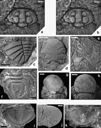
(a–b) Primaspis sp., NMW 2015.31G.56, cranidium: (a) internal mould, dorsal view; (b) annotated internal mould to show outline of occipital spines. (c–d) Pliomerina sulcifrons (Weber Reference Weber1948): (c) NMW 2008.34G.155.2, pygidium, dorsolateral view; (d) NMW 2015.31G.57, incomplete cranidium, internal mould, dorsal view. (e, i, k) Asaphidae gen. et. sp. indet.: (e) NMW 2015.31G.10, hypostome, internal mould, ventral view; (i) NMW 2015.31G.2, pygidium, internal mould, dorsal view; (k) NMW 2015.31G.3, pygidium, largely testate, dorsal view. (f–h, j) Sphaerexochus conusoides Koroleva, Reference Koroleva1959: (f) NMW 2008.34G.158, pygidium, exfoliated, dorsal view; (g) NMW 2008.34G.157, cranidium, partly exfoliated, dorsal view; (h) NMW 2015.31G.59, cranidium, exfoliated, dorsal view; (j) NMW 2008.34G.157, cranidium, partly exfoliated, lateral view. From sample F-524 (a–b) and sample F-1007 (c–k), base of the Mayatas Formation, Upper Ordovician, Katian. Scale bars=1 mm (a–d, i); 3 mm (e, k); 2 mm (f–h, j).

Material. One internal mould of a cranidium (NMW 2015.31G.56). Sample F-524.
Description. Cranidium is incomplete, and preservation as an internal mould exaggerates the width of the furrows. Projected maximum width (tr.) around double the length (sag., excluding occipital spines). Glabella convex (tr., sag.), tapering forwards; maximum width (tr.) occurs at around mid-length (sag.) of L1, approximately equal to length of glabella plus occipital ring. Glabella with two clearly developed pairs of lateral lobes and a third, small, indistinct pair. L1 pair inflated, tops roughly level with median lobe lying between them; subcircular in outline, slightly longer (sag.) than wide (tr.). L2 inflated, lying below median lobe; around two-thirds of the length and width of L1; subcircular. S1 deep and wide at inner end, very shallow at outer end so that L1 and L2 are almost fused laterally; outer half transverse, inner part curved backwards at an angle of around 45°. S2 deep and wide, shallowing at its outer end, angled back at around 45°. S1, S2 and occipital furrow connected by moderately shallow longitudinal furrow, which is widest (tr.) immediately in front of occipital furrow and narrows forwards. L3 lacking in convexity, defined by S2 and indistinct, shallow S3. Median lobe sub-parallel-sided opposite L1, tapering opposite L2, sub-parallel-sided opposite L3, then widening out slightly over short (sag.) distance to bluntly truncated anterior margin. Median lobe strongly convex opposite L1 and L2, gently convex anterior of L2 and sloping down to anterior margin of glabella. Front of median lobe falls away steeply to shallow and wide (sag.) anterior border furrow, which separates it from very short (sag.), vertical anterior border. Occipital ring damaged and incomplete, relatively short (tr.) and apparently of uniform length medially and laterally. Faint external moulds mark the position of a pair of occipital spines, which are initially directed straight backwards at an angle of around 60° from the anterolateral corners of the occipital ring, with their proximal ends tapering and curving gently inwards (Fig. 9b). In between the occipital spines, a small number of small tubercles or very short spines lie close to posterior margin of occipital ring. Fixigenae opposite L1 convex, of similar width (tr.) to L1; fall away steeply to axial furrows and towards posterior margin. Anteriorly, fixigenae more gently convex, sloping down to anterolateral margins. Palpebral lobes not visible. Details of external ornament not generally preserved, although the right fixigena shows traces of medium, densely distributed tubercles.
Remarks. Primaspis was originally erected as a subgenus of Acidaspis (Richter & Richter, Reference Richter and Richter1917), with Odontopleura primordialis (Barrande, Reference Barrande1846) as type species. The first detailed study and diagnosis (Prantl & Přibyl Reference Prantl and Pribyl1949) focused on Bohemian species and retained the subgeneric status of Acidaspis (Primaspis). Whittington's (Reference Whittington1956) re-diagnosis established Primaspis as a separate genus, although he noted that the North American species differed from the type species, principally in lacking occipital spines. Přibyl & Vaněk (Reference Pribyl and Vaňek1965) proposed the subgenus Primaspis (Meadowtownella) for those Anglo-Scandinavian and North American species lacking spines on the occipital ring, although some authors (e.g., Bruton Reference Bruton1966, Reference Bruton1968; Siveter Reference Siveter1989) felt these differences were insufficient to warrant subgeneric status. However, with the exception of a small number of Bohemian species (Šnajdr Reference Šnajdr1984), all other known species of Primaspis differ from P. primordialis in lacking occipital spines, and Ramsköld & Chatterton (Reference Ramsköld and Chatterton1991) placed these within Meadowtownella, raised to generic status. A formal diagnosis of Meadowtownella was presented by Conway & Botting (Reference Conway and Botting2012), and the majority of species historically assigned to Primaspis are now placed within it. A second subgenus of Primaspis erected by Přibyl & Vaněk (Reference Pribyl and Vaňek1965), Chlustinia, is now given generic status and appears to be a rare Bohemian endemic, restricted to three named species (Šnajdr Reference Šnajdr1984) and one tentatively-assigned pygidium (Mergl Reference Mergl2014) from the Prague Basin.
Four species remaining in Primaspis are the type species and three other Bohemian species with occipital spines (Šnajdr, Reference Šnajdr1984). Mergl (Reference Mergl2014) described odontopleurids from a Katian/Hirnantian boundary sequence in the Prague Basin, and summarised older odontopleurid occurrences in the region, noting that Primaspis is restricted to four formations, which are Sandbian to lowermost Katian in age (see Fatka et al. Reference Fatka, Lerosey-Aubril, Budil and Rak2013, fig. 1 for an updated Ordovician stratigraphy of the Prague Basin). The Primaspis described here differs from P. primordialis (Letná Formation), in having a much narrower median glabellar lobe, which is roughly equal in width (tr.) to the lateral lobes lying either side of it, whilst that of P. primordialis is around double the width of the lateral lobes. Also, the anterior portion of the fixigena, opposite L2, is relatively wider (tr.) than in the type species, and the first lateral glabellar lobe (L1) is more circular in outline, as opposed to elongate suboval. Primaspis oxitron Šnajdr, Reference Šnajdr1984 from the Libeň Formation also has a wider (tr.) median glabellar lobe than the Kazakhstanian species, and a much denser ornament of tubercles than that suggested by the traces of ornament preserved on the new specimen. Primaspis tremenda (Barrande, Reference Barrande1852) from the Zahořany Formation also has a relatively wide (tr.) median glabellar lobe, and longitudinal glabellar furrows which are shallow and weakly impressed, as compared to the deep, prominent furrows of the Kazakhstanian species. All three of these Bohemian Primaspis species have a first lateral glabellar lobe (L1) that is more elongate (sag.) and less circular in outline than the new specimen from Kazakhstan. The single holotype specimen of Primaspis propiofan Šnajdr, Reference Šnajdr1984 (Vinice Formation) is a poorly preserved external mould of a complete exoskeleton, and it is difficult to make a comprehensive comparison with the cranidium from his published photograph. However, as with the other Bohemian species, the median glabellar lobe appears to be relatively wider than that of the Primaspis figured here, and P. propiofan has a dense ornament of ‘minute vertical spines’ on the cranidium, of which there is no evidence on the specimen from Kazakhstan.
In summary, the new Primaspis cranidium from Kazakhstan likely represents a new species, but the single, partially effaced sclerite is considered an inadequate basis on which to found a new taxon.
Order Phacopida Salter, Reference Salter1864
Suborder Cheirurina Harrington & Leanza, Reference Harrington and Leanza1957
Family Pliomeridae Raymond, Reference Raymond1913
Subfamily Pliomerinae Raymond, Reference Raymond1913
Genus Pliomerina Chugaeva, Reference Chugaeva1958
Type species. Pliomera martelli Reed, 1917, from the Shihtian Formation (Llanvirn) of Pupiao, western Yunnan Province, China.
Pliomerina sulcifrons (Weber, Reference Weber1948)
1948 Pliomera (Encrinurella) sulcifrons Weber, p. 74, pl. X, figs 16–17
1958 Pliomerina sulcifrons (Weber); Chugaeva, p. 100, pl. X, figs 14–16
Material. Two damaged cranidia (NMW 2015.31G.57–58), and two pygidia (NMW 2008.34G.155.2, 2015.31G.54.2). Sample F-1007.
Description. The two partial cranidia comprise most of the glabella, plus part of the occipital ring in one specimen. It is uncertain whether the specimens are conspecific, as one glabella is narrower than the other; estimated maximum width is marginally shorter than glabella length (sag., excluding axial ring), as opposed to glabella width 110 % of length in the other specimen. Both specimens have similarly proportioned glabellar lobes. L4 makes up half of total glabella length (sag., excluding axial ring) in both specimens; L3 and L2 are of similar length at their proximal ends but L3 expands more to become longer and wider (tr.) than L2 abaxially; L1 is shorter and slightly narrower (tr.) than L2. S3 is relatively longer (tr.) in the narrower specimen, extending 40 % of maximum glabella width in from glabella margin, and is directed more strongly obliquely backwards. In the other specimen, S3 reaches 24 % of glabella width and is transverse abaxially, curving backwards just at its proximal end. S1 and S2 close to transverse in both cranidia, just slightly angled backwards. Occipital ring partially preserved in one specimen, comprising 13 % total glabella length, and tapering strongly abaxially. Both cranidia are internal moulds, but preserve some evidence of ornament. A triangular patch of large, closely-spaced tubercles covers the median anterior part of L4, the apex of the triangle extending back to between one-third and one-half total length (sag.) of L4 (Fig. 9d). The broader cranidium retains a small area of cuticle, covering the distal ends of L1 and L2 and ornamented with small, irregularly distributed tubercles; traces of these tubercles are preserved on the internal mould over the entire glabella surface. The narrower cranidium preserves no evidence of tubercles apart from the large ones on L4, although the entire glabellar surface has a dense covering of fine granules.
The better preserved of the two Pliomerina pygidia recovered cannot be fully exposed, as it lies underneath one of the best Amphilichas cranidia (Fig. 9c). Pygidial length 58 % of estimated maximum width. Axis width 25 % of anterior pygidial width, tapering backwards. Axis with five axial rings and conical terminal piece, which comprises 36 % of axis length. First axial ring longest (sag.) and widest (tr.); remaining axial rings of similar length (sag.) to each other, but successively narrower moving backwards. Pleural field with five pleurae directed outwards and backwards. In posterior view, first pleura slopes gently down from axis to fulcrum, then directed much more steeply downwards distal of fulcrum. Other pleurae also directed steeply downwards, but follow an even curve from axial furrow to lateral border. Interpleural furrows moderately deep, becoming successively narrower (exsag.) backwards; fourth interpleural furrow shallower but clearly incised.
Remarks. Whether the two fragmentary cranidia are conspecific is uncertain, as one is narrower than the other and appears to lack the widespread ornament of small tubercles found on the other. However, both cranidia have similarly proportioned glabellar lobes, with L4 making up half of preoccipital glabellar length, L2 and L3 of similar length adaxially, and L1 much smaller. The distal end of S3 occurs at the anterolateral corners of the glabella in both, at the junction of the preglabellar and axial furrows. Large tubercles form a dense triangular patch at the anterior end of L4 in both specimens.
Pliomerina sulcifrons (Weber, Reference Weber1948) has the most similar glabella to the two described here, in terms of overall length/width proportions and relative size of the glabellar lobes. The specimens figured by Chugaeva (Reference Chugaeva1958) from the upper Sandbian Anderken Formation of Chu–Ili in southern Kazakhstan differ in having S3 straighter and more steeply declined backwards. However, Weber's (Reference Weber1948) original specimens of Pliomera (Encrinurella?) sulcifrons from the Anderken Formation have S3 furrows with a comparable curve and orientation to the Lake Atansor specimens. Chugaeva (Reference Chugaeva1958) doesn't mention a patch of large tubercles in her description (just describing the cranidia as tuberculate overall), although these are visible on one of her photos (pl. 10, fig. 14).
Chugaeva (Reference Chugaeva1958) described two additional Pliomerina species from the Ordovician of Kazakhstan: P. unda Chugaeva from the Otar Beds (Upper Ordovician, lower Katian) and P. dulanensis Chugaeva from the Dulankara Formation (Upper Ordovician, lower to middle Katian). P. unda differs from P. sulcifrons in having a shorter (sag.) anterior glabellar lobe and L1 that is much shorter (sag.) and more constricted at its proximal end. P. dulanensis has similarly proportioned glabellar lobes to the cranidia described here, but the glabella is relatively wider (around 1.2 times wide as long) than both, and its pygidium has a rectangular terminal piece rather than a subtriangular one.
Outside Kazakhstan, Pliomerina is known from the western Yunnan Province (Reed Reference Reed1917); North and Northwest Mongolia, China (Zhou & Fortey Reference Zhou and Fortey1986; Zhang & Jell Reference Zhang and Jell1987; Chu et al. Reference Chu, Lin and Zhang1979; Zhou et al. Reference Zhou, Li and Qu1982); Inner Mongolia, China (Zhou & Zhou Reference Zhou and Zhou2006); South Korea (Kobayashi Reference Kobayashi2006; Turkey (Dean Reference Dean1973; Dean & Zhou Reference Dean and Zhou1988); Australia (Webby Reference Webby1971; Edgecombe et al. Reference Edgecombe, Banks and Banks1999a); and the Argentinian precordillera (Edgecombe et al. Reference Edgecombe, Chatterton, Waisfeld and Vaccari1999b). However, as Edgecombe et al. (Reference Edgecombe, Chatterton, Waisfeld and Vaccari1999b) observed, many species are based on only a few specimens and are in need of revision.
Family Cheiruridae Salter, Reference Salter1864
Subfamily Sphaerexochinae Öpik, Reference Öpik1937
Genus Sphaerexochus Beyrich, Reference Beyrich1845
Type species. Sphaerexochus mirus Beyrich, Reference Beyrich1845, from the Liteň Formation (Silurian) of the Czech Republic (by original designation).
Remarks. We follow Jell & Adrain (Reference Jell and Adrain2002) in considering Korolevium Přibyl & Vaněk in Přibyl et al., Reference Pribyl, Vaněk and Pek1985, a junior subjective synonym of Sphaerexochus.
Sphaerexochus conusoides Koroleva, Reference Koroleva1959
1958 Sphaerexochus hisingeri Chugaeva, p. 88, pl. 9, figs 13–22, text fig. 7.
1997 Sphaerexochus kasachstanicus Koroleva (nomen nudum), p. 2, figs 1–6
1997 Sphaerexochus ovalis Koroleva (nomen nudum), p. 4, figs 7, 8
1997 Sphaerexochus conusoides indigenus Koroleva (nomen nudum), p. 6, figs 12–14
1997 Sphaerexochus kasachstanicus varius Koroleva (nomen nudum), p. 7, figs 15, 16
2011 Sphaerexochus conusoides Koroleva; Ghobadi Pour et al., p. 174, figs 2.8, 9.
Material. Fifty cranidia (NMW 2008.34G.157, 2015.31G.59–107), four pygidia (NMW 2008.34G.158, 2015.31G.108–110) and four damaged pygidia (2015.31G.111–114) from sample F-1007.
Description. Cranidium with very inflated, subcircular glabella, which has three pairs of glabellar furrows (Fig. 9g, h, j). S2 and S3 faintly expressed next to axial furrow, but fade out quickly.
Pygidium description based on exfoliated specimens, with small areas of remnant cuticle. No testate pygidia are present. Pygidium wider than long, with length (sag.) 0.81 of width (tr.) in only specimen preserving entire length and width (Fig. 9f). Axis widest at first axial ring, equal to 0.38 to 0.40 of maximum pygidium width (tr.); tapering gently posteriorly. Axis with two clearly defined axial rings; third axial ring very poorly differentiated from terminal piece by very faint, shallow furrow. Terminal piece subtriangular, outlined by very shallow, wide furrows. Three pairs of pleural ribs, separated by wide and shallow interpleural furrows, and tipped with spines. First pleura transverse for half its length (tr.), then strongly curved back with its tip directed slightly in towards the midline. Second pleura angled strongly posteriorly with its tip strongly curved inwards towards midline. Third pleura directed almost straight backwards, with a gentle curve in towards the midline that almost brings the tips of its spines together. Spines damaged in most specimens; anterior two spines on one specimen taper to bluntly rounded tips. Gaps between spines rounded and relatively wide proximally, narrower distally. Ornament of medium to coarse pits on exfoliated specimens, generally widely spaced but more densely distributed in some areas; e.g., along embayment between posterior pair of spines. Pitting also visible on fragments of remnant cuticle.
Remarks. In a conference proceedings, Koroleva (Reference Koroleva1997) recognised five Sphaerexochus species from Kazakhstan, including a new sub-species of S. conusoides, S. conusoides indigenus, although all taxa were based on very few specimens. The Lake Atansor specimens figured here show that there is variation in the outline and proportions of cranidia, and we therefore do not think there are sufficient differences to distinguish distinct species in Koroleva's material, and they are likely conspecific with S. conusoides. In addition, the four additional taxa cannot be considered as valid because they were not reproduced in a widely available publication as identical copies (ICZN Article 8.1.3.1 and 8.1.3.2). Specimens from the upper Sandbian Anderken Formation of the Chu–Ili terrane, assigned to S. hisingeri Warburg, by Chugaeva (Reference Chugaeva1958) are very similar to Koroleva's S. conusoides specimens, and are considered conspecific.
4. Acknowledgments
The authors acknowledge support from Amgueddfa Cymru – National Museum Wales. The Editor, Petr Budil and a further two anonymous reviewers are thanked for their comments on the submitted manuscript, which greatly improved this paper.