Introduction
Major depressive disorder (MDD) is a chronic, recurring and severe mental illness. Identification of endophenotypes – illness biomarkers that are independent of the clinical state, heritable and follow a familial association – is therefore pivotal for deeper insights into the aetiology of MDD and early prevention strategies (Hasler et al. Reference Hasler, Drevets, Manji and Charney2004).
Emerging evidence highlights aberrant neurocognitive processing of emotional faces as a candidate endophenotype for MDD. Patients with MDD show greater attention to and recognition of negative (fearful and/or sad) relative to positive (happy) facial expressions (for review, see Bourke et al. Reference Bourke, Douglas and Porter2010). This is accompanied by hyper-activity to negative faces in limbic regions including amygdala, fusiform gyrus, insula and subgenual anterior cingulate cortex (sgACC) as well as hypo-activity in the medial, inferior and dorsolateral prefrontal cortex (PFC), supragenual ACC, insula and superior temporal cortex (Fitzgerald et al. Reference Fitzgerald, Laird, Maller and Daskalakis2008; Stuhrmann et al. Reference Stuhrmann, Suslow and Dannlowski2011). Patients also show hypo-activity to happy faces in limbic regions, fusiform gyrus and insula (Stuhrmann et al. Reference Stuhrmann, Suslow and Dannlowski2011). This imbalance in neural activity is accompanied by reduced functional coupling (FC) (i.e. correlation of regional neural activity over time) between nodes of a cortico-limbic network including amygdala, medial PFC (mPFC) and pregenual ACC (pgACC) during negative face processing (Anand et al. Reference Anand, Li, Wang, Wu, Gao, Bukhari, Mathews, Kalnin and Lowe2005; Chen et al. Reference Chen, Suckling, Ooi, Fu, Williams, Walsh, Mitterschiffthaler, Pich and Bullmore2008; Matthews et al. Reference Matthews, Strigo, Simmons, Yang and Paulus2008; Dannlowski et al. Reference Dannlowski, Ohrmann, Konrad, Domschke, Bauer, Kugel, Hohoff, Schöning, Kersting, Baune, Mortensen, Arolt, Zwitserlood, Deckert, Heindel and Suslow2009; Erk et al. Reference Erk, Mikschl, Stier, Ciaramidaro, Gapp, Weber and Walter2010; Kong et al. Reference Kong, Chen, Tang, Wu, Driesen, Womer, Fan, Ren, Jiang, Cao, Blumberg, Xu and Wang2013). This disruption of cortico-limbic FC is associated with greater depression severity (Matthews et al. Reference Matthews, Strigo, Simmons, Yang and Paulus2008; Dannlowski et al. Reference Dannlowski, Ohrmann, Konrad, Domschke, Bauer, Kugel, Hohoff, Schöning, Kersting, Baune, Mortensen, Arolt, Zwitserlood, Deckert, Heindel and Suslow2009) and longer illness course (Dannlowski et al. Reference Dannlowski, Ohrmann, Konrad, Domschke, Bauer, Kugel, Hohoff, Schöning, Kersting, Baune, Mortensen, Arolt, Zwitserlood, Deckert, Heindel and Suslow2009). Limbic over-responsiveness and defective prefrontal top-down control of emotional reactivity to negative faces have therefore been hypothesized to constitute a pathophysiological mechanism in MDD (Phillips et al. Reference Phillips, Ladouceur and Drevets2008).
Negative face processing bias can be observed after clinical remission of depression. Remitted patients show greater attention to and recognition of negative facial expressions (Bhagwagar et al. Reference Bhagwagar, Cowen, Goodwin and Harmer2004; Joormann & Gotlib, Reference Joormann and Gotlib2007; LeMoult et al. Reference LeMoult, Joormann, Sherdell, Wright and Gotlib2009) and hyper-activity to negative faces in the amygdala (Neumeister et al. Reference Neumeister, Drevets, Belfer, Luckenbaugh, Henry, Bonne, Herscovitch, Goldman and Charney2006; Victor et al. Reference Victor, Furey, Fromm, Ohman and Drevets2010), dorsolateral PFC (dlPFC) and caudate (Norbury et al. Reference Norbury, Selvaraj, Taylor, Harmer and Cowen2010; Thomas et al. Reference Thomas, Elliott, McKie, Arnone, Downey, Juhasz, Deakin and Anderson2011). They also display more negative FC (i.e. anticorrelations) between amygdala and ventral frontal regions including the orbitofrontal cortex during negative face processing (Goulden et al. Reference Goulden, McKie, Thomas, Downey, Juhasz, Williams, Rowe, Deakin, Anderson and Elliott2012). However, studies in healthy first-degree relatives of patients with MDD are scarce and yielded conflicting results. While one study reported faster recognition of fearful expressions in healthy relatives (Le et al. Reference Le, Cowen and Harmer2007), other studies – including a study by our group in healthy monozygotic (MZ) twins at familial risk for MDD – found no such facial expression recognition bias (Mannie et al. Reference Mannie, Bristow, Harmer and Cowen2007; Miskowiak et al. Reference Miskowiak, Glerup, Vestbo, Harmer, Reinecke, Macoveanu, Siebner, Kessing and Vinberg2015). Nevertheless, our high-risk MZ twins did show increased attentional vigilance towards subliminally processed fearful faces and general facial expression recognition difficulties (Miskowiak et al. Reference Miskowiak, Glerup, Vestbo, Harmer, Reinecke, Macoveanu, Siebner, Kessing and Vinberg2015). Functional MRI investigations of first-degree relatives have also produced equivocal results; fearful faces produced dlPFC hypo-activity but no aberrant amygdala reactivity in a study of young off-spring of MDD patients (Mannie et al. Reference Mannie, Taylor, Harmer, Cowen and Norbury2011), while amygdala hyper-activity to negative faces was reported in another study (Monk et al. Reference Monk, Klein, Telzer, Schroth, Mannuzza, Moulton, Guardino, Masten, McClure-Tone, Fromm, Blair, Pine and Ernst2008). Our MZ high-risk twins showed increased fronto-parietal reactivity and stronger anticorrelations between nodes of a distributed fronto-limbic-parietal network during emotional face processing (Miskowiak et al. Reference Miskowiak, Glerup, Vestbo, Harmer, Reinecke, Macoveanu, Siebner, Kessing and Vinberg2015). This suggests that aberrant neural response to emotional faces in individuals at familiar risk for MDD represents compensatory mechanisms for heightened sensitivity to negative faces. However, given the scarcity of studies there is a need for additional studies in healthy individuals at familial risk.
Complementing our previous study in 30 MZ twins (Miskowiak et al. Reference Miskowiak, Glerup, Vestbo, Harmer, Reinecke, Macoveanu, Siebner, Kessing and Vinberg2015), we conducted an independent study with a similar design in 42 healthy, never-depressed dizygotic (DZ) twins with or without co-twin history of depression (high-risk v. low-risk groups). This enables insight into neurocognitive markers of familial risk in an ‘intermediate high-risk group’ with more shared environment than singletons but less similar genetic makeup than MZ twins. We hypothesized that the DZ high-risk twins compared with DZ low-risk twins would display negative bias in behavioural and neural response to emotional faces, as reflected by: (i) greater vigilance to and/or recognition of fearful faces, (ii) greater neural response to fearful faces in prefrontal and parietal regions without changes in amygdala activity and (iii) stronger negative FC between amygdala and the PFC and ACC during emotional face processing. Given the lower genetic load in the DZ high-risk twins, we hypothesized that the magnitude of differences in neural and behavioural response to emotional faces between high-risk and low-risk DZ twins would be smaller than those observed in MZ twins (Miskowiak et al. Reference Miskowiak, Glerup, Vestbo, Harmer, Reinecke, Macoveanu, Siebner, Kessing and Vinberg2015).
Materials and methods
Participants and recruitment
Forty-two healthy, never-depressed DZ twins from same-gender twin-pairs were included in the study as a part of a high-risk study elucidating risk factors for affective disorder in a large twin cohort (N = 234) approved by the Danish Ministry of Health, Danish Scientific Ethics Committee and Data Protection Agency (see Vinberg et al. Reference Vinberg, Miskowiak and Kessing2013). In brief, the original cohort was recruited through record linkage between the Danish Twin Registry, the Danish Psychiatric Central Research Register and the Danish Civil Registration System and took part in a cross-sectional baseline study in 2003–2005 (see Christensen et al. Reference Christensen, Kyvik and Kessing2006, Reference Christensen, Kyvik and Kessing2007). Participants were then followed for a 7-year period with contact every 6 months and telephone interviews in 2010–2012, on which occasion they were asked to participate in the present fMRI study. The included 22 DZ twins had a co-twin history of hospital admission for major depression (high-risk), while the remaining 20 twins had no first-degree family history of psychiatric illness (low-risk).
Experimental design
Participants attended one test session at the Danish Research Center for Magnetic Resonance. They completed a set of questionnaires for measurement of their mood and subjective state, personality traits and coping style, which was followed by fMRI assessing neural response to emotional faces. After the scan, participants completed a faces dot-probe and a facial expression recognition task on a test computer. The experimenters were blinded to risk status and blinding was maintained throughout data management and analysis.
Emotional face processing task during fMRI
Neural response to emotional faces was assessed with an incidental face processing task from the Emotional Test Battery (ETB; P1Vital Oxford). Pictures of happy or fearful faces were projected from a computer using e-prime software (version 1.2) onto an opaque screen at the foot end of the scanner bed, which participants viewed through an angled mirror. Happy and fearful faces were presented in blocks of 25 s. Each block consisted of 10 faces displayed for 200 ms followed by a fixation cross shown for 2300 ms. Face blocks were interleaved by 16 s inter-blocks with a central fixation cross. There were four blocks of each emotion condition and eight inter-blocks, which lasted together 5 min 28 s. During this time, participants performed a gender discrimination task by pressing the keys of a response pad with their right middle and index fingers for ‘male’ and ‘female’, respectively. Responses were recorded and used for the calculation of mean reaction times (RT) and accuracy.
Behavioural tasks outside the scanner
Faces dot-probe task
Vigilance to happy and fearful facial expressions was investigated with a faces dot-probe task from the ETB. Pairs of faces consisting of an emotional and a neutral expression or two neutral expressions of the same person were presented on the computer. An equal number of three types of face pairs were presented: happy-neutral, fearful-neutral and neutral-neutral in masked (subliminal) and unmasked (supraliminal) conditions. In the masked condition, the emotional faces were shown for 17 ms immediately replaced by a neutral face mask for 83 ms, while face pairs were shown for 100 ms in the unmasked condition. In each trial, one face was immediately replaced by two dots presented vertically (:) or horizontally (. .). Participants indicated the orientation of the dots by pressing labelled keys on the keyboard. There were 192 trials, consisting of 64 masked and unmasked happy-neutral pairs, 64 masked and unmasked fear-neutral pairs, 64 neutral-neutral pairs. Eight blocks of unmasked trials and eight blocks of masked trials (with 12 trials per block) were presented in an alternating order [for more details see (Murphy et al. Reference Murphy, Downham, Cowen and Harmer2008)]. RT for correct responses and accuracy were recorded. Outcome measures were vigilance to masked and unmasked fearful and happy faces, as reflected by differences in RTs for dots replacing neutral v. emotional probes.
Facial expression recognition task
The facial expression recognition task from the ETB involved presentation of faces on a computer screen expressing happiness, surprise, sadness, fear, anger or disgust. Stimuli from Ekman & Friesen (Reference Ekman and Friesen1979) were presented in randomized order for 500 ms immediately replaced by a blank screen. Participants determined each expression by pressing the corresponding key on the keyboard. The faces were morphed at 10% steps in shape and texture differences between a neutral face (0%) and a full emotion face (100%), for more details see (Harmer et al. Reference Harmer, Shelley, Cowen and Goodwin2004). There were 250 stimuli presentations consisting of four examples of every emotion at each intensity level and a neutral face for every emotion. RT for correct responses, accuracy and misclassifications were recorded. Outcome measures were accuracy of facial expression recognition and RTs for correctly identified expressions.
Mood and subjective state
Mood and subjective state were assessed with the State and Trait Anxiety Inventory (STAI) (Spielberger, Reference Spielberger1983), the Beck Depression Inventory (BDI; Beck et al. Reference Beck, Erbaugh, Ward, Mock and Mendelsohn1961) and visual analogue scales of relevant mood states (happiness, sadness, arousal, anxiety, dizziness and nausea). Neuroticism was assessed with the Eysenck Personality Questionnaire (Eysenck & Eysenck, Reference Eysenck and Eysenck1975) and coping styles with the Coping Inventory for Stressful Situations (Endler & Parker, Reference Endler and Parker1999). The number of severe stressful life events (LEs) in the lifetime before were obtained at baseline (2003–2005) with a Danish version of the questionnaire by Kendler et al. (Reference Kendler, Kessler, Walters, MacLean, Neale, Heath and Eaves1995). Participants completed the Kendler LE questionnaire annually in the follow-up period between baseline and the present study (see Vinberg et al. Reference Vinberg, Miskowiak and Kessing2013).
Magnetic resonance imaging
MRI data were collected with a 3 Tesla Siemens Trio MR scanner using an eight-channel head array coil. Blood-oxygen level dependent (BOLD)-sensitive fMRI applied a T2*-weighted gradient echo spiral echo-planer imaging sequence with a repetition time (TR) with a duration of 2.49 s, a flip angle of 90° and an echo time of 30 ms. A total of 128 brain volumes were collected in a single fMRI session, and each of these consisted of 42 slices with a slice thickness of 3 mm and a field of view (FOV) of 256 × 256 mm2, using a 64 × 64 grid. High-resolution three-dimensional T1-weighted spin echo images were obtained after the first session of BOLD fMRI (T1 = 800; TE = 3.93; TR = 1540 ms, flip angle 90°; 256 × 256 FOV; 192 slices).
Statistical analysis of demographic, behavioural and mood data
Visual analogue scale ratings of mood, coping styles and behavioural performance on the facial expression recognition test were analysed with repeated measures analysis of variance with mood ratings/coping styles/behavioural performance as the within-subject factors and group as the between-subjects factor. We used Greenhouse-Geisser correction for non-sphericity when appropriate. Significant interactions were followed up by simple main effect analyses (t tests for normally distributed data and Mann–Whitney U tests for non-normally distributed data). Bech Depression Inventory and STAI ratings, demographic variables, neuroticism and behavioural performance on the dot-probe test (fear and happiness vigilance scores) were compared between groups using independent sample t tests for normally distributed data and Mann–Whitney U tests for non-normally distributed data. Signal detection theory was applied to obtain a measure of accuracy for facial expression recognition corrected for response tendency (d′) (Grier, Reference Grier1971). The alpha-level used to determine significance was p < 0.05 (two-tailed). Statistical analyses were performed with the Statistical Package for Social Sciences (SPSS; version 22; IBM Corporation, Armonk, New York, USA).
fMRI data analysis
We investigated the hypothesis that high-risk twins would show greater fronto-parietal response but no differential amygdala response to fearful faces with whole-brain exploratory analysis and region of interest (ROI) analysis, respectively. The hypothesis that high-risk twins display stronger negative FC between amygdala and prefrontal regions during emotional face processing was investigated with psychophysical interaction (PPI) analyses (details below).
Functional MRI data processing was performed with the FMRI Expert Analysis Tool (FEAT; version 6.00) part of FMRIB's Software Library (FSL, www.fmrib.ox.ac.uk/fsl). Pre-processing included image realignment, non-brain removal, spatial normalization to an MNI (Montreal Neurologic Institute) template and spatially smoothing (Gaussian kernel, 5 mm full-width-half-maximum). The time series in each session were high pass-filtered (to maximum 0.008 Hz). Two experimental conditions – ‘fearful faces’ and ‘happy faces’ – were modelled separately by convolving trials with a canonical hemodynamic response function (Boynton et al. Reference Boynton, Engel, Glover and Heeger1996). At the group level, all analyses employed a full mixed-effects approach (Woolrich et al. Reference Woolrich, Behrens, Beckmann, Jenkinson and Smith2004), a valid method with low type I errors rates (<5%) (Eklund et al. Reference Eklund, Nichols and Knutsson2016). Given the exploratory nature of the study, Z (Gaussian T) statistic images were thresholded using clusters determined by Z > 2.0 and a cluster significance of p < 0.05 corrected for multiple comparisons at a cluster level, consistent with the approach in our MZ twin study (Miskowiak et al. Reference Miskowiak, Glerup, Vestbo, Harmer, Reinecke, Macoveanu, Siebner, Kessing and Vinberg2015). Three face contrasts were chosen: fear > happy, happy > fear, fear and happy > baseline (inter-blocks with a central fixation cross). For brain regions in which significant group × task interactions were observed in the whole-brain analyses, cluster maxima were localized using Talairach coordinates (Talairach & Tournoux, Reference Talairach and Tournoux1988). The mean percent signal change in these clusters to fearful and happy faces (v. baseline) was extracted for illustrative purposes.
Amygdala response was investigated by creating ROIs for the left and right amygdala in standard space with mri3dX (http://www.idoimaging.com/program/160), which uses a stored representation of the Talairach Daemon Database (Lancaster et al. Reference Lancaster, Woldorff, Parsons, Liotti, Freitas, Rainey, Kochunov, Nickerson, Mikiten and Fox2000). Mean percent BOLD signal change for each participant to fearful and happy faces was computed in left and right amygdala separately and compared between the groups. We applied Bonferroni correction to adjust for the number of comparisons of signal change from these two amygdala ROIs (p ⩽ 0.05/2: p ⩽ 0.025).
Functional connectivity analysis was performed by extracting for each participant a deconvolved time series for the emotional face blocks v. baseline cluster identified within the anatomical left and right amygdala, respectively, obtained by using the amygdala structural mask for small volume correction in FSL (thresholded at Z > 2.0, p < 0.05, corrected for multiple comparisons at a cluster level). These time courses were then entered in two separate PPI analyses with the functional left amygdala cluster and the functional right amygdala cluster as the seed region, respectively, along with the two psychological regressors (fear, happy) and the two PPI regressors (fear × time series, happy × time series). These contrast images were then entered into the group level (high-risk v. low-risk twins) using a mixed-effects analysis across the whole brain to identify brain areas in which regional activity co-varied stronger with that of the left and right amygdala in one of the two groups during fear blocks, happy blocks and emotional face blocks in general. Z-statistic images were thresholded at Z > 2.0, with a cluster threshold of p < 0.05, including a multiple-comparison correction. We tested for a linear relationship between significant clusters of differential connectivity between the groups and the behavioural response to fear with Pearson's correlation analyses for PPI standardized betas v. fear vigilance/recognition accuracy scores.
Results
Participant characteristics and mood
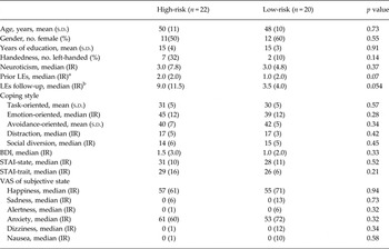
Demographic information, mood and coping scores for the high-risk (N = 22) and low-risk groups (N = 20) are presented in Table 1. The two groups were well-matched for age, gender and education levels (p ⩾ 0.6), handedness (p ⩾ 0.1), neuroticism (p ⩾ 0.4), severe LEs in the lifetime before the baseline assessment (p ⩾ 0.07), mood and subjective state (p ⩾ 0.2) and coping style (p ⩾ 0.3). There was a strong trend towards more stressful LEs in the 7-year period prior to the fMRI study in high-risk twins (Mann–Whitney U = 101, p = 0.054).
Demographic information and mood ratings on the test day for high-risk and control

Groups. Means and standard deviation (s.d.) are displayed for normally distributed data, while medians and interquartile ranges (IR) are displayed for non-normally distributed data.
a Number of severe LEs in the lifetime before the original baseline assessment in 2003–2005.
b Stressful life events in the 7 years prior to the present study. LE, life event; s.d., standard deviation; IR, interquartile range.
Behavioural results
Gender discrimination and faces dot-probe
Gender discrimination during fMRI scanning showed no behavioural differences between risk groups (p ⩾ 0.3).
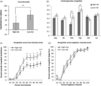
Dot-probe data were lost for one participant (high-risk) due to technical difficulties; the results were therefore analyzed for 41 participants (21 high-risk, 20 low-risk). High-risk twins displayed less vigilance to unmasked (supraliminal) fearful faces than the low-risk group (Mann–Whitney U = 115, Z = 2.5, p = 0.01) (Fig. 1 a). Post-hoc comparisons of response times with dots replacing fearful v. neutral faces within each group separately showed that the low-risk group displayed significant fear vigilance (i.e. faster responses to dots replacing fearful faces; t = 3.32, df = 19, p = 0.004), whereas the high-risk group showed neither bias towards nor away from fearful faces (p ⩾ 0.4; i.e. greater indifference or ‘attention control’). There was a trend towards lower vigilance to unmasked happy faces in the high-risk v. low-risk groups (t = 1.9, df = 39, p = 0.07). No differences between groups were found in speed to masked emotional faces (p ⩾ 0.5) or accuracy in any condition (p ⩾ 0.1).
Behavioural data. (a). Fear vigilance in the faces-dot probe test. High-risk twins displayed reduced supraliminal vigilance to fearful v. neutral faces, as reflected by longer response times for dots replacing unmasked fearful v. neutral faces. (b) Accuracy in the recognition of emotional facial expressions corrected for participants response tendency (d′). High-risk twins showed a specific reduction in the recognition of fearful and happy expressions compared with low-risk twins in the absence of general differences in accuracy across the other emotions. (c) Recognition of fearful and happy facial expressions across the 10 emotion intensity levels. High-risk twins showed reduced recognition of moderate to high intensity fear but no differential accuracy for happiness across the different intensity levels compared with low-risk twins. Values represent the mean scores. Error bars represent the s.e.m. One star indicates p < 0.05, two stars indicate p < 0.01 and three stars indicate p < 0.001.

Facial expression recognition
There was a main effect of emotion on facial expression recognition accuracy [F(5,200) = 15.0, p < 0.001], reflecting greater accuracy for positive expressions (happy and surprise) than negative (anger, disgust, fear, sadness) expressions across the entire cohort (Wilcoxon signed-rank test: Z = 5.2, p < 0.001). High-risk twins showed reduced recognition of fear and happiness compared with low-risk twins [F(5,200) = 3.3, p = 0.01; U = 113 Z = 2.7, p = 0.01; happy: U = 133, Z = 2.2, p = 0.03] in the absence of differences in overall emotion recognition accuracy (p ⩾ 0.1) (Fig. 1). Examination of fear recognition accuracy across the ten intensity levels revealed reduced recognition of moderate and high intensity fear in high-risk v. low-risk twins [F(5.4,215) = 2.4, p = 0.04; 50%: U = 159, Z = 2.0, p = 0.046; 60%: U = 141, Z = 2.1, p = 0.04; 70%: U = 130, Z = 2.3, p = 0.02; 80%: U = 129, Z = 2.3. p = 0.02; 90%: U = 132, Z = 2.3, p = 0.02; 100%: U = 89, Z = 3.4, p = 0.001], see Fig. 1. In contrast, the recognition of happiness showed no interaction between group and intensity level (p ⩾ 0.2). Speed of facial expression recognition showed no differences between groups (p ⩾ 0.3).
Functional brain activation during face processing
Whole-brain fMRI analysis
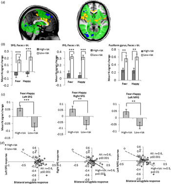
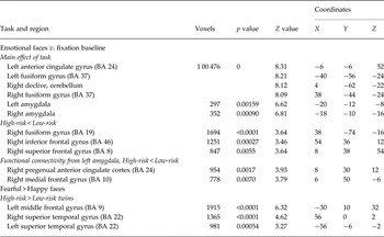
Emotional faces (v. baseline) activated a network of prefrontal and occipito-parietal regions across all participants (Fig. 2, green network; for peak cluster activations see Table 2). High-risk twins showed reduced neural response to all emotional faces (v. baseline) in the superior frontal gyrus (SFG), inferior frontal gyrus (IFG) and occipital regions of the face processing network compared with low-risk twins (Fig. 2, blue clusters; for peak cluster activations, see Table 2).
Whole-brain analyses. (a) Neural response to emotional (happy and fearful) faces v. baseline across all participants (main effect of task; marked with green), areas showing reduced response to emotional faces v. baseline in high-risk twins (group × task interaction; marked with blue), and regions showing greater response to fearful v. happy faces in high-risk v. low-risk twins (group × task interaction; marked with yellow). (b) Extraction of BOLD signal change from the regions showing a group × task interaction for emotional faces v. baseline revealed reduced activity to emotional faces v. baseline in high-risk (N = 22) compared with low-risk twins (N = 20) within the inferior and superior frontal and fusiform gyri. (c) Extraction of BOLD signal change from the regions showing a group × task interaction for fearful v. happy faces revealed specifically greater fear-associated activity in high-risk v. low-risk twins. Values represent mean percentage signal change. Error bars represent the s.e.m. One star indicates p < 0.05, two stars indicate p < 0.01 and three stars indicate p < 0.001. Increased MFG and SFG response to fearful v. happy faces correlated negatively with fear-specific bilateral amygdala response in the high-risk group and across the entire cohort. The solid and dotted lines denote the linear trends for the correlation in high-risk twins and across the entire cohort, respectively. Please note that the ROI was non-independent and thus the effect sizes may be exaggerated (Kriegeskorte et al. Reference Kriegeskorte, Simmons, Bellgowan and Baker2009).

Peak cluster activation in brain regions of increased BOLD response (whole-brain analyses with Z = 2.0, p = 0.05, cluster-corrected and within the amygdala in the analyses using the structural amygdala for small volume correction) during processing of fearful and happy faces v. fixation: (i) across all participants (main effect of task) and (ii) in high-risk v. low-risk twins and (iii) peaks in clusters showing negative functional coupling (anticorrelations) with amygdala in high-risk v. low-risk twins (Low-risk > High-risk) in whole-brain PPI analyses (Z = 2.0, pP = 0.05, cluster-corrected)

MNI coordinates (x, y, z) refer to the point of peak activation within each cluster identified using this threshold. BA: Brodmann area.
Whole-brain analysis of neural response to fearful v. happy faces revealed no main effect of task across the entire cohort (i.e. no regions were activated more by one v. the other emotion). However, high-risk twins showed greater activity to fearful v. happy faces within the left MFG (BA 9) and bilateral superior temporal gyri (STG; BA 22) (Fig. 2, yellow clusters; for peak cluster activations, see Table 2).
Amygdala ROI
Fearful and happy faces produced significant bilateral amygdala activation across all participants (p < 0.001), but without differences between high-risk and low-risk groups (p ⩾ 0.9 and p ⩾ 0.2, respectively). There was a moderate to strong negative correlation between bilateral amygdala response to fearful v. happy faces and the fear-specific MFG and SFG activity identified in the whole-brain analysis across the entire cohort [MFG: r(40) = −0.6, p ⩽ 0.001; right SFG: r(40) = −0.6, p ⩽ 0.001; left SFG: r(40) = −0.6, p ⩽ 0.001] and within the high-risk group [MGF: r(20) = −0.5, p = 0.01; right SFG: r(20) = −0.6, p = 0.003] (see Fig. 2).
Functional connectivity of the amygdala
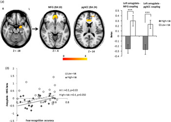
Whole-brain PPI analysis with the left amygdala functional cluster (ROI; emotional faces v. baseline) as the seed region revealed aberrant functional coupling with the MFG (BA 10) and pgACC (BA 24) during emotional face blocks (v. baseline) in high-risk v. low-risk twins (see Fig. 3; for peaks of the group differences, see Table 2). Post-hoc paired t tests of the standardized betas in these clusters within each group revealed significant negative FC (anticorrelations) between left amygdala and the mPFC and pgACC clusters in high-risk twins (amygdala–mPFC coupling: t = 3.1, df = 21, p = 0.01; amygdala–pgACC coupling: t = 2.8, df = 21, p = 0.01), whereas low-risk twins co-activated these regions (t = 2.9, df = 19, p = 0.01 and t = 2.8, df = 19, p = 0.01, respectively) (see Fig. 3). Whole-brain PPI analysis showed no difference between groups in FC from the right amygdala functional cluster.
Whole-brain PPI analysis with left amygdala functional cluster (emotional faces v. baseline across all participants) as the seed region showed different functional coupling (FC) of the left amygdala with MFG (BA 10) and pgACC (BA 24) in high-risk compared with low-risk twins (amygdala–MFG coupling: t = 4.22, df = 40, p ⩽ 0.001; amygdala–pgACC coupling: t = 3.93, df = 40, p ⩽ 0.001); Whereas low-risk twins showed positive FC of the left amygdala with MFG (BA 10) and pgACC (BA 24), high-risk twins showed negative FC (anticorrelations) between these regions. Amygdala-MFG anticorrelations correlated with reduced recognition of fear in high-risk twins and across the entire cohort. Values represent the mean standardized betas in the clusters. Error bars represent the s.e.m. One star indicates p < 0.05, two stars indicate p < 0.01 and three stars indicate p < 0.001. The solid and dotted lines denote the linear trends for the correlation in high-risk twins and across the entire cohort, respectively.

Exploratory post-hoc analyses revealed that stronger amygdala-mPFC anticorrelations were associated with decreased fear recognition accuracy across the entire cohort [r(40) = 0.3, p = 0.03) and within the high-risk group [r(20) = 0.4, p = 0.050] (Fig. 3). While stronger amygdala-pgACC co-activation correlated with greater fear vigilance in the low-risk group [r(18) = 0.6, p = 0.001], this association was absent in the high-risk group (p ⩾ 0.2).
Exploratory correlations between stressful LEs and the behavioural and fMRI changes
There was no collinearity between recent stressful life events and risk status (variation inflation factor = 1.1). Given the strong trend towards more recent stressful LEs in high-risk twins, we therefore conducted post-hoc exploratory Pearson's correlations to investigate whether observed neurocognitive differences in high-risk v. low-risk groups were associated with the degree of adversity. More LEs correlated weakly with decreased fear vigilance across the entire sample [r(40) = −0.3, p = 0.046] but not in the high-risk group alone (p ⩾ 0.1). In contrast, no correlation occurred between LEs and the recognition of fearful or happy expressions (p ⩾ 0.3). Notably, entering LEs as a covariate in the statistical models did not alter the observed effects of risk status on fear vigilance or emotion recognition (p ⩽ 0.046) and revealed no additional effects of LEs (p ⩾ 0.13).
More LEs also correlated with increased fear-specific activity in the identified STG (but not mPFC) clusters across the entire cohort [left STG: r(34) = 0.4, p = 0.04; right STG: r(34) = 0.4, p = 0.03], which was reduced to a trend-level in high-risk twins [r(15) = 0.5, p = 0.059 and r(15) = 0.4, p = 0.10, respectively]. Finally, more LEs were associated with stronger amygdala-mPFC anticorrelations across the entire cohort [r(34) = −0.3, p = 0.04; for amygdala-pgACC: p = 0.09], which was reduced to a trend-level in high-risk twins [r(15) = −0.4, p = 0.09].
Discussion
This study investigated cognitive and neural response to emotional faces in healthy, never-depressed DZ twins at high v. low familial risk for depression. In contrast with our hypothesis, high-risk twins showed less fear vigilance and reduced recognition of fearful and happy facial expressions compared with low-risk twins. This was accompanied by reduced neural response within fronto-occipital regions to emotional faces and greater response to fearful v. happy faces in medial prefrontal and superior temporal regions. While low-risk twins showed co-activation between the left amygdala and the mPFC and pgACC, high-risk twins displayed anticorrelations between these regions, which correlated with their decreased recognition of fear. These effects occurred in the absence of differences between groups in mood, neuroticism, subjective state or coping styles.
Our finding of less fear vigilance and lower sensitivity to emotional – particularly fearful – faces in DZ high-risk twins contrasts with the increased attention to and recognition of negative facial expressions in MDD (for review, see Bourke et al. Reference Bourke, Douglas and Porter2010) and no or subtle negative face bias in other healthy individuals at familial risk (Le et al. Reference Le, Cowen and Harmer2007; Mannie et al. Reference Mannie, Bristow, Harmer and Cowen2007; Miskowiak et al. Reference Miskowiak, Glerup, Vestbo, Harmer, Reinecke, Macoveanu, Siebner, Kessing and Vinberg2015). Several studies report a bias away from threat (i.e. attention avoidance of threat) in post-traumatic stress disorder (PTSD) in the presence of imminent threat or stress (e.g. Bar-Haim et al. Reference Bar-Haim, Holoshitz, Eldar, Frenkel, Muller, Charney, Pine, Fox and Wald2010; Wald et al. Reference Wald, Lubin, Holoshitz, Muller, Fruchter, Pine, Charney and Bar-Haim2011) and in anxious children (for review, see Pine & Fox, Reference Pine and Fox2015). Our DZ high-risk twins showed no sign of attention avoidance of threat but rather more indifference or possibly greater attention control than low-risk twins (i.e. no bias toward or away from fear). Given the correlation between a higher number of recent stressful LEs and more attention control across the entire cohort and a strong trend toward more stressful LEs in the high-risk group, it could be speculated that that high-risk twins’ greater attention control was acquired partially through their recent life stress. Such attention control may confer resilience – a positive adaptation to stress or adversity that makes these individuals less vulnerable to depression (Haglund et al. Reference Haglund, Nestadt, Cooper, Southwick and Charney2007; Rutter, Reference Rutter2012). Consistent with this assumption, strengthened attention control after attention control training seems to contribute to symptom reduction in PTSD (Badura-Brack et al. Reference Badura-Brack, Naim, Ryan, Levy, Abend, Khanna, McDermott, Pine and Bar-Haim2015).
The pattern of neural activity that confers resilience to depression is still unclear. Disruption of FC between amygdala and ventral PFC during negative emotional processing is hypothesized to play a central role in the pathophysiology of MDD (Kong et al. Reference Kong, Chen, Tang, Wu, Driesen, Womer, Fan, Ren, Jiang, Cao, Blumberg, Xu and Wang2013). Therefore, our DZ high-risk twins’ distinct cortico-limbic anticorrelations during emotional face processing – which correlated with greater attention control in the presence of fearful faces – may be a compensatory mechanism or marker of resilience that keeps them from getting depressed despite their familial vulnerability. This is similar to the greater amygdala-pgACC anti-correlations during emotional face processing in MZ twins at familial risk for depression (Miskowiak et al. Reference Miskowiak, Glerup, Vestbo, Harmer, Reinecke, Macoveanu, Siebner, Kessing and Vinberg2015). In keeping with this, combat-exposed resilient individuals who did not develop post-traumatic stress showed increased medial prefrontal top-down regulation of amygdala response to emotional faces (Shin et al. Reference Shin, Wright, Cannistraro, Wedig, McMullin, Martis, Macklin, Lasko, Cavanagh, Krangel, Orr, Pitman, Whalen and Rauch2005). We found in an exploratory posthoc analysis that more stressful LEs were associated with stronger cortico-limbic anti-correlations, suggesting that prefrontal top-down control may be strengthened through adversity. Together, these findings point to greater cortico-limbic top-down regulation of emotional reactivity as a common compensatory mechanism across distinct at-risk populations.
The decreased fronto-occipital response to emotional faces in our DZ high-risk twins contrasts with exaggerated response in these regions to negative faces in MDD (Surguladze et al. Reference Surguladze, Brammer, Keedwell, Giampietro, Young, Travis, Williams and Phillips2005; Suslow et al. Reference Suslow, Konrad, Kugel, Rumstadt, Zwitserlood, Schöning, Ohrmann, Bauer, Pyka, Kersting, Arolt, Heindel and Dannlowski2010) and to emotional faces in MZ high-risk twins (Miskowiak et al. Reference Miskowiak, Glerup, Vestbo, Harmer, Reinecke, Macoveanu, Siebner, Kessing and Vinberg2015). Further, the increased fear-specific activity in our DZ high-risk twins within the mPFC, STG and insula is opposite to reduced superior temporal and insula activity to negative faces in MDD patients (Fitzgerald et al. Reference Fitzgerald, Laird, Maller and Daskalakis2008). The correlation between greater fear-related mPFC-STG activity and more LEs suggests that this compensatory mechanism may be strengthened by exposure to stress in twins who remained healthy despite their familial risk.
A strength of the study was the thorough longitudinal assessments of participants over several years prior to this study, which enabled inclusion of only healthy, never-depressed twins. A limitation was the modest sample size (N = 42) and hence possibly limited generalizability. Another potential limitation is the relatively high age (50 years) of our high-risk twins. The participants had thus passed the major risk periods for MD onset despite their familial vulnerability and stressful LEs, suggesting that they exhibited resilience rather than vulnerability to depression. We could not conduct direct comparisons of neural activity between MZ and DZ high-risk twins to disentangle genetic makeup from shared environment because of differences in the scanning parameters between the studies. It was a limitation that we did not assess alexithymia since alexithymia is associated with decreased neural activity in a broad emotion processing network to emotional stimuli (e.g. Kret & Ploeger, Reference Kret and Ploeger2015) and could have influenced our results. Another limitation was that we did not assess childhood trauma since early life stress influences neural activity and functional connectivity in the emotion processing network (Grant et al. Reference Grant, Wood, Sreenivasan, Wheelock, White, Thomas, Knight and Deshpande2015). The study is merely associative in nature given the cross-sectional design, thus highlighting a need for prospective studies to clarify whether the neurocognitive differences in DZ high-risk twins indeed confer resilience. Functional connectivity is also merely a correlational measure, which precludes a strong inference regarding the causal direction in the connectivity. Finally, cluster-extent-based thresholding in fMRI analysis has limited spatial informativeness when clusters span multiple anatomical regions due to liberal statistical thresholds (Woo et al. Reference Woo, Krishnan and Wager2014). Despite the relatively liberal cluster-extent-based statistical threshold in our exploratory whole-brain analysis (Z = 2.0, p < 0.05), the identified clusters with differential activity between groups were spatially specific. Nevertheless, our results should be regarded as exploratory in nature.
In conclusion, this exploratory study delineates neural and cognitive changes in DZ twins at familial risk for depression. We observed less fear vigilance and reduced recognition of negative and positive facial expressions, which was accompanied by cortico-limbic anti-correlations and decreased fronto-occipital activity to emotional faces. These findings contribute to the understanding of the neural and cognitive mechanisms of depression and resilience.
Acknowledgements
The study was funded by the Danish Council for Independent Research and the Lundbeck Foundation. K.W.M.’s salary is partially funded by the Lundbeck Foundation (R93-A8635) and the Weimann Stipend. H.R.S. is supported by a Grant of Excellence on the control of actions ‘ContAct’ from the Lundbeck Foundation (R59-A5399). The Simon Spies Foundation is acknowledged for donation of the Siemens Trio Scanner.
Declaration of interest
K.W.M. has received consultancy fees within the last 3 years from Lundbeck and Allergan; M.V. has been a consultant for Eli Lilly, Lundbeck; Servier and Astra Zeneca; C.J.H. has received consultancy fees from P1vital ltd, Servier, Eli-Lilly, is a company director of Oxford Psychologists ltd. and has also received grant income from GSK, Lundbeck, Servier and Astra Zeneca; H.R.S. was within the past 3 years received honoraria as reviewing editor for Neuroimage, as speaker for Biogen Idec Denmark A/S and scientific Advisor for Lundbeck; L.V.K. has within the last 3 years been a consultant for Lundbeck, AstraZeneca and Servier. All other authors report no biomedical financial interests or potential conflicts of interest.
Ethical standards
The authors assert that all procedures contributing to this work comply with the ethical standards of the relevant national and institutional committees on human experimentation and with the Helsinki Declaration of 1975, as revised in 2008.