INTRODUCTION
Multiple infections by different parasites are commonly observed within single host individuals (Cox, Reference Cox2001; Alizon et al. Reference Alizon, de Roode and Michalakis2013), and such coexisting parasites may interact to either enhance or reduce the pathologic effects of infection (Alizon et al. Reference Alizon, de Roode and Michalakis2013; Budischak et al. Reference Budischak, Sakamoto, Megow, Cummings, Urban and Ezenwa2015). Co-infection also can influence host survival (Klemme et al. Reference Klemme, Louhi and Karvonen2016). For example, survival of laboratory mice infected with Trypanosoma brucei increased when the mice were experimentally infected with a second, less virulent, genotype (Balmer et al. Reference Balmer, Stearns, Schötzau and Brun2009). In another set of experimental infections of mice, two helminth species and one bacterial strain affected each other's virulence in co-infection, a result of direct competition for host resources and of interaction with the host immune system (Budischak et al. Reference Budischak, Sakamoto, Megow, Cummings, Urban and Ezenwa2015). While within-host parasite interactions may influence parasite virulence, and therefore, the consequences of parasitism, co-infections in wild populations remain understudied.
In one-host-one-parasite systems, the evolution of virulence shifts the trade-off between parasite replication and host mortality; high host mortality can outweigh the benefits to the parasite of increased parasite transmission (de Roode et al. Reference de Roode, Yates and Altizer2008; Alizon et al. Reference Alizon, de Roode and Michalakis2013). However, co-infections can make such trade-offs irrelevant by causing parasite virulence to increase beyond the transmission-host mortality threshold (de Roode et al. Reference de Roode, Helinski, Anwar and Read2005). For example, red crossbills (Loxia curvirostra) co-infected with two species of malaria parasites, Plasmodium relictum and Plasmodium ashfordi, develop higher parasitaemias of both infections than birds infected with either one of these parasite species alone (Palinauskas et al. Reference Palinauskas, Valkiūnas, Bolshakov and Bensch2011; Dimitrov et al. Reference Dimitrov, Palinauskas, Iezhova, Bernotienė, Ilgūnas, Bukauskaitė, Zehtindjiev, Ilieva, Shapoval, Bolshakov, Markovets, Bensch and Valkiūnas2015). Increased virulence is not the only possible outcome of co-infection. Apparent competition is often detected in co-infections, as one parasite infection can interact through the host immune system to influence the probability of acquiring a subsequent infection (Mideo, Reference Mideo2009). For instance, field voles (Microtus agrestis) are more or less likely to acquire new infections over time depending on pre-existing parasite infections, regardless of their body condition (Telfer et al. Reference Telfer, Lambin, Birtles, Beldomenico, Burthe, Paterson and Begon2010).
Here, we ask how two common protozoan parasites influence each other's occurrence in a host population of yellow-breasted chats (Parulidae, Icteria virens) in North America. Avian haemosporidian parasites (Order Haemosporida; Genera Plasmodium, Haemoproteus and Leucocytozoon) are protozoans transmitted by blood-feeding dipterans, replicating in the tissues of their vertebrate hosts, and undergoing sexual reproduction and sporulation in the midgut of their invertebrate dipteran vectors (Valkiūnas et al. Reference Valkiūnas, Anwar, Atkinson, Greiner, Paperna and Peirce2005). Experimental infections indicate that haemosporidian parasites reduce host activity, reproductive success and longevity (Atkinson et al. Reference Atkinson, Dusek, Woods and Iko2000; Palinauskas et al. Reference Palinauskas, Valkiūnas, Bolshakov and Bensch2008, Reference Palinauskas, Valkiūnas, Kriћanauskienė, Bensch and Bolshakov2009; Knowles et al. Reference Knowles, Nakagawa and Sheldon2009, Reference Knowles, Wood and Sheldon2010; Asghar et al. Reference Asghar, Hasselquist, Hansson, Zehtindjiev, Westerdahl and Bensch2015). The principal vectors for Plasmodium parasites are Culicidae mosquitos; Haemoproteus parasites are transmitted by biting midges (Ceratopogonidae: Culicoides) and hippoboscid flies (Hippoboscidae); Leucocytozoon parasites are vectored by Simuliidae black-flies (Valkiūnas, Reference Valkiūnas2004).
Trypanosomes (Class Kinetoplastida; Order Trypanosomatida) are transmitted by a variety of vectors, including mosquitoes, mites, black flies, hippoboscids and midges (Votýpka et al. Reference Votýpka, Lukeš and Oborník2004; Van Dyken et al. Reference Van Dyken, Bolling, Moore, Blair, Beaty, Black and Foy2006; Černý et al. Reference Černý, Votýpka and Svobodová2011; Ivkovic et al. Reference Ivkovic, Kesic, Mihaljevic and Kudela2013). In contrast to haemosporidian parasites, which are transmitted to the vertebrate host by inoculation of sporozoites along with the vector's saliva, trypanosomes actively reach the host's blood stream by crossing mucosal membranes (Desser et al. Reference Desser, McIver and Jez1975). Metacyclic trypomastigotes, the trypanosome infective stage from invertebrate to vertebrate host, may develop in the insect's hindgut, although Trypanosoma culicavium infecting Culex spp. mosquitos develop first in the midgut, and later migrate to the stomodeal valve of the insect host (Votýpka et al. Reference Votýpka, Szabova, Radrova, Zidkova and Svobodová2012), showing that development strategies in the invertebrate vector are variable. Transmission to the vertebrate host can happen via abraded skin, with parasites entering the host through the epidermis, or via an oral route, whereby birds feed on infected vectors and the parasites enter the bloodstream by crossing the mucosa of the bird's oral cavity, oesophagus or crop (Baker, Reference Baker1956a ; Votýpka and Svobodová, Reference Votýpka and Svobodová2004). Although little is known about the pathogenicity of trypanosome infections in birds, these parasites are reported to cause subclinical infections of low parasitaemia in avian hosts (Baker, Reference Baker1956b ; Valkiūnas et al. Reference Valkiūnas, Bairlein, Iezhova and Dolnik2004; Zídková et al. Reference Zídková, Cepicka, Szabová and Svobodová2012), with extremely variable prevalence across host species, ranging from zero to as high as 100% (Greiner et al. Reference Greiner, Bennett, White and Coombs1975a ; Kirkpatrick and Lauer, Reference Kirkpatrick and Lauer1985; Kirkpatrick and Suthers, Reference Kirkpatrick and Suthers1988; Sehgal et al. Reference Sehgal, Jones and Smith2001).
Yellow-breasted chats are the largest of the North American warblers, although their taxonomic placement is not fully resolved (Lovette et al. Reference Lovette, Pérez-Emán, Sullivan, Banks, Fiorentino, Córdoba-Córdoba, Echeverry-Galvis, Barker, Burns, Klicka, Lanyon and Bermingham2010). They typically inhabit dense shrublands and second-growth forests (Morris et al. Reference Morris, Porneluzi, Haslerig, Clawson and Faaborg2013; Reidy et al. Reference Reidy, Thompson and Kendrick2014). Individuals breed across most of the USA, Southern British Columbia and Alberta, as well as Northern Mexico; the winter range extends from Mexico to Panama. Yellow-breasted chats that we have sampled across Missouri, Tennessee and Mexico are mainly parasitized by one specialist lineage of haemosporidian parasite, designated in our laboratory as Plasmodium sp. OZ08 (GenBank accession HM222485). Surveys of blood parasites in North America have detected trypanosomes in blood films of I. virens (Stabler et al. Reference Stabler, Holt and Kitzmiller1966; Greiner et al. Reference Greiner, Bennett, White and Coombs1975b ; Kirkpatrick and Suthers, Reference Kirkpatrick and Suthers1988). During a recent investigation of white blood cell frequencies in birds infected with haemosporidian parasites (Ellis et al. Reference Ellis, Kunkel and Ricklefs2014), we detected trypanosome parasites in the blood films of yellow-breasted chats sampled in the Ozarks plateau of Southern Missouri. Accordingly, in this study, we sought to determine whether: (1) occurrences of haemosporidian and trypanosome parasites are associated in this host population; (2) co-infections exhibit an age or sex bias; and (3) co-infections affect host condition, using, as proxies, regenerative anaemia and white blood cell counts.
MATERIALS AND METHODS
Blood sampling
Yellow-breasted chats were caught using standard mist-net protocols. Sampling was originally conducted to survey haemosporidian parasites of avian assemblages in the Ozarks plateau of southern Missouri (91.03°W, 37.12°N), during the summer of the years 1999–2002 and again in 2011 (Ricklefs et al. Reference Ricklefs, Swanson, Fallon, MartÍnez-AbraÍn, Scheuerlein, Gray and Latta2005; Svensson-Coelho et al. Reference Svensson-Coelho, Blake, Loiselle, Penrose, Parker and Ricklefs2013; Ellis et al. Reference Ellis, Collins, Medeiros, Sari, Coffey, Dickerson, Lugarini, Stratford, Henry, Merrill, Matthews, Hanson, Roberts, Joyce, Kunkel and Ricklefs2015). All sampling was covered under state and federal permits, with field protocols approved by an Institutional Animal Care and Use Committee (IACUC) at the University of Missouri-St. Louis. For each individual captured, a small blood sample was taken through brachial venipuncture and stored in Longmire's lysis buffer (Longmire et al. Reference Longmire, Maltbie and Baker1997) and a thin blood film was prepared in the field. We fixed all blood smears with methanol on the same day, and stained them with Giemsa within 30 days, following a standard staining protocol (Valkiūnas, Reference Valkiūnas2004). Individuals were identified as after-hatch year (AHY) and hatch-year (HY) birds, which we refer to as adults and juveniles, respectively. All birds were released at the site of capture.
Molecular detection and genotyping of parasites
We extracted DNA from blood samples using a standard isopropanol precipitation protocol, preceded by enzymatic protein digestion and precipitation with ammonium acetate. Detection and genotyping of haemosporidian infections have been described elsewhere (Ricklefs et al. Reference Ricklefs, Swanson, Fallon, MartÍnez-AbraÍn, Scheuerlein, Gray and Latta2005; Svensson-Coelho et al. Reference Svensson-Coelho, Blake, Loiselle, Penrose, Parker and Ricklefs2013; Ellis et al. Reference Ellis, Collins, Medeiros, Sari, Coffey, Dickerson, Lugarini, Stratford, Henry, Merrill, Matthews, Hanson, Roberts, Joyce, Kunkel and Ricklefs2015). Detection and genotyping of trypanosome parasites were based on a nested PCR assay that amplifies a 770 bp fragment of the 18S SSU rRNA gene sequence, adapted from Valkiūnas et al. (Reference Valkiūnas, Iezhova, Carlson and Sehgal2011). The outer PCR assay used the primers Tryp763 and Tryp1016, with 10 µL of master mix containing 0·6 µ m of each primer, 200 µ m of each dNTP, 1·2 mm of MgCl2, 8 mm of Tris–HCl, 40 mm of KCl, 0·1 µL Taq DNA polymerase and 2 µL of DNA template. The inner PCR assay used 0·5 µL of DNA template from the outer reaction, in a 20 µL reaction volume with the primers Tryp99 and Tryp957. The PCR master mix for the inner reaction contained 0·6 µ m of each primer, 200 µ m of each dNTP, 1·2 mm of MgCl2, 8 mm of Tris–HCl, 40 mm of KCl, 0·5 µL of Taq DNA polymerase. Thermocycler conditions for the outer essay were: initial denaturation at 95 °C for 5 min, 5 cycles of 95 °C for 1 min, 45 °C for 30 s and 65 °C for 1 min, followed by 35 cycles of 95 °C for 1 min, 50 °C for 30 s, 72 °C for 1 min and a final extension at 65 °C for 10 min. On the inner PCR assay, thermocycler conditions were: initial denaturation of 96 °C for 3 min, 25 cycles of 96 °C for 30 s, 58 °C for 1 min and 72 °C for 30 s, and a final extension at 72 °C for 7 min. We measured the DNA concentration and purity from all samples that tested negative, and confirmed that negative results were not due to extraction failures. We used the product of the inner PCR assay for bidirectional sequencing with BigDye Terminator v3.1 using ABI PRISM® 3100 DNA Analyser (Applied Bio Systems®, USA) at the University of Missouri-St. Louis. DNA sequences were edited and analysed on CLC Main Workbench version 7.0 (QIAGEN, http://www.clcbio.com).
Molecular sexing of hosts
For all individuals whose sex we could not determine based on plumage differences observed in the field (N = 100), we used molecular sexing based on the PCR amplification of CDH1 genes found in avian sex chromosomes (Fridolfsson and Ellegren, Reference Fridolfsson and Ellegren1999). Primers were 2550F and 2718R, in a PCR master mix with a final volume of 10 and 1 µL of genomic DNA. The master mix contained 0·3125 units of Taq DNA polymerase, 1·5 µL of 10× buffer, 2·5 mm of each dNTP, 1·625 mm of MgCl2 and 0·5 µ m of each primer. Thermal cycling conditions were as follows: 2 min at 94 °C, followed by 31 cycles of 45 s at 94 °C, 45 s at 48 °C, 45 s at 72 °C and final elongation for 10 min at 72 °C. Sex was determined by the number of bands after running 8 µL of amplicon on a 2% agarose gel for 40 min.
Microscopy
We selected slides for microscopic viewing based on the PCR detection of parasites. We analysed five slides for each of the following groups of individuals: (1) negative for both haemosporidian and trypanosome parasites; (2) positive for both haemosporidian and trypanosome parasites; (3) positive only for haemosporidian parasites; and (4) positive only for trypanosome parasites (four slides analysed). We examined blood slides using an Olympus 224 CX31 light microscope, equipped with a Leica ICC50 HD digital camera and Leica Application Suite, version 4.4.0, imaging software. We took 100 pictures, corresponding to 100 fields at high magnification (×1000), and used ImageJ to count the number of cell nuclei on each picture (Gering and Atkinson, Reference Gering and Atkinson2004; Girish and Vijayalakshmi, Reference Girish and Vijayalakshmi2004). We manually recorded the numbers of heterophils, eosinophils, basophils, lymphocytes, thrombocytes, as well as the number of polychromatic erythrocytes (PE) and the number of mature erythrocytes infected with haemosporidian parasites. Infections by haemosporidian parasites can lead to elevated counts of heterophils (Ellis et al. Reference Ellis, Kunkel and Ricklefs2014), which could be costly to the host due to tissue damage related to inflammatory responses (Ots et al. Reference Ots, MurumAgi and HOrak1998). Decreased numbers of lymphocytes could signal immunosuppression, as these cells are responsible for the production of antibodies against infective agents (Ots et al. Reference Ots, MurumAgi and HOrak1998). PE are erythrocytes prematurely released, or just released, from the bone marrow into the blood stream. PE have basophilic cytoplasm and cell nuclei showing clumps of condensed chromatin. Elevated levels of PE may signify regenerative anaemia, which is a condition caused by either blood loss or haemolysis (Samour, Reference Samour, Harrison and Lightfoot2006; Atkinson et al. Reference Atkinson, Saili, Utzurrum and Jarvi2014). The proportion of each white blood cell type was calculated as the number of cells per 104 non-PE.
Statistical analyses
We used Fisher's exact tests and log linear tests for two-way and three-way contingency analysis, respectively. We applied generalized linear models with a binomial error distribution to determine the effects of individual parasite lineages on the presence/absence of a second parasite. The first of these models tested the effects of haemosporidian lineages on the occurrence of trypanosome parasites. In this model, presence/absence of trypanosome parasites in each individual host was the response variable, and the presence/absence of each lineage of haemosporidian parasite was a multi-level categorical predictor. The second model followed the same reasoning, but with trypanosome lineages as the predictor variable and the presence/absence of haemosporidian parasites as the response variable. Blood cell count data were represented as proportions of cells relative to 104 non-PE. We used ANOVA and the post hoc Tukey's HSD to compare haematological parameters determined through microscopy between groups of birds with single and co-infections. All analyses were performed in R (R Core Team, 2015).
RESULTS
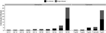
We screened 153 individual yellow-breasted chats, of which 52 were infected with haemosporidian parasites, 40 were infected with trypanosomes and 23 harboured co-infections with lineages of both parasites. Genotyping identified ten haemosporidian lineages, with 33 (63%) of the infections being Plasmodium lineage OZ08 and 7 (13%) Plasmodium lineage OZ01 (Fig. 1). We observed five trypanosome lineages, 29 (72·5%) of which were assigned to the lineage Tryp01, which closely matches (nucleotide pairwise difference of 1·6%) a SSU rRNA sequence of Trypanosoma bennetti (Genbank accession AJ223562), first isolated from an American kestrel (Falco sparverius) (Haag, Reference Haag1998). The lineage Tryp04 also matches T. bennetti AJ223562 (nucleotide pairwise difference of 2·2%). Tryp02 and Tryp03 do not match any identifiable morphotype sequence available in Genbank. The trypanosome lineages Tryp01–Tryp03 have an average nucleotide pairwise difference of 2%; Tryp04 differs by an average of 6·5% from Tryp01–Tryp03. Sequences for new trypanosome lineages were deposited in Genbank: accession numbers KX179915–KX179918.
Prevalence of lineages of and haemosporidian (left) and trypanosome (right) parasites retrieved from yellow-breasted chats sampled in the Ozarks (MO).

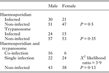
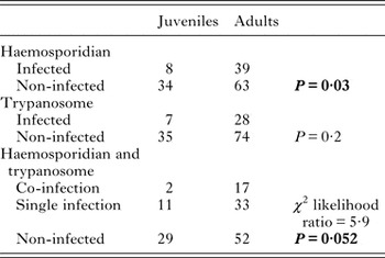
We determined the sex of 149 individuals, of which 81 were males and 68 were females. Sexes did not differ in the prevalence of single or co-infections (Table 1). Overall, 102 individuals were identified adults, and 42 as juveniles. Adults were more likely than juveniles to be infected with haemosporidian parasites; however, we found no effect of age on the occurrence of trypanosome infections (Table 2). We had determined in the field the age of 30 out of the 33 individuals infected with the specialist Plasmodium lineage OZ 08, and 28 of these were adults. Additionally, only ten adults harboured infections by lineages other than OZ 08. Co-infections were more common in adults than in juveniles, though the difference was only marginally significant (Table 2).
Number of males and females of yellow-breasted chats (Icteria virens) harbouring single infections and co-infections of haemosporidian and trypanosome parasites

P represents significance value from a Fisher's exact test, or, when specified, by the χ 2 likelihood ratio from a log-linear test.
Number of hatch year (juveniles) and after hatch year (adults) of yellow-breasted chats (Icteria virens) harbouring single infections and co-infections of haemosporidian and trypanosome parasites

P represents significance value from a Fisher's exact test, or, when specified, by the χ 2 likelihood ratio from a log-linear test. Significant differences are indicated in bold type.
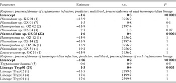
An individual I. virens carrying one parasite infection was almost four times more likely to be carrying a second type of parasite (Fisher's exact test, P = 0·0004, odds ratio = 3·9, CI = 1·7–8·9). The Plasmodium lineage OZ08, the most common haemosporidian lineage infecting yellow-breasted chats (Fig. 1), was positively associated with the occurrence of trypanosome infections in individual hosts. That is, if an individual was infected with OZ08, it would more likely be infected with any given lineage of trypanosome parasite (Table 3). The Plasmodium lineage OZ04, recovered from only three individuals, showed a positive, marginally significant relationship with trypanosome infections; two of the three hosts infected with OZ04 also were infected with trypanosome parasites (Table 3). When we looked at the effects of trypanosome lineages on the probability of harbouring haemosporidian infections, we found that Tryp01, the most frequent trypanosome lineage, increased the probability of an individual host also being infected with a haemosporidian parasite (Table 3).
Estimate, standard error (s.e.) and P-value for the two generalized linear models determining the effects of individual parasite lineages on the presence/absence of a second parasite. Number of individuals infected with each lineage is represented in parentheses

We ran the same analyses treating lineages with less than five infections as either haemosporidian or trypanosome infections and we found no differences compared with the results presented on the table.
Counts of PE, white blood cells and erythrocytes infected with haemosporidian parasites are summarized in online Table S1 of the Supplemental Material. Yellow-breasted chats that tested negative for both haemosporidian and trypanosome infections had lower levels of PE compared with individuals with either single or co-infections (F = 11·9; P < 0·001, online Tables S1 and S2 of the Supplemental Material). Individuals infected with both Plasmodium spp. and Trypanosoma spp. had levels of haemosporidian-infected erythrocytes comparable with individuals infected only with Plasmodium spp. parasites (t-test = −0·4366, d.f. = 4, P = 0·7). The proportion of heterophils, eosinophils, basophils, lymphocytes, monocytes and thrombocytes did not differ among individuals that were uninfected, co-infected or singly infected.
DISCUSSION
We found a positive association between haemosporidian and trypanosome infections, meaning that individuals infected by one parasite are more likely to carry a second protozoan infection. Specifically, the Plasmodium lineage OZ08 was positively associated with the trypanosome lineage Tryp01. A relationship between haemosporidian and trypanosome parasites was similarly observed in Alaskan passerine birds, where Trypanosoma infections were positively associated with Leucocytozoon and Haemoproteus infections, but not with Plasmodium infections (Oakgrove et al. Reference Oakgrove, Harrigan, Loiseau, Guers, Seppi and Sehgal2014). These associations are unlikely to result solely from these parasites being co-vectored, as the transmission routes of avian trypanosomes and some avian haemosporidian parasites may differ (Baker, Reference Baker1956a , Reference Baker c ; Molyneux and Robertson, Reference Molyneux and Robertson1974; Votýpka and Svobodová, Reference Votýpka and Svobodová2004). We also found that trypanosome infections cause comparable levels of regenerative anaemia as Plasmodium infections, which provides evidence of disease related to Trypanosoma.
An association between trypanosome and haemosporidian parasites also has been reported in Eurasian sparrowhawks (Accipeter nisus) and common buzzards (Buteo buteo) (Svobodová et al. Reference Svobodová, Weidinger, Peške, Volf, Votýpka and Voříšek2015). For individuals of these raptor species, co-infections involving Trypanosoma and Leucocytozoon parasites were more frequent than co-infections involving Trypanosoma and the other two haemosporidian parasite genera (Svobodová et al. Reference Svobodová, Weidinger, Peške, Volf, Votýpka and Voříšek2015). Blackflies can vector the Trypanosoma and Leucocytozoon parasites of sparrowhawks and buzzards, but Haemoproteus and Plasmodium parasites require other vectors (Svobodová et al. Reference Svobodová, Weidinger, Peške, Volf, Votýpka and Voříšek2015). The co-infections by Trypanosoma and Leucocytozoon in sparrowhawks and buzzards could be due to co-transmission by blackflies. However, we find it unlikely that co-transmission is responsible for the observed association between Trypanosoma and Plasmodium in yellow-breasted chats. First, although we have not identified the vectors of trypanosomes for yellow-breasted chats, T. culicavium isolated from Culex mosquitoes is the only known case of avian trypanosome parasites potentially transmitted by mosquitoes (Votýpka et al. Reference Votýpka, Szabova, Radrova, Zidkova and Svobodová2012). Second, Trypanosoma–Plasmodium co-infection might not be survived by mosquito vectors. Experimental infections of avian haemosporidians in Culicidae vectors show that infections with a single parasite species reduce mosquito survival rates, even in naturally occurring (i.e. co-evolved) parasite–vector associations (see Ferguson and Read, Reference Ferguson and Read2002; Valkiūnas et al. Reference Valkiūnas, Kazlauskienė, Bernotienė, Bukauskaitė, Palinauskas and Iezhova2014).
Empirical evidence suggests that the association of haemosporidian and trypanosome infections could be even stronger than what we observed, since trypanosome parasites are difficult to detect in the peripheral blood of birds (Kirkpatrick and Suthers, Reference Kirkpatrick and Suthers1988; Apanius, Reference Apanius1991). Experimental infections of Trypanosoma avium in canaries demonstrated that, upon ingestion of infected louse-flies, metacyclic trypomastigotes first migrated to the host lymph nodes (Baker, Reference Baker1956c ). Within a few days post-infection, and after growing in size in the lymph nodes, individual trypanosomes could be found in the peripheral blood, where they remained for 2 to 3 months before migrating to the bone marrow (Baker, Reference Baker1956c ; Molyneux and Robertson, Reference Molyneux and Robertson1974; Valkiūnas et al. Reference Valkiūnas, Bairlein, Iezhova and Dolnik2004; Votýpka and Svobodová, Reference Votýpka and Svobodová2004). Thus, the narrow window of detection of the parasite in the peripheral blood suggests that we likely underestimated the prevalence of trypanosome parasites in our study population. However, our samples were collected during the summer months, when trypanosomes are known to reappear in the peripheral blood of infected birds, and when haemosporidian infections also relapse (Valkiūnas et al. Reference Valkiūnas, Bairlein, Iezhova and Dolnik2004). Additionally, detection bias in our molecular methods might have resulted in underestimating the strength of the association between haemosporidian and trypanosome parasites; in the presence of co-infections of either type of parasite (i.e. multiple haemosporidians or multiple trypanosomes infecting the same individual host), a PCR reaction may amplify one parasite over the other (Pérez-Tris and Bensch, Reference Pérez-Tris and Bensch2005). However, this bias does not affect the probability of detecting one of two or more infections of either type of parasite.
We detected five lineages of Trypanosoma parasites infecting a single host population. In samples from 71 bird species caught in African rainforests, Sehgal et al. (Reference Sehgal, Jones and Smith2001) found eight trypanosome lineages, four of which were found infecting olive sunbirds (Cyanomitra olivacea). In an assemblage of 49 bird species in Alaska, only T. avium was detected (Oakgrove et al. Reference Oakgrove, Harrigan, Loiseau, Guers, Seppi and Sehgal2014). As trypanosomes are known to have low host specificity (Bennett et al. Reference Bennett, Earle and Squires-Parsons1994; Sehgal et al. Reference Sehgal, Jones and Smith2001), each of the lineages identified in this study might infect multiple bird species in the Ozarks region. Our findings also suggest high genetic diversity of Trypanosoma lineages, which might correspond to species other than T. bennetti depending on analyses of morphological characters and further genetic markers.
The lack of support for sex-biased incidence of infection is consistent with the findings of previous studies. For example, Kirkpatrick and Suthers (Reference Kirkpatrick and Suthers1988) analysed blood films of 697 individuals from 59 bird species and found no differences between males and females in the prevalence of Plasmodium, Haemoproteus and Trypanosoma, or co-infections amongst the three genera of parasites. In our study, haemosporidian infections were found more often in adults than juveniles, which is consistent with at least three hypotheses: (1) mortality due to haemosporidian infections is higher among juveniles, leading to detection bias favouring infections in older individuals; (2) infections accumulate with age, meaning that older birds have been exposed more to infected vectors than juveniles; (3) most infections in adults correspond to relapses of chronic infections acquired in previous years. Hypothesis (1) seems improbable, given the high local abundance of yellow-breasted chats. Some combination of (2) and (3) more likely explains the higher prevalence of infections in adults. Previous studies have demonstrated that older birds are not only more likely to be infected than juveniles (Svobodová et al. Reference Svobodová, Weidinger, Peške, Volf, Votýpka and Voříšek2015), but also that they are more likely to harbour specialist rather than generalist parasites, possibly reflecting differences in the ability of specialist vs generalist parasites to infect hosts that they share (Medeiros et al. Reference Medeiros, Ellis and Ricklefs2014).
Haemosporidian parasite relapse in spring is an adaptation of the parasite to seasonal host susceptibility, availability of naïve hosts and vector availability (Cornet et al. Reference Cornet, Nicot, Rivero and Gandon2014; Pérez-Rodríguez et al. Reference Pérez-Rodríguez, de la Hera, Bensch and Pérez-Tris2015). Additionally, in chronically infected birds in temperate regions, trypanosomes spend the winter in the bone marrow, and only migrate to the peripheral blood during spring and summer months (Baker, Reference Baker1956a , Reference Baker c ). Kirkpatrick and Suthers (Reference Kirkpatrick and Suthers1988) found HY North American birds to have half the haemosporidian and trypanosome infections as AHY individuals. The difference between our findings and those of Kirkpatrick and Suthers (Reference Kirkpatrick and Suthers1988) may be due to the different methods used to detect trypanosomes; they surveyed trypanosomes using blood cultures, which boost the detectability of these parasites when they are uncommon in the peripheral blood (Stabler et al. Reference Stabler, Holt and Kitzmiller1966; Kirkpatrick and Lauer, Reference Kirkpatrick and Lauer1985). Thus, although PCR methods have high sensitivity when it comes to detection of blood parasites, it is possible that we failed to detect some developing trypanosome infections in juvenile birds.
While the pathogenicity of some avian haemosporidian lineages is fairly well understood (Valkiūnas, Reference Valkiūnas2004), little is known about the consequences of Trypanosoma infections for birds. The similar white blood cell levels of infected and uninfected birds is consistent with previous studies, in which white blood cell responses to blood parasite infections occurred in some populations of hosts, but not others (Ricklefs and Sheldon, Reference Ricklefs and Sheldon2007). Ellis et al. (Reference Ellis, Kunkel and Ricklefs2014) found that haemosporidian-infected birds of the Ozarks presented higher levels of heterophils and lymphocytes than non-infected individuals. The difference in results between this study and Ellis et al. (Reference Ellis, Kunkel and Ricklefs2014) could be due to the latter being a broader analysis of the effects of any haemosporidian infection on the white blood cell profiles of several host species, whereas this study focused on Plasmodium-infected individuals from one population of a single host species.
We found evidence for regenerative anaemia associated with trypanosome parasites on the same level as that caused by haemosporidian infections. Elevated numbers of PE in trypanosome-infected individuals is evidence of disease. Although we found a statistically significant difference in PE between trypanosome-infected and uninfected individuals, the number of slides available for analysis was limited, and we interpret these results as evidence that trypanosome infections have the potential to cause disease. More importantly, we recommend that future studies investigating the effect of avian trypanosome infections include in their design some measure of anaemia. Although reports are scarce, ours is not the first to associate avian Trypanosoma parasites with disease-like symptoms in birds. Experimental infections of Trypanosoma bouffardi in canaries revealed myocarditis, nephritis, congestion of the spleen, and myofibril degeneration in infected subjects (Molyneux et al. Reference Molyneux, Cooper and Smith1983). However, experimental studies of infection of Trypanosoma in canaries have not detected multiplication of the parasite (Baker, Reference Baker1956c ; Votýpka and Svobodová, Reference Votýpka and Svobodová2004), which suggests that canaries are not natural hosts for the parasites (Chatterjee and Ray, Reference Chatterjee and Ray1971). When Trypanosoma avium was experimentally inoculated in red-whiskered bulbuls (Otocompsa jocosa), Chatterjee and Ray (Reference Chatterjee and Ray1971) observed trypomastigotes multiplying by binary fission in the blood and bone marrow. However, the evidence of multiplication by binary fission does not make the pathogenicity of avian trypanosomes straightforward to understand, as parasites are not multiplying within the erythrocytes, as are Plasmodium schizonts. Moreover, comparable numbers of PE between single and co-infected individuals suggest that either haemosporidian or trypanosome parasites have suppressed virulence in co-infections.
In conclusion, we have demonstrated a positive association between Plasmodium and Trypanosoma infections in yellow-breasted chats, with evidence of pathogenicity of trypanosomes in these hosts. The association between these two protozoans could arise from a negative effect of one parasite on the host's immune response to the other. Future studies might focus on determining whether such an association remains during host migration and wintering. The basic life cycle of avian trypanosome parasites and their pathogenicity in vertebrate hosts remains poorly understood. However, the evidence presented here sheds some light on the mechanisms of within-host interactions between trypanosomes and haemosporidians, two common parasites of birds.
SUPPLEMENTARY MATERIAL
The supplementary material for this article can be found at http://dx.doi.org/10.1017/S0031182016001384.
ACKNOWLEDGEMENTS
We thank Patricia G. Parker for laboratory support. Fieldwork was facilitated by Steven Latta, Alicia Burke, Kristen Bird, Elyse Coffey, Melanie Kunkel and the Missouri Department of Conservation.
FINANCIAL SUPPORT
Laboratory work was funded by the Curators of the University of Missouri to R. E. R.