

Introduction
The cytokines are low-molecular weight proteins that are produced by a variety of cells and tissuesReference Moldoveanu, Shephard and Shek1–Reference Weinstock, Konig, Harnischmacher, Keul, Berg and Northoff6. They are released locally when cells are perturbed by a variety of challenges ranging from minor to severeReference Moldoveanu, Shephard and Shek1–Reference Weinstock, Konig, Harnischmacher, Keul, Berg and Northoff6. In the case of the latter, they play a key role in the modulation of inflammation and as a reaction to an infection or tissue injuryReference Moldoveanu, Shephard and Shek1–Reference Weinstock, Konig, Harnischmacher, Keul, Berg and Northoff6. The cytokine response is complex and multifaceted, utilizing endocrine, paracrine and autocrine actions to facilitate intracellular communicationsReference Moldoveanu, Shephard and Shek1–Reference Weinstock, Konig, Harnischmacher, Keul, Berg and Northoff6. The resulting interactions between various tissues, cells and signalling pathways can recruit many immune cell types, such as monocytes, lymphocytes and neutrophils to aid in the repair process to tissue injuryReference Moldoveanu, Shephard and Shek1–Reference Weinstock, Konig, Harnischmacher, Keul, Berg and Northoff6. Cytokines can also modulate the inflammatory and immune response by exerting inhibitory effects on immune cells, resulting in restrictions on growth, differentiation and functionReference Moldoveanu, Shephard and Shek1–Reference Weinstock, Konig, Harnischmacher, Keul, Berg and Northoff6. Prior studies of horses have reported that a variety of perturbations, such as obesity, ageing and exercise, cause increases in the expression of mRNA for interferon-γ (IFN-γ), tumour necrosis factor-α (TNF-α), interleukin (IL)-6 and a number of other cytokinesReference Adams, Katepalli, Kohler, Reedy, Stilz and Vick7–Reference Liburt, McKeever, Streltsova, Manso, Gordon and Horohov13.
TNF-α is an acute pro-inflammatory cytokine that has been used as a general marker of inflammationReference Moldoveanu, Shephard and Shek1–Reference Weinstock, Konig, Harnischmacher, Keul, Berg and Northoff6. TNF-α plays a role in the response to muscle damage, muscle proteolysis and impaired skeletal muscle glucose uptakeReference Moldoveanu, Shephard and Shek1–Reference Weinstock, Konig, Harnischmacher, Keul, Berg and Northoff6, Reference Malm14. IFN-γ is a major pro-inflammatory cytokine synthesized by T-helper 1 and natural killer (NK) cellsReference Elenkov15. It acts on stimulated macrophages, T-cytotoxic cells, NK cells and B lymphocytes, and is involved in nitric oxide synthesis, antibody production and antiviral activityReference Elenkov15, Reference Ijzermans and Marquet16. IL-6 has been documented to function in the multifaceted cascade of events involved in the onset and modulation of inflammationReference Moldoveanu, Shephard and Shek1, Reference Pedersen and Hoffman-Goetz2. In that role, there are conflicting studies suggesting that IL-6 has both anti-inflammatory and pro-inflammatory rolesReference Moldoveanu, Shephard and Shek1–Reference Weinstock, Konig, Harnischmacher, Keul, Berg and Northoff6. However, when it comes to its biological role during exercise, the preponderance of information on the effect of IL-6 demonstrates that it is an important signalling protein in the integrated control of glucose homeostasis in muscleReference Moldoveanu, Shephard and Shek1–Reference Weinstock, Konig, Harnischmacher, Keul, Berg and Northoff6, Reference Nieman, Nehlsen-Cannarella, Fagoaga, Henson, Utter and Davis18–Reference Steensberg, Keller, Starkie, Osada, Febbraio and Pedersen21. This role that IL-6 plays in glucose homoeostasis has led researchers to classify IL-6 as a ‘myokine’ or a muscle cytokineReference Pedersen and Hoffman-Goetz2, Reference Malm14.
The role of the cytokines in the modulation of the inflammatory response to exercise has been well documented in a large number of basic and applied research papersReference Moldoveanu, Shephard and Shek1–Reference Steensberg, Keller, Starkie, Osada, Febbraio and Pedersen21. On an applied level, many of those experiments have had the goal of understanding the mechanisms behind exercise-induced increases in the mediators of the inflammatory response. Even with a greater understanding of the mechanism behind the inflammatory response, the elusive objective of many studies has been to discern the fine line between minor muscle inflammation that evokes an adaptive response versus an excessive increase in markers of inflammation associated with more frank muscle damage, loss of function and performanceReference Moldoveanu, Shephard and Shek1–Reference Steensberg, Keller, Starkie, Osada, Febbraio and Pedersen21. However, it should be noted that the cytokines not only play a role in inflammation, they also play an important role in pathways impacting multiple physiological responses to exerciseReference Moldoveanu, Shephard and Shek1, Reference Pedersen and Hoffman-Goetz2. These pathways include those affecting a variety of neuroendocrine mechanisms associated with the control of peripheral and central mechanisms affecting exercise performanceReference Moldoveanu, Shephard and Shek1, Reference Pedersen and Hoffman-Goetz2. Peripherally, the cytokines interact in the complex control of peripheral blood flow as well as metabolic pathways used to fuel exerciseReference Moldoveanu, Shephard and Shek1, Reference Pedersen and Hoffman-Goetz2. Centrally, they appear to have a role in the control of cardiovascular and respiratory functionReference Moldoveanu, Shephard and Shek1, Reference Pedersen and Hoffman-Goetz2. The widespread nature of the role the cytokines play in the response to exercise suggests that their moderate increase, evoked by acute exercise, is appropriate and part of the protective mechanism dealing with the physiological challenge of exertionReference Moldoveanu, Shephard and Shek1, Reference Pedersen and Hoffman-Goetz2. Excessive exercise intensity, duration and resistance can cause significant increases in cytokines, resulting in post-exercise muscle soreness with a spectrum of symptoms that have been classified as delayed onset muscle soreness (DOMS)Reference Moldoveanu, Shephard and Shek1, Reference Pedersen and Hoffman-Goetz2. In its extreme, DOMS can result in the classic signs of inflammation including heat, pain, swelling and loss of function.
These ergolytic effects of post-exercise inflammation can be countered through the use of non-steroidal anti-inflammatory drugs (NSAID), including various pharmaceutical and nutraceutical anti-inflammatory countermeasuresReference Streltsova, McKeever, Liburt, Manso, Gordon and Horohov12, Reference Liburt, McKeever, Streltsova, Manso, Gordon and Horohov13, Reference Tobin, Chay, Kamerling, Woods, Weckman and Blake22. Recent studies of humans and horses have examined the administration of pharmaceutical and nutraceutical substances (food extracts having anti-inflammatory properties) to block the inflammatory response to very intense exertionReference Streltsova, McKeever, Liburt, Manso, Gordon and Horohov12, Reference Liburt, McKeever, Streltsova, Manso, Gordon and Horohov13. Among those pharmaceutical interventions, there is a long list of NSAIDs commonly given to both humans and horsesReference Tobin, Chay, Kamerling, Woods, Weckman and Blake22, Reference Cho23. In the case of horses, the most utilized NSAID is phenylbutazoneReference Tobin, Chay, Kamerling, Woods, Weckman and Blake22. Phenylbutazone is a very potent analgesic and anti-inflammatory agentReference Tobin, Chay, Kamerling, Woods, Weckman and Blake22. However, its systemic effects can also alter the normal exercise-induced response of the endocrine, cardiovascular and respiratory systemsReference Tobin, Chay, Kamerling, Woods, Weckman and Blake22–Reference Beluche, Bertone, Anderson and Rohde34. Interestingly, no study to date has examined the effect of phenylbutazone on the cytokine response to exertion in the horse. Therefore, this study tested the hypothesis that phenylbutazone would alter the exercise-induced increase in cytokines measured in blood.
Materials and methods
Animals and dosing
The Rutgers University Institutional Animal Care Review Board approved all methods and procedures used in this experiment. Nine healthy, unfit Standardbred mares (aged 10 ± 4 years, ~500 kg) were utilized for three different trials (standing control (CON, n = 9); exercise with phenylbutazone (EX-bute, 2 g given orally 2 h before the graded exercise test (GXT), n = 9); and exercise with water (EX-water, n = 9). The EX-water GXTs were conducted as part of a separate trailReference Streltsova, McKeever, Liburt, Manso, Gordon and Horohov12 that was conducted months before the subsequent two rounds. The CON and EX-bute GXTs utilized seven of the nine horses used in the original nutraceutical experiment, with the addition of two mares that had been held in reserve and were housed and cared for, even though they were part of the group used in the original experiment. All mares were accustomed to the laboratory and running on the treadmill before the start of the experiment.
During the study, the horses were housed as a group on a dry lot. Each mare was fed approximately 2.5 kg of total mixed ration hay cubes (Eckenberg Farms™, Mattawa, WA, USA) twice a day (~5 kg d− 1 total) at about 07.00 and 15.00 hours. Water and grass hay were provided ad libitum. The ration was formulated to meet the energy guidelines of the National Research Council (1989) for adult horses conducting periodic exercise. Mares were not fed on the morning of the exercise test.
Graded exercise test
The GXT used previously published methodsReference Streltsova, McKeever, Liburt, Manso, Gordon and Horohov12, Reference Liburt, McKeever, Streltsova, Manso, Gordon and Horohov13 with the mares running on a high-speed horse treadmill (Sato I; Equine Dynamics, Inc., Lexington, KY, USA) at a fixed 6% grade. The GXTs started at an initial speed of 4 m s− 1 for 1 min. Speed was then increased to 6 m s− 1, followed by incremental increases of 1 m s− 1 every 60 s (omitting 5 m s− 1) until the horses reached fatigue. Fatigue was defined as the point where the horse could not keep up with the treadmill despite humane encouragement.
Blood sampling and real-time polymerase chain reaction
Blood samples (30 ml) collected during all three trials were obtained via venepuncture into PAXgene™ Blood RNA Tubes (Qiagen, Inc., Santa Clarita, CA, USA) before exercise, immediately at the end of the test and at 30 min, 1, 2, 4 and 24 h post-GXT for the measurement of mRNA expression of IFN-γ, TNF-α and IL-6. Samples collected for the CON and EX-bute trials were also assayed for IL-1 and IL-10.
Total RNA was subsequently isolated from the tubes using spin columns according to the manufacturer's instructions. The RNA was quantified using a spectrophotometer (BioPhotometer; Eppendorf, Hamburg, Germany). In all cases, optical density260/280 ratios were greater than 1.9, and RNA yields were greater than 50 μg ml− 1. One microgram of RNA was reverse transcribed into cDNA in an 80 μl reaction containing 20 units of AMV (avian myeloblastosis virus) RT, 0.5 μg of oligo dT primers, 40 units of RNAsin and 5 mM of MgCl2 (Promega, Madison, WI, USA). Cytokine-specific cDNA was then amplified and quantitated by ‘real-time’ PCR (ABI Systems 7500 Sequence Detection System, Foster City, CA, USA) using the Taq thermostable DNA polymerase and primers based on the sequences for equine cytokines and β-glucuronidase (β-gus). Specific primers and FAM (6-carboxy-fluorescein)-labelled probes for each cytokine and β-gus, provided as Assay-by-Design kits (ABI), were added to 25 μl reactions in 96-well microplates containing the Taq polymerase (12.5 μl of Universal Master Mix; ABI) and 5 μl of cDNA. The following PCR conditions were employed: 95°C for 10 min followed by 40 cycles of 95°C for 15 s and 60°C for 60 s, as recommended by the manufacturer.
Differences in RNA isolation and cDNA construction between samples were corrected using β-gus as an internal control for each sampleReference Breathnach, Sturgill-Wright, Stiltner, Adams, Lunn and Horohov35. Relative differences in cytokine mRNA expression resulting from exercise were determined by relative quantification. Relative quantification provides accurate comparison between the initial levels of target cDNA in a sample without requiring that the exact copy number be determinedReference Livak and Schmittgen36. The pre-exercise samples were selected as the calibrator, and the change in cytokine gene expression post-expression relative to the calibrator was then determined for each sample.
Statistical analysis
Results are expressed as means ± standard error of the mean. For comparison by group and time, a two-way ANOVA for repeated measures was used with the a priori level of statistical significance set at P < 0.05. Post hoc comparisons were made using the Student–Newman–Keuls test (SigmaStat 2.0; SPSS, Inc., Chicago, IL, USA).
Results
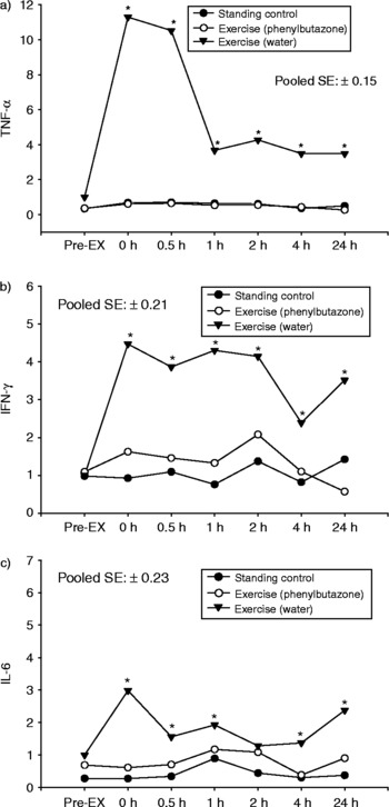
Exercise caused increases (P < 0.05) in mRNA expression for TNF-α, IFN-γ and IL-6 (Fig. 1) that remained elevated through the 24 h collection point. There were no changes (P>0.05) in mRNA expression for TNF-α, IFN-γ and IL-6 cytokines during the CON and EX-bute trials. IL-1 and IL-10 were measured only during the CON and EX-bute rounds of the experiment. There were no alterations (P < 0.05) across time, or due to exercise, in these two cytokines; therefore, those data are not shown.
Graphs present mean (a) TNF-α, (b) IFN-γ and (c) IL-6 mRNA expression. There was a significant effect of exercise on mRNA expression for TNF-α, IFN-γ and IL-6 mRNA when horses were administered water, but no effect of exercise (P>0.05) when horses were given phenylbutazone or during standing control. An asterisk (*) denotes a significant (P < 0.05) difference between groups at a given sample point.

Discussion
The cytokine responses to exercise in the horses of the present study appear to be similar to those documented for other species such as rats, mice and humansReference Moldoveanu, Shephard and Shek1–Reference Steensberg, Keller, Starkie, Osada, Febbraio and Pedersen21, Reference Helge, Stallknecht, Pedersen, Galbo, Kiens and Richter37–Reference Haahr, Pedersen, Fomsgaard, Tvede, Diamant and Klarlund39. Studies of humans have documented exercise-induced increases in plasma concentrations of TNF-α, IL-1-β, IL-6, IL-1ra, IL-10 and IFN-γ during and after exercise, with the magnitude and duration of the response influencing the intensity, mode (eccentric vs. concentric) and duration of the exercise challengeReference Moldoveanu, Shephard and Shek1–Reference Steensberg, Keller, Starkie, Osada, Febbraio and Pedersen21, Reference Helge, Stallknecht, Pedersen, Galbo, Kiens and Richter37–Reference Haahr, Pedersen, Fomsgaard, Tvede, Diamant and Klarlund39. Other papers have documented that a variety of exercise challenges cause increases in urinary concentrations of TNF-α, IL-1-β, IL-6, IL-2 receptors and IFN-γReference Moldoveanu, Shephard and Shek1, Reference Pedersen and Hoffman-Goetz2.
Studies of horses have reported up-regulation of mRNA for TNF-α in horses during clinical situations where inflammation is present (e.g. arthritis, laminitis and obesity)Reference Adams, Katepalli, Kohler, Reedy, Stilz and Vick7–Reference Liburt, McKeever, Streltsova, Manso, Gordon and Horohov13, Reference Horohov, Keadle, Pourciau, Littlefield-Chabaud, Kamerling and Keowen40, Reference Horohov, Dimock, Guirnalda, Folsom, McKeever and Malinowski41. This has also been shown to increase following experimentally induced inflammation where horses have been challenged with joint lesions or where they have been challenged with pathogens during vaccine developmentReference Adams, Katepalli, Kohler, Reedy, Stilz and Vick7–Reference Liburt, McKeever, Streltsova, Manso, Gordon and Horohov13, Reference Horohov, Keadle, Pourciau, Littlefield-Chabaud, Kamerling and Keowen40, Reference Horohov, Dimock, Guirnalda, Folsom, McKeever and Malinowski41. Intense exercise has also been shown to cause an increase of TNF-α in humansReference Kimura, Suzui, Nagao and Matsumoto3, Reference Haahr, Pedersen, Fomsgaard, Tvede, Diamant and Klarlund39 and horsesReference Streltsova, McKeever, Liburt, Manso, Gordon and Horohov12, Reference Liburt, McKeever, Streltsova, Manso, Gordon and Horohov13. Results have been mixed regarding the effects of exercise on IFN-γ. Most studies of humans and species other than horses have shown that excessively intense or long exercise causes an inhibition of IFN-γ production, with some papers suggesting that this leads to immunosuppressionReference Kimura, Suzui, Nagao and Matsumoto3, Reference Suzuki, Nakaju, Yamada, Totsuka, Sato and Sugawara5, Reference Weinstock, Konig, Harnischmacher, Keul, Berg and Northoff6, Reference Haahr, Pedersen, Fomsgaard, Tvede, Diamant and Klarlund39. However, it has also been reported that gene expression for IFN-γ, as well as the concentration of IFN-γ in plasma, is increased during the recovery from exertionReference Kimura, Suzui, Nagao and Matsumoto3, Reference Moyna, Acker, Fulton, Weber, Goss and Robertson17, Reference Baum, Muller-Steinhardt, Liesen and Kirchner38. Baum et al. Reference Baum, Muller-Steinhardt, Liesen and Kirchner38 observed increases in IFN-γ mRNA expression 24 h after moderate exercise, but marked decreases in IFN-γ concentration levels 30 min after exhaustive exercise in humans, and thus suggested that the IFN-γ response is affected by the type of exercise performed. Interestingly, studies of horses have demonstrated that intense, short-term exercise causes an increase in IFN-γ mRNA expression in the immediate period following exerciseReference Streltsova, McKeever, Liburt, Manso, Gordon and Horohov12, Reference Liburt, McKeever, Streltsova, Manso, Gordon and Horohov13.
The second finding of the present study was that phenylbutazone blocked the exercise-induced increase in cytokinesReference Adams, Katepalli, Kohler, Reedy, Stilz and Vick7–Reference Liburt, McKeever, Streltsova, Manso, Gordon and Horohov13 seen in horses. This is a new and novel finding, since most studies of phenylbutazone that focused on exercise used in vitro experiments to examine the anti-inflammatory effects on more general systemic actions during exertion. Mechanistically, NSAIDs affect the inflammatory pathways in a variety of ways, with direct actions on cellular function and indirect actions on signalling between cells through endocrine, paracrine, autocrine and cytokine pathways. It is well documented that acute exercise induces changes in many components of the immune systemReference Liao, Zhou, Ji and Zhang42–Reference Simon44, including increased expression of pro-inflammatory cytokines such as IL-1β, IL-6 and TNF-α. These cytokines are known to be involved in the regulation of the immune response and inflammation. Although the production of pro-inflammatory cytokines is believed to benefit the host defence system, the overproduction of these cytokines may lead to inflammation, subsequently causing muscle damage, weakness and increased risk of infectionReference Meksawan, Venkatraman, Awad and Pendergast45. There is also a systemic anti-inflammatory response tightly regulated by cytokines and characterized by changes in circulating leukocyte receptor and functional activityReference Peake, Nosaka and Suzuki46. It has been proposed that while a single bout of strenuous exercise produces a transient pro-inflammatory response, exercise training suppresses this inflammatory process, thereby contributing to the beneficial effects of habitual physical activityReference Kasapis and Thompson43.
While anti-inflammatory medications and nutritional supplements have often been used to limit exercise-induced inflammation and soreness, the limited data available indicate that anti-inflammatory drugs largely have minimal influence on inflammatory responses to eccentric exerciseReference Peterson, Trappe, Mylona, White, Lambert and Evans47, Reference Rhind, Gannon, Shephard and Shek48 and could lead to increased expression of some inflammatory markersReference Nieman, Henson, Dumke, Oley, McAnulty and Davis49. Phenylbutazone is routinely used in the horse for treatment of soft tissue inflammation and can be detected in plasma samples of racehorsesReference Dirikolu, Woods, Boyles, Lehner, Harkins and Fisher50, Reference Dodman, Blondeau and Marini51, Reference Barker52 as well as in their immediate environmentReference Barker52, Reference Authie, Garcia, Popot, Toutain and Doucet53. By inhibiting the cyclooxygenase enzyme system, which is responsible for synthesis of prostanoids such as PGE2, phenylbutazone markedly reduces prostanoid-dependent swelling, oedema, erythema and hypersensitivity to pain in inflamed tissuesReference Beretta, Garavaglia and Cavalli54, Reference Takada, Bhardwaj, Potdar and Aggarwal55. Like other NSAIDs, phenylbutazone inhibits TNF-induced NF-κB activation and IL-6 productionReference Komatsu, Yaju, Chiba and Okumoto56. While the precise mechanism responsible for these effects remains unknown, this probably involves an non-cyclooxygenase activity of the drugReference Rhind, Gannon, Shephard and Shek48.
The above speculation has focused on mechanisms behind the anti-inflammatory actions of phenylbutazone and how it can be a beneficial pharmacological intervention to counter the ergolytic effects of severe post-exercise inflammation. However, evoking a modulated up-regulation in inflammatory messengers may also be important for evoking an adaptive response to exercise. Many of the signalling pathways associated with the cytokine response to minor tissue repair are needed for increases in muscle strength and sizeReference Moldoveanu, Shephard and Shek1, Reference Pedersen and Hoffman-Goetz2. To that end, the increase in mRNA expression for the cytokines measured in the present study was rapid and of a magnitude consistent with what some have termed a ‘beneficial inflammatory response’ to acute exertion. Thus, one should caution against the prophylactic misuse of NSAIDs, as tone may actually inhibit those processes associated with tissue repair and muscle hypertrophy, as well as other cytokine-influenced systemic effects that are part of the normal exercise-induced response of the endocrine, cardiovascular and respiratory systems.
Acknowledgements
The authors would like to thank the graduate and undergraduate students from Rutgers University and the Rutgers Equine Science Center for their contributions to this project. The support for the present study was provided by the United States Department of Defense, US Army Natick Soldier Center, the New Jersey State Equine Initiative and the New Jersey Agricultural Experimentation Station. None of the authors of this paper has a financial or personal relationship with other people or organizations that could inappropriately influence or bias the content of the paper.