Significant outcomes
• Extracellular glycine and glutamate levels in the striatum were measured in sandy (sdy) mice.
• Basal extracellular levels of glycine and glutamate were higher in sdy mice.
• These results suggest that dysbindin might regulate glycine and glutamate release.
Limitations
• We did not compare extracellular glycine and glutamate levels in sdy mice after methamphetamine (METH) administration with those after saline administration.
Introduction
Glycine is a co-agonist of an N-methyl-d-aspartate receptor (NMDAR) d-serine/glycine site, and it regulates glutamatergic neurotransmission (Reference Yang and Svensson1). Several studies have reported an association between glycine and schizophrenia (Reference Javitt2). Glycine levels are increased in the temporal lobes in schizophrenics, and altered plasma glycine levels are also reported in patients with schizophrenia. These plasma glycine levels are inversely correlated with negative symptoms (Reference Hons, Zirko, Ulrychova, Cermakova, Doubek and Libiger3). Mice with reduced NMDAR glycine affinities have been reported to show behavioural abnormalities related to both the negative symptoms and the cognitive deficits of schizophrenic patients (Reference Labrie, Lipina and Roder4). Administration of glycine and d-serine has been found to attenuate deficits of prepulse inhibition caused by MK-801, an NMDAR antagonist (Reference Kanahara, Shimizu and Ohgake5). Both clinical and preclinical studies have suggested a relationship between schizophrenia and glycine; however, the underlying mechanisms remain uncertain.
The possible genetic bases of schizophrenia have received increased attention in recent years, and dysbindin-1 (DTNBP1: dystrobrevin-binding protein 1), which encodes dysbindin protein, is a leading candidate gene for schizophrenia (Reference Allen, Bagade and McQueen6). Reduced expression of dysbindin was found in the post-mortem brains of patients with schizophrenia (Reference Talbot, Eidem and Tinsley7,Reference Weickert, Straub and McClintock8), and genetic variations in dysbindin-1 impact human intelligence and memory functions (Reference Hashimoto, Noguchi and Hori9,Reference Hashimoto, Noguchi and Hori10). Thus, dysbindin-1 has attracted attention as a likely candidate gene that may mediate individual vulnerability to schizophrenia (Reference Olgiati, Mandelli and Lorenzi11).
Sdy mice have a large deletion in the dysbindin-1 gene, and this deletion results in almost total loss of the dysbindin protein. Sdy mice show behavioural abnormalities related to schizophrenia, including impairments of both long-term and working memory (Reference Hattori, Murotani and Matsuzaki12,Reference Takao, Toyama and Nakanishi13). Moreover, sdy mice show marked reductions in the ratio of neural differentiation in the dentate gyrus (Reference Nihonmatsu-Kikuchi, Hashimoto and Hattori14). Sdy mice also show significant hypersensitivity to dopamine and serotonin (Reference Kobayashi, Umeda-Yano, Yamamori, Takeda, Suzuki and Hashimoto15), and the increases in extracellular dopamine levels after potassium stimulation in sdy mice are lower than those in wild-type mice (Reference Nagai, Kitahara and Shiraki16). Dysbindin is also associated with the regulation of glutamate release and its signalling at the cellular level (Reference Numakawa, Yagasaki and Ishimoto17). However, no study to date has reported the extracellular amino acid levels in sdy mice, at least to our knowledge. Therefore, we investigated the possible relationship between dysbindin activity and glycine, a regulator of glutamatergic neurotransmission, in the present study. We measured the extracellular glycine levels in the striatum of sdy mice using in vivo microdialysis methods. We additionally measured the extracellular levels of glutamate, which is closely related to the pathophysiology of schizophrenia and which has been reported to be influenced by dysbindin. In addition, we measured changes in these amino acids after METH administration because dysbindin is reportedly related to METH psychosis, a model for schizophrenia (Reference Kishimoto, Ujike and Motohashi18).
Materials and methods
Animals
Male sdy mice weighing between 26.5 and 35.3 g were obtained from The Jackson Laboratories (Bar Harbor, ME, USA). Body weights varied because we did not have adequate numbers of sdy mice. However, to compensate for this variability, we used age- and weight-matched wild-type mice in the experiment. We used DBA/2J strain mice as wild-type mice. The animals were housed in a temperature-controlled environment (22 ± 1°C) with free access to food and water and were maintained on a 12-h light–dark cycle (light phase: 06:30–18:30 h). The features of sdy mice have been described previously (Reference Hattori, Murotani and Matsuzaki12). The mice were allotted a 10-day acclimation period before beginning the experiments. All procedures were approved by the Hokkaido University School of Medicine Animal Care and Use Committee and were in compliance with the Hokkaido University School of Medicine Guide for the Care and Use of Laboratory Animals.
Drug
METH (Dainippon Sumitomo Pharma Co. Ltd., Osaka, Japan) was dissolved in saline to a final concentration of 0.3 mg/ml. Mice were administered 0.1 ml/10 g (3.0 mg/kg) of METH intraperitoneally (i.p.). We have reported that administration of high-dose METH (2.5 mg/kg) increased extracellular dopamine levels and, consequently, induced delayed increases in extracellular glutamate levels (Reference Abekawa, Ito, Nakagawa, Nakato and Koyama19,Reference Ito, Abekawa and Koyama20). However, a low dose of METH (1.0 mg/kg) did not induce the same increases in rats. Thus, we used 3.0 mg/kg as the dose of METH in this study.
In vivo microdialysis procedures
All experiments were performed in awake and freely moving mice. METH was administered i.p. 100 min after the first dialysate samples were collected. Extracellular glycine and glutamate concentrations were analysed using a high-performance liquid chromatography system (HPLC) (Eicom Corp., Kyoto, Japan).
Under pentobarbital anaesthesia (50 mg/kg i.p.), AG-4 guide cannulae (Eicom Corp.) leading to the surface of the striatum were implanted into the mouse brain with the aid of a stereotaxic apparatus. The following coordinates relative to the bregma were used for cannulae implantation: A + 0, ML + 2.0, and DV + 2.5 mm (Reference Paxinos and Franklin21). Dialysis probes with a 0.22 mm outer diameter (A-I-02; Eicom Corp.) were then inserted into the guide cannulae such that 2 mm of each probe was exposed to the striatum tissue. The mice were housed individually following these procedures. Perfusion with artificial cerebrospinal fluid (CSF; 145 mM NaCl, 3.0 mM KCl, 1.3 mM CaCl2, and 1.0 mM MgCl2) at a flow rate of 2 μl/min was started 24 h after surgery. Following an initial 2-h perfusion period, dialysate samples were collected and placed in sample vials containing 50 μl of 0.05 M acetic acid every 20 min for a total of 380 min.
To determine changes in amino acid concentrations, we used identical equipment and a previously described protocol (Reference Abekawa, Ito and Koyama22), except that a 10 μl aliquot of the o-phthaldialdehyde derivatisation reagent was added to 30 μl of dialysate and allowed to react for 2.5 min. Afterwards, 30 μl of the reactant was injected into an HPLC system.
Statistical analysis
Basal levels were defined as the average of absolute values from five consecutive samples (−80 to 0 min) before METH injections. Differences in absolute values at baseline between the wild-type mice and the sdy mice were analysed using an unpaired t-test (two-tailed). To investigate differences between the wild-type mice and the sdy mice in the extracellular glycine and glutamate concentrations after METH treatment over time (0–280 min), a repeated measures analysis of variance (ANOVA) for absolute values was used. When the interaction was found to be significant, post hoc comparisons were made using an unpaired t-test. The area under the curve (AUC) showing the changes from baseline for the 0–280-min period was compared between the sdy mice and the wild-type mice. Differences in the AUC between the sdy mice and the wild-type mice were analysed using an unpaired t-test (two-tailed). We calculated the AUC using the equation of Chefer and Shippenberg (Reference Chefer and Shippenberg23). Differences were considered significant at p < 0.05.
Results
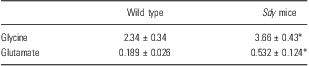
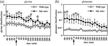
Basal extracellular levels of glycine and glutamate in the striatum of the sdy mice were significantly higher than those of the wild-type mice (Table 1). For the extracellular glycine concentrations in the striatum, a two-way ANOVA with repeated measures (0–280 min) indicated a significant main effect of genotype [F(1, 13) = 5.355, p = 0.0377] and a significant main effect of time after METH treatment [F(14, 182) = 6.123, p < 0.0001]. There was a significant interaction between genotype and time after METH treatment [F(14, 182) = 2.120, p = 0.0126]. The extracellular glycine levels of the sdy mice were significantly higher than those of the wild-type mice (unpaired t-test: 20, 40, 80–120, and 180 min, p < 0.05) (Fig. 1a). A two-way ANOVA with repeated measures (0–280 min) indicated a significant main effect of time after METH treatment on striatal extracellular glutamate concentrations [F(14, 182) = 7.156, p < 0.0001] and a significant interaction between genotype and time after METH treatment [F(14, 182) = 4.079, p < 0.0001]. There was no significant main effect of genotype [F(1, 13) =3.819, p = 0.0725]. Extracellular glutamate levels in the sdy mice were significantly higher than those of the wild-type mice (unpaired t-test: 20 and 40 min, p < 0.05) (Fig. 1b). The AUC of extracellular glutamate levels in the sdy mice (−36.7 ± 10.1) was significantly lower than that in the wild-type group (−7.4 ± 4.5) (unpaired t-test, p = 0.042). On the other hand, the AUC of extracellular glycine levels was not different between the sdy mice and the wild-type mice (−215.4 ± 55.0 and −122.1 ± 23.3, respectively) (unpaired t-test, p = 0.210).
Basal extracellular levels of glycine and glutamate in the striatum of the wild-type group and sdy mice group
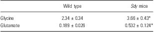
The glycine and glutamate concentrations of the dialysate samples are expressed as absolute values (fmol/20 min fraction). Values represent mean ± SEM. n = 6 (wild-type group), n = 9 (sdy mice group).
*p < 0.05 compared with the wild-type group.
Effect of acute METH (3.0 mg/kg i.p.) treatment on extracellular glycine and glutamate concentrations. The glycine (a) and glutamate (b) concentrations of the dialysate samples are expressed as absolute values (fmol/20 min fraction). Values represent the mean ± SEM. Arrows represent the time of METH injection. N = 6 (wild-type group), N = 9 (sdy mice group). *P < 0.05 compared with the wild-type group.

Discussion
This study shows that extracellular glycine and glutamate levels in the striatum of sdy mice, which lack dysbindin protein owing to a deletion in the dysbindin-1 gene by mutation, are abnormally increased compared with wild-type mice. This is the first study to show increased extracellular levels of these two amino acids in the brain of mice lacking dysbindin. Because both amino acids act on NMDARs of the brain as agonists, and because glutamate stimulates other subtypes of glutamate receptors (Reference Yang and Svensson1), in vivo receptor function of NMDAR and other receptor subtypes may be persistently elevated in sdy mice. Abnormal glutamate elevation may cause neurodegeneration, such as apoptosis, in sdy mice resembling the pathogenesis of schizophrenia (Reference Abekawa, Ito, Nakagawa, Nakato and Koyama19), although apoptosis in sdy mice has not been reported.
The mechanisms of elevated basal levels of extracellular glycine in the striatum of sdy mice are not clear. To our knowledge, no study to date has investigated glycine in sdy mice. Dysbindin interacts with various proteins, such as Munc 18-1 and snapin, and may regulate synaptic vesicle exocytosis (Reference Feng, Zhou and He24,Reference Hikita, Taya and Fujino25). These interactions alone do not provide evidence for the mechanisms of elevated glycine.
Glycine acts as a co-agonist at the strychnine-insensitive glycine-recognition site (glycine B site) of the NMDAR complex. In addition, glycine acts on inhibitory glycine receptors (glycine A site), which exist not only in the spinal cord and brainstem, but also in the cerebral cortex and hippocampus (Reference Legendre26). Accordingly, increased extracellular glycine may act on both glycine sites (A and B). Administration of glycine transporter inhibitors increases extracellular glycine levels in the brain (Reference Perry, Falcone and Fell27) and induces various behaviours (Reference Labrie, Clapcote and Roder28,Reference Shimazaki, Kaku and Chaki29). Direct agonists of the NMDAR glycine site and glycine transporter inhibitors induce anxiogenic-like behaviours (Reference Labrie, Clapcote and Roder28), which are also shown by sdy mice (Reference Hattori, Murotani and Matsuzaki12). These anxiogenic behaviours, induced by drugs acting on glycine sites, may be mediated by effects on the glycine B site. On the other hand, although sdy mice show social withdrawal (Reference Hattori, Murotani and Matsuzaki12,Reference Feng, Zhou and He24), glycine transporter inhibitors do not affect social interaction (Reference Shimazaki, Kaku and Chaki29). Therefore, it is possible that the anxiogenic-like behaviours in sdy mice are caused by elevated extracellular glycine levels in the brain, likely through the glycine B site.
In this study, basal levels of extracellular glutamate in the striatum were elevated in sdy mice. Glutamate is an important neurotransmitter at excitatory synapses. Glutamate acts on ionotropic glutamatergic receptors and metabotropic glutamatergic receptors (Reference Gaspar, Bustamante, Silva and Aboitiz30). A previous study reported that tissue glutamate concentrations in various brain regions, including the striatum, of sdy mice were not different from those of wild-type mice (Reference Hattori, Murotani and Matsuzaki12). Thus, increased extracellular glutamate levels in the striatum of sdy mice are not because of increased synthesis or decreased metabolism of glutamate, but may reflect increased release of glutamate. A previous in vitro study reported data inconsistent with the results of this study. That study showed that knockdown of dysbindin with small interfering RNA reduced basal glutamate levels and high-KCl-evoked glutamate release in cortical cultures in vitro (Reference Numakawa, Yagasaki and Ishimoto17). The reason for this inconsistency between in vivo and in vitro results is unclear. High-dose METH reportedly induces delayed increases in extracellular glutamate levels in the prefrontal cortex, nucleus accumbens, and striatum of rats (Reference Abekawa, Ito, Nakagawa, Nakato and Koyama19,Reference Ito, Abekawa and Koyama20,Reference Abekawa, Ohmori and Koyama31). In contrast to previous reports, high-dose METH did not increase extracellular glutamate levels in the striatum of sdy or wild-type mice. Instead, decreased extracellular glutamate was observed in the sdy mice but not the wild-type mice. Because high-dose METH increased extracellular dopamine levels in the striatum of sdy and wild-type mice similarly (data not shown), it is unlikely that the effect of high-dose METH was much lower in sdy mice than in wild-type mice. Unknown underlying mechanisms mediate this abnormal response. Nevertheless, this reduction in extracellular glutamate by high-dose METH seems similar to the reduction in evoked glutamate release observed in cortical cultures in vitro (Reference Numakawa, Yagasaki and Ishimoto17). Accordingly, sdy mice show bidirectional responses of increased basal levels and stimulus (METH)-evoked decreases in extracellular glutamate levels in the striatum.
Compared with wild-type mice, sdy mice show decreases in the social interaction test (Reference Hattori, Murotani and Matsuzaki12,Reference Feng, Zhou and He24) and NMDAR hypofunction in the prefrontal cortex (Reference Karlsgodt, Robleto and Trantham-Davidson32). Because the NMDAR antagonist MK-801 decreases social interaction (Reference Dyck, Guest, Sookram, Basu, Johnson and Mishra33) and increases extracellular glutamate levels in the brain (Reference Ito, Abekawa and Koyama20), decreased social interaction in sdy mice may be related to hypofunction of NMDARs and increased extracellular glutamate levels. However, until now, no study has reported the effect of glutamate transporter inhibitors, which increase extracellular glutamate levels, on social withdrawal and anxiogenic-like behaviour, both observed in sdy mice (Reference Hattori, Murotani and Matsuzaki12,Reference Feng, Zhou and He24). In addition, glial glutamate and aspartate transporter (GLAST) knockout mice do not show abnormal social interaction but do demonstrate a lesser preference for a novel social stimulus (Reference Karlsson, Tanaka, Saksida, Bussey, Heilig and Holmes34). Therefore, the functional significance of the persistent elevation of basal levels of extracellular glutamate in the striatum of sdy mice remains unclear.
Some limitations to our present study must be noted. Our data show that higher extracellular glycine and glutamate levels in the sdy mice at baseline gradually decreased to control levels following METH administration, although decreases in extracellular glycine levels after METH were not statistically significant. However, we did not compare extracellular glutamate levels in sdy mice after METH administration with those after saline administration. Thus, we cannot conclude that METH decreased extracellular glutamate levels in the sdy mice. Furthermore, in this study, we used pentobarbital as anaesthesia on the day before the dialysis experiment. Pentobarbital facilitates GABAA receptor function (Reference Nestler, Hyman and Malenka35). We cannot completely exclude the possibility that pentobarbital had some influence on glutamatergic neurotransmission. We will use another anaesthetic and allow the animals more time to recover after the operation in future work.
The present study is the first of its kind to report higher basal extracellular glycine and glutamate levels in the striatum of sdy mice, suggesting that dysbindin regulates glycine and glutamate release. Further studies are needed to examine the effects of dysbindin on neurotransmitter release in other brain regions, including the dorsolateral prefrontal cortex and hippocampus, in which post-mortem studies have also identified abnormal expression of dysbindin in schizophrenia (Reference Talbot, Eidem and Tinsley7,Reference Weickert, Straub and McClintock8).
Acknowledgements
This work was supported in part by a Grant-in-Aid for Scientific Research, No. 17790800 (Y.K.), from the Japanese Ministry of Education, Culture, Sports, Science and Technology. We are grateful for the professional assistance of language editors in preparing the manuscript for publication. Authors’ contributions: Conception, design, and acquisition of data: Y.K., R.H., T.I., and T.K. Analysis and interpretation of data: Y.K., R.H., T.A., A.K., and S.H. Writing of the paper: Y.K., R.H., T.I., T.A., S.H., and T.K. Final approval of the version to be published: Y.K., R.H., T.I., T.A., A.K., S.H., and T.K. Declaration of Interest: The authors declare no conflict of interest.