I. INTRODUCTION
Non-dispersive as well as dispersive double-crystal (DC) monochromators are quite common at synchrotron sources. However, in the case of neutrons, at present one can find only several scattering instruments in the world which use non-dispersive DC setting for neutron monochromatization (e.g. HB-1A Triple-Axis Spectrometer at High-Flux Isotope Reactor in ORNL, Double-Crystal Diffractometer at NIST for USANS, 4F1, and 4F2 spectrometers in LLB Saclay). Such monochromator with two parallel crystals produces monochromatic beam running parallel to incident white beam. For the change of the neutron wavelength such monochromator does not need a movable (rotating) table and the position of the sample is fixed. The problem occurs when using mosaic crystals. Owing to the double reflection process monochromatic beam current is much smaller than in the case of one crystal monochromator. For one mosaic monochromator, the neutron current is proportional to its peak reflectivity r (r < 1). Then for DC monochromator it is proportional roughly to r 2 and the situation becomes dramatically inconvenient when r ≈ 0.5 or less. The situation is even much worse in the case of dispersive or quasi-dispersive DC (n, ± m) setting employing mosaic crystals and the obtained monochromatic current is rather small since the individual space-phase elements of the crystals do not match as it is in the case of the non-dispersive (n, − n) setting. Therefore, it is commonly accepted opinion that a dispersive DC setting is not convenient for neutron monochromatization because of a low monochromatic neutron current and thus a low luminosity of the scattering instrument that is really true in the case of mosaic crystals. However, in the case of bent-perfect crystals (e.g. Si or Ge), the situation is much different and some advantages of the dispersive DC setting can be found. The general formula for the reflection probability (often called as peak reflectivity) of neutron passing through a homogeneously deformed perfect crystal has been derived in the form as (Kulda, Reference Kulda1984; Mikula et al., Reference Mikula, Kulda, Vrána and Chalupa1984)
 $$r\lpar R_{\rm M} \rpar =\lcub 1 - {\rm exp}\, \lsqb \!- Q_{hkl} \lpar \partial \Delta \theta /\partial s_0 \rpar ^{ - 1} \rsqb \rcub \comma \;$$
where (∂Δθ/∂s 0) is the rate of change of the Bragg angle on the flight path Δs 0 in the crystal in the incident beam direction and Q hkl is the kinematical reflectivity of the crystal unit volume. For cylindrically curved monochromator and symmetric reflection geometry (∂Δθ/∂s 0) has been derived in the form (Mikula et al., Reference Mikula, Kulda, Vrána and Chalupa1984, Reference Mikula, Kulda, Lukáš, Vrána and Wagner1994)
 $$\eqalign{ \partial \Delta \theta /\partial s_0&=\lpar 1/R_{\rm M} \rpar \lpar 1/{\rm cos}\, \theta _{\rm M} \rpar \cr & \quad\times\lsqb 1 - \lpar 1+\sigma \rpar \, {\rm sin}\, \theta _{\rm M} \, \, {\rm sin}\, \theta _{\rm M} \rsqb \comma} \;$$
where R M is the bending radius, θ M the Bragg angle, and σ the Poisson constant of the crystalline material. An example of calculated peak reflectivities for the used reflections of Si crystal, which perfectly fit with the experiment (Mikula et al., Reference Mikula, Kulda, Vrána and Chalupa1984, Reference Mikula, Chalupa, Kulda, Vrána, Sedláková and Michalec1985) is shown in Figure 1. It can be seen from Figure 1, that when the curvature of the crystals is not drastic, the reflection probability of both bent-perfect crystals can be close to 1 and there is a small and acceptable loss of neutrons because of the double-reflection process. Furthermore, curved crystal instruments are known by the fact that they do not use Soller collimators and the resolution can be controlled through the bending of the crystals. Possible employment of the focusing does not necessarily imply focusing in real space, but it can also cover focusing in scattering which is very often used in powder diffractometry, namely, for strain/stress measurements in polycrystalline materials (Vrána et al., Reference Vrána, Lukáš, Mikula and Kulda1994; Wagner et al., Reference Wagner, Mikula and Lukáš1994; Popovici and Yelon, Reference Popovici and Yelon1995; Wimpory et al., Reference Wimpory, Mikula, Šaroun, Poeste, Li, Hoffman and Schneider2008; Seong et al., Reference Seong, Em, Mikula, Saroun and Kang2010). DC arrangements with the bent-perfect crystals can provide variety of performances and some of them have been already proposed for exploitation in neutron diffractometry. With two bent crystals in successive reflection, the double-crystal rocking curve is generally much broader in comparison with the flat perfect crystals. However, when the phase-space windows of both bent crystals match, the DC rocking curve is narrow because it is related only to the convolution of the reflectivity curves and thus the intensity of the doubly reflected beam is high. This matching of the phase-space windows can be even achieved with the bent crystals having different d-spacings by a proper adjustment of individual curvatures of the crystals (Popovici et al., Reference Popovici, Stoica, Chalupa and Mikula1988; Popovici and Yelon, Reference Popovici and Yelon1995). Popovici and Yelon (Reference Popovici and Yelon1995) point out the possibility of a rather sharp focusing (up to 1 mm spot) by means of successive reflections on three Si(111) crystals (mosaic + bent + bent) in the (+1, −1, −1) setting, however, with a rather low luminosity. Another proposal of using the antiparallel (+n, +n) setting of two bent Cu(200) crystals (copper plates of small mosaic spread of about 3′) achieving the spot width of 2.5 mm for residual strain/stress instrument was reported in Popovici and Yelon (Reference Popovici and Yelon1994). Very promising results of an extensive investigation of (n, −m) combination of a vertically focusing mosaic Ge(331) crystal in combination with a horizontally bent-perfect Si(111) crystal were published in papers of Mikula et al. (Reference Mikula, Vrána, Choi, Kim, Oh, Sung, Lee, Tanaka, Muslih and Niimura2002, Reference Mikula, Vrána, Wagner, Choi, Moon, Lee and Inoue2004). Here, it was clearly demonstrated both real-space focusing and focusing in scattering on a standard polycrystalline γ-Fe sample (pin of the diameter of 2 mm) at λ = 0.183 nm. Furthermore, it has been found that for an optimum curvature of the bent Si(111), full-width at half-maxima (FWHMs) of the γ-Fe diffraction profiles in the 2θ S-angular scale for both parallel and antiparallel settings were practically the same. This fact can have enormous impact in powder diffractometry and namely for residual strain/stress measurements when permitting to measure two strain components simultaneously. Of course, that in the DC combination of mosaic and bent-perfect crystal because of a large mosaicity of the mosaic crystal, the matching of the phase-space elements is not as good as it can be in the case of two bent crystals. Recently, we have dealt with the investigation of the properties of the (+n, −m) setting of the bent Si(111) + Si(220)-sandwich for neutron monochromatization and its possibility to be employed in neutron diffractometry. Some results of the preliminary investigations with one, two, and four 1.3 mm thick bent Si(220) slabs in the form of the sandwich were published in Mikula et al. (Reference Mikula, Vrána, Šaroun, Em, Seong and Woo2012). On the basis of the obtained promising results, we continued extensive investigations in this direction when using a sandwich of Si(111)/Si(220) crystal slabs as the second component of the DC setting and making the DC monochromator more versatile.

II. EXPERIMENTAL ARRANGEMENT
For this experiment, we used neutron optics diffractometer installed at the 10 MW research reactor LWR-15. The diffractometer operates at the fixed neutron wavelength of λ = 0.162 nm, which is provided by a cylindrically bent-perfect Si(111) crystal of the dimensions of 200 × 40 × 4 mm3 (length × width × thickness). Schematic sketch of the diffractometer performance is drawn in Figure 2. Depending on the experimental requirements, the diffractometer can operate in two- or three-axis mode. First it should be pointed out that the radius of the curvature of the first Si(111) crystal of the DC setting was fixed (R I ≈ 10 m), which was not changed during the experiment. The second bent-perfect Si(111) or Si(220) crystals used in the sandwich had the dimensions of 200 × 40 × 2 and 200 × 40 × 1.3 mm3, respectively. It should be pointed out that because of a negligible attenuation of the silicon for the neutron wavelength of λ = 0.163 nm, both slabs in the sandwich can be considered transparent and the diffraction can take place at any depth of the slab if the Bragg condition is fulfilled. For neutron detection either a point detector or scintillation camera (SC) or neutron imaging plate (IP) were used. The sample could be put on the third axis of the instrument which is at the distance of 50 cm from the sandwich. The distance between the first Si(111) and sandwich Si(111)/Si(220) crystals was fixed and equal to 180 cm. No Soller collimator was installed before and after the first Si(111) crystal. The focal length of the first bent Si(111) crystal was about 130 cm and the convergent rays of the extracted monochromatic beam from Si(111) crossed somewhere between the bent Si(111) crystal and the bent Si(111)/Si(220) sandwich. On the other hand, the focusing is poor because the divergence of the incident polychromatic beam on the Si(111) crystal was 40′. To be sure that the point detector (5 cm diameter) can register all diffracted neutrons, we used a Cd window of 20 × 20 mm2 in front of the sandwich (see Figure 2).
(Color online) Sketch of the diffraction performance as used for the experimental investigations for λ = 0.163 nm and for Bragg angles θ 111 = 15° and θ 220 = 25.1°.

III. EXPERIMENTAL RESULTS
A. (1, − 1) diffraction performance with the active Si(111) slab from the sandwich
It is well known, that in the case of parallel (1, −1) setting of the same crystal slabs, the setting is fully non-dispersive only in the case of flat perfect crystals. But in the case of bent-perfect crystals because of a non-negligible divergence of the beam reflected by the first crystal as well as its thickness, the second bent crystal situated at some distance from the first one cannot collect and diffract all neutrons impinging on it (as it is in the case of flat crystals), because the divergent neutrons find on the second bent crystal a different Bragg condition. This effect takes place for any curvature of the second crystal and becomes stronger with increasing the distance between the crystals. Also, DC rocking curves become wider (see Figures 3 and 4). On the other hand, it improves considerably the collimation of the double-reflected beam and simultaneously, monochromatic neutron current correspondingly decreases.
(Color online) Rocking curves of the Si(111) bent crystal (different R 2, t = 2 mm) with respect to Si(111) (R 1 = 12 m, t = 4 mm) in the parallel (1, − 1) setting.

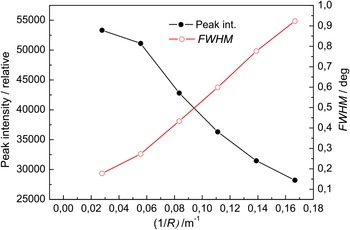
(Color online) Dependences of the peak intensity and FWHM (see the rocking curves in figure 3) on the curvature of the second crystal Si(111) of the DC (1, − 1) setting.

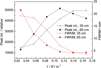
Furthermore, the parameters of the diffraction profiles of the beam after the second reflection and registered by an SC at the distance of 35 and 65 cm from the sandwich were measured. Figure 5 shows the obtained results. It can be seen from Figure 5 that FWHM of the beam profile decreases and the peak intensity increases (up to saturation) with the increase of the curvature. It is mainly brought about by increasing the effective mosaicity of the second crystal. However, when comparing the FWHM dependences corresponding to the distances of 35 and 65 cm, it can be seen that there is only small focusing effect slightly dependent on the curvature.
(Color online) Parameters of the diffraction profiles (FWHM and the peak intensity) as registered at the peak positions of the rocking curves (see Figure 3) by an SC at the distances of 35 and 65 cm from the sandwich.

In order to see the resolution properties of the double-reflected beam, as a next step we carried out diffraction on standard polycrystalline sample [α-Fe(211) pin of the diameter of 2 mm] put to the monochromatic beam at the distance of 50 cm from the second Si(111) slab. Figure 6 shows an example of the diffraction profile taken by IP for the bending radius of the Si(111) crystal slab of 6 m (1/R = 0.167 m−1). Figure 7 shows FWHM of the corresponding diffraction profiles for three curvatures of the Si(111) crystal. The best FWHM = 0.0116 rad was achieved for minimum curvature of the Si(111) slab of 0.056 m−1. In this case, the diameter of the sample introduces to the resolution uncertainty only a small amount. As the FWHM represents Δ(2θ) uncertainty of the scattering angle, the derived value of Δd/d ≈ 0.006 (d is the lattice constant) in our case shows, that this double-crystal monochromator performance can be successfully used in a powder diffractometry, at least for residual strain/stress measurements. For macro-strain/stress measurements, one can work even with a released resolution up to a factor two. However, it is recommended to do the resolution release by the employment of a thicker (e.g. 4 mm) Si(111) slab which can correspondingly increase the luminosity of the instrument. A simple release of the resolution by changing the curvature of the crystal has only an effect on the resolution but no positive effect on luminosity (see Figures 4 and 5). Resolution properties of the monochromatic beam are also documented in the following Figures 8 and 9, where edge refraction effects obtained from several samples are shown. The samples were installed behind the second crystal and the IP was put at 50 cm from the samples. The inspection of Figure 9 reveals that in the case of imaging on the group of office staples the refraction maxima and minima from individual staples (of the group) partly overlap and therefore, the refraction contrast is much smaller in comparison with the individual office staple, or at the end of the group.
Diffraction profile from α-Fe(211) pin of the diameter of ϕ = 2 mm taken by IP at the distance 40 cm from the pin.

(Color online) FWHM of the diffraction profiles imaged by IP from the α-Fe(211) pin for three curvatures of the Si(111) slab in the sandwich.

Refraction effects on 2 mm Fe and 17 mm brass edges.

(Color online) Refraction effects on mutually separated one office staple, hypodermic needle, and a group of office staples.

B. (n, − m) diffraction performance with the active Si(220) slab from the sandwich
By a simple changing of the Bragg angle on the sandwich the bent Si(220) slab can be brought to be operative. Even though that in this case the Si(220) slab from the sandwich is active, there are no second-order neutrons because thanks to the first bent Si(111) crystal, the related Si(222) reflection is forbidden. Figure 10 shows an example of the related DC rocking curve and Figure 11 (similar to Figure 5) shows the dependences of the parameters of the double-diffracted beam profiles (the FWHM and the peak intensities) behind the sandwich taken by SC at two different distances. Similar to Figure 5, it can be seen that the focusing effect is evident. Then, at the distance of 50 cm from the sandwich a polycrystalline sample (2 mm α-Fe pin) was situated into the beam and the profiles of the diffracted beam were studied. Figure 12 shows examples of the diffraction profiles taken for the radius of curvature of the Si(220) slab of R = 9 m and Figure 13 roughly summarizes the FWHM dependence of the diffraction profiles on the Si(220) curvature. Scattering angle for the sample α-Fe(211) reflection was 2θ S = 88°. If we take into account the diameter of the α-Fe pin, 50 µm pixel dimension of IP and the distance of IP from the pin, we arrive for the used curvatures at very low divergence Δ(2θ S) of about 1.5 × 10⁻³ rad, which documents a high-resolution property of the DC performance.
Rocking curve of the Si(220) bent crystal (R 2 = 12 m, t = 1.3 mm) with respect to the Si(111) premonochromator (R 1 = 12 m, t = 4 mm) in the parallel (n, − m) setting.

(Color online) Parameters of the diffraction profiles (FWHM and the peak intensity) as registered at the peak positions of the rocking curves by an SC at the distances of 10 and 45 cm as a function of the curvature for the active Si(220) slab in the sandwich.

Diffraction profile related to α-Fe(211) reflection obtained from the pin of the diameter of ϕ = 2 mm and imaged by IP. The distance of the IP from the pin was 40 cm.

(Color online) FWHM of the diffraction profiles imaged by IP situated at the distance of 40 cm from the 2 mm α-Fe pin for three curvatures of the Si(220) slab.

IV. CONCLUSION
Some properties of neutron diffractometer with the DC monochromator system with the sandwich consisting of two different Si-crystals on the second axis have been introduced. Inspection and comparison of Figures 7 and 13 reveal that the DC performance with the Si(220) crystal slab provides much better angular resolution than the one with the Si(111) slab, however, delivers correspondingly less monochromatic beam current on the polycrystalline sample. In both alternatives, the DC setting provides a focused neutron beam of a sufficient current at the sample position that can be used in powder diffractometry, of course, at a reasonably high-flux neutron source. For the sake of comparison with the profiles shown in Figures 6 and 12, Figure 14 shows the diffraction profile from α-Fe(211) pin installed about 20 cm before the sandwich, i.e. in the monochromatized beam coming from the 4 mm thick Si(111) premonochromator. The influence of the quasi-dispersivity of the presented double-crystal monochromator design on the intensity related to the diffraction profiles as well as on its FWHM is evident. This newly designed performance can be at least successfully exploited in diffractometers dedicated to residual strain/stress measurements when permitting strain/stress scanning (e.g. in the vicinity of welds) with good luminosity and worse resolution performance or microstrain studies (e.g. microstrain behavior in samples under the external load) on the basis of the peak profile analysis with the other high-resolution performance. For small curvatures of the crystal slabs in the sandwich the DC system can be also used for edge refraction studies when providing a highly monochromatic and horizontally highly angularly correlated beam with a good spatial coherence. In principle, one can use more than two (e.g. three or four) different slabs in the sandwich. It should be pointed out that the monochromatic beam properties after the DC diffraction depend on several parameters, namely the distance between the premonochromator and the sandwich, chosen neutron wavelength, the thickness of the slabs in the sandwich and their curvatures. Therefore, for the design of individual DC diffractometer configuration and its full optimization, Monte Carlo simulations are desirable (Šaroun and Kulda, Reference Šaroun and Kulda2009). In the present case, the instrument could not be fully optimized because of using a fixed curvature of the first Si(111) crystal slab. Furthermore, because of the given experimental conditions, the distance between the bent crystals was also fixed and rather large (1.7 m). When all these parameters in connection with a possibility of an employment of vertical focusing are optimized, the luminosity of the presented experimental setup could be by one order of magnitude higher. One should keep in mind that in this case one works with open beams without any Soller collimators and the DC setting behaves as a self-collimation focusing assembly producing also a highly monochromatized beam. Therefore, a high resolution is thus expected in both Δ(2θ) (for an optimum curvature of the crystal slab in the sandwich) as well as Δλ/λ. Even at our medium-power neutron source when using a high-resolution position sensitive detector instead of IP (having rather low efficiency), the time expected for the collection of the data related to an individual diffraction profile is expected to be <20 min.
Diffraction profile from the α-Fe(211) pin of ϕ = 2 mm situated 20 cm before the sandwich and imaged by IP at the distance of 40 cm.

ACKNOWLEDGEMENTS
The related measurements were carried out at the CANAM infrastructure of the NPI ASCR Rez. Bragg diffraction optics investigations are in the Czech Republic supported by GACR project no. 14-36566G, by the ESS project LM2010011: “Contribution to Partnership in Large Research Infrastructure of Pan-European Importance” as well as by the project of EU-FP7-NMI3 II: “Integrated Infrastructure Initiative for Neutron Scattering and Muon Spectroscopy”, 2009–2015.