Introduction
Rhinoplasty is a facial plastic surgical procedure indicated for improvement of nasal aesthetics and airway function. Lateral osteotomy is a fundamental step in rhinoplasty operations, and is responsible for a significant number of intra- and post-operative complications, including bleeding, eyelid oedema and peri-orbital ecchymosis.Reference Erişir and Tahamiler1 Trauma to the angular artery during osteotomy and inadequate local haemostasis may lead to excessive intra-operative bleeding, which can increase surgical time, the risk of morbidities and post-operative recovery time.Reference Cochran and Landecker2–Reference Hwang, Lee, Kim, Kim and Kang4
Unfortunately, there are insufficient data regarding the role of angular artery damage on ecchymosis. In order to investigate the role of angular artery damage, a study was performed on fresh frozen cadavers.
Materials and methods
This investigation was carried out in the anatomy laboratories at the TOBB University of Economics and Technology Faculty of Medicine in Ankara, Turkey. Twenty fresh frozen cadaver heads (40 hemifaces) were studied. The ‘high-low-high’ lateral osteotomy was performed by the course participants, who were moderately experienced in rhinoplasty (as the course was on advanced rhinoplasty).
A 4 mm guarded osteotome was used for the lateral osteotomies. In all cadavers, lateral osteotomies were carried out bilaterally, after the creation of a subperiosteal tunnel. Following completion of the lateral osteotomy, a skin incision was made in the midline dorsum. The dermis and subcutaneous tissues were carefully dissected, taking care not to damage the angular artery. Overlying tissues were cut and retracted to show the course of the angular artery. The lateral osteotomy line and the angular artery were dissected, and any damage to the artery was noted. All evaluations were made by the authors.
All cadavers were provided by an American Association of Tissue Banks registered body donor organisation; namely, Science Care, Phoenix, Arizona, USA.
Two of the cadaver heads were injected with red polymer (Biodur® S14) into the common carotid artery for better arterial identification. Both common carotid arteries were dissected and irrigated with saline solution until the fluid became clear. Then the arteries were injected with S14, similar to the method described by Yu and colleagues.Reference Yu, Weng, Wang, Mu and Li5
Results
The angular artery was not damaged in any of the cadavers. The angular artery was always lateral to the line of lateral osteotomy.
Discussion
During rhinoplasty, osteotomies are responsible for a significant amount of intra- and post-operative complications, including bleeding, eyelid oedema and peri-orbital ecchymosis.Reference Erişir and Tahamiler1 There are different and contradicting reports on the incidence of peri-orbital oedema and ecchymosis after rhinoplasty surgery. Oedema and ecchymosis after rhinoplasty may occur in different degrees according to some avoidable or preventable, and unavoidable factors, such as: coagulation disorders, excess subcutaneous fat tissue, the surgical procedure performed, periosteal elevation, the type of osteotomy, the operation duration, location differences of the angular artery and drug use prior to surgery.Reference Kılıc, Tuncel, Comert and Sencan6–Reference Saedi, Sadeghi and Fekri8
Trauma to the angular artery during osteotomy, and inadequate local haemostasis, may lead to excessive intra-operative bleeding, which can increase surgical time, the risk of morbidities and post-operative recovery time.Reference Cochran and Landecker2–Reference Hwang, Lee, Kim, Kim and Kang4 The purpose of periosteal elevation is to decrease damage to the periosteum and displace the angular artery from the osteotomy site, providing protection to the angular artery in order to prevent injury and subsequent bleeding.Reference Ong, Farhood, Kyle and Patel9–Reference Berinstein, Bane, Cupp, DeMarco and Hunsaker11
El-Sisi et al.Reference El-Sisi, Abdelwahab and Most12 have claimed that periosteal elevation does not protect the angular artery, and that a sweeping injury could affect the surrounding tissue, and may cause more oedema and ecchymosis. Kara et al.Reference Kara, Kara and Yaylali13 and Al Afraj et al.Reference Al-Arfaj, Al-Qattan, Al-Harethy and Al-Zahrani14 have stated that the creation of subperiosteal tunnels carries a greater risk of damaging the overlying vessels and causes additional soft tissue trauma.Reference Saedi, Sadeghi and Fekri8
• Lateral osteotomy is a fundamental step in rhinoplasty operations, and is responsible for numerous intra- and post-operative complications
• Angular artery trauma during osteotomy is generally considered a contributor to post-operative eyelid oedema and ecchymosis
• This study was carried out on fresh frozen cadaver heads (40 hemifaces)
• The angular artery was not damaged in any of the cadavers
• The angular artery was always lateral to the line of lateral osteotomy
• Angular artery trauma cannot be considered a major contributory cause of post-operative eyelid oedema or peri-orbital ecchymosis
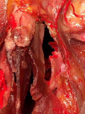
It has also been argued that angular artery trauma is one of the main causes of peri-orbital oedema and ecchymosis. However, this argument lacks evidence and there are no supporting data. As can be seen in Figure 1, it is very difficult to damage the angular artery; the lateral osteotomy cut is more medial to the angular artery, or the angular artery is more lateral to the osteotomy site.
Angular artery in a fresh frozen cadaver injected with red polymer (Biodur S14); the location of the angular artery and the lateral osteotomy site are seen.

Conclusion
This study found that the ‘high-low-high’ lateral osteotomy does not damage or traumatise the angular artery. The ecchymosis and oedema are related to other factors.
Acknowledgements
The authors are grateful to all the generous people who donated their bodies for medical education and research, and to Prof Selcuk Tunalı from the Anatomy Department of TOBB Ankara University for the cadaveric injections and preparations. This study was performed on cadavers provided (by an American Association of Tissue Banks registered body donor organisation – Science Care, Phoenix, Arizona, USA) for a cadaver course on endoscopic sinus surgery. The authors performed the study after the course; the cadavers were allowed to be used for free after the course by the Turkish Education and Health Foundation.
Competing interests
None declared