Introduction
Several longitudinal magnetic resonance imaging (MRI) studies of first-episode psychosis (FEP) have investigated the possibility of a progressive deterioration in brain morphology in schizophrenia during early stages of illness (Pantelis et al. Reference Pantelis, Yucel, Wood, Velakoulis, Sun, Berger, Stuart, Yung, Phillips and McGorry2005; DeLisi, Reference DeLisi2008). Grey matter (GM) volume loss or ventricular volume increase over time have been reported in some (Cahn et al. Reference Cahn, Hulshoff Pol, Lems, van Haren, Schnack, van Der Linden, Schothorst, van Engeland and Kahn2002; Kasai et al. Reference Kasai, Shenton, Salisbury, Hirayasu, Lee, Ciszewski, Yurgelun-Todd, Kikinis, Jolesz and McCarley2003a, Reference Kasai, Shenton, Salisbury, Hirayasu, Onitsuka, Spencer, Yurgelun-Todd, Kikinis, Jolesz and McCarleyb; Nakamura et al. Reference Nakamura, Salisbury, Hirayasu, Bouix, Pohl, Yoshida, Koo, Shenton and McCarley2007; Théberge et al. Reference Théberge, Williamson, Ayoama, Drost, Manchanda, Malla, Northcott, Menon, Neufeld, Rajakumar, Pavlosky, Densmore, Schaefer and Williamson2007; Koo et al. Reference Koo, Levitt, Salisbury, Nakamura, Shenton and McCarley2008; van Haren et al. Reference van Haren, Hulshoff Pol, Schnack, Cahn, Brans, Carati, Rais and Kahn2008; Sun et al. Reference Sun, Stuart, Jenkinson, Wood, McGorry, Velakoulis, van Erp, Thompson, Toga, Smith, Cannon and Pantelis2009; Mané et al. Reference Mané, Falcon, Mateos, Fernandez-Egea, Horga, Lomeña, Bargalló, Prats-Galino, Bernardo and Parellada2009) but not all such MRI investigations (Keshavan et al. Reference Keshavan, Haas, Kahn, Aguilar, Dick, Schooler, Sweeney and Pettegrew1998; Ho et al. Reference Ho, Andreasen, Nopoulos, Arndt, Magnotta and Flaum2001; Lieberman et al. Reference Lieberman, Chakos, Wu, Alvir, Hoffman, Robinson and Bilder2001; Puri et al. Reference Puri, Hutton, Saeed, Oatridge, Hajnal, Duncan, Chapman, Barnes, Bydder and Joyce2001; Wood et al. Reference Wood, Velakoulis, Smith, Bond, Stuart, McGorry, Brewer, Bridle, Eritaia, Desmond, Singh, Copolov and Pantelis2001; DeLisi et al. Reference DeLisi, Sakuma, Maurizio, Relja and Hoff2004; Zipursky et al. Reference Zipursky, Christensen and Mikulis2004; DeLisi & Hoff, Reference DeLisi and Hoff2005; Whitworth et al. Reference Whitworth, Kemmler, Honeder, Kremser, Felber, Hausmann, Walch, Wanko, Weiss, Stuppaeck and Fleischhacker2005; Price et al. Reference Price, Cercignani, Bagary, Barnes, Barker, Joyce and Ron2006).
Up until now, no longitudinal MRI studies of FEP have used population-based designs, which enable the recruitment of cases of incident psychosis from well-defined catchment areas, and healthy controls from exactly the same neighbourhood. Such epidemiological designs minimize limitations of sample size, allow the inclusion of patients from the entire spectrum of psychosis severity, and increase confidence that the samples of patients and healthy controls are representative of the population from the environmental setting whence they are recruited (Tanskanen et al. Reference Tanskanen, Ridler, Murray, Haapea, Veijola, Jääskeläinen, Miettunen, Jones, Bullmore and Isohanni2008).
We previously reported results of a population-based, voxel-based morphometry (VBM) study, which compared a relatively large sample of FEP cases with a group of asymptomatic controls recruited exactly in the same area. Significant GM volume reductions in FEP subjects relative to controls emerged in regions previously highlighted as critical to the pathophysiology of schizophrenia, namely the prefrontal cortex, superior temporal gyrus (STG), insula and hippocampal area (Schaufelberger et al. Reference Schaufelberger, Duran, Lappin, Scazufca, Amaro, Leite, de Castro, Murray, McGuire, Menezes and Busatto2007). In the present report, we describe the results of a longitudinal study in which repeated morphometric MRI data were acquired after a median period of 15 months in a subsample of the above group of FEP cases and controls.
In a hypothesis-driven fashion, we predicted that changes in ventricle size and GM volume indicating a progressive course would be present in the FEP patients compared with controls, involving most prominently the frontal and temporal regions implicated both in the previous longitudinal MRI studies of FEP (DeLisi, Reference DeLisi2008) and in our own case-control investigation of GM abnormalities in FEP at baseline (Schaufelberger et al. Reference Schaufelberger, Duran, Lappin, Scazufca, Amaro, Leite, de Castro, Murray, McGuire, Menezes and Busatto2007). Given the inclusion of cases from the entire spectrum of psychosis severity in our study, we also aimed to test the hypothesis that morphological changes in the above brain structures indicating a progressive course would be present in subjects with non-remitting illness compared with remitting cases of schizophrenia/schizophreniform disorder.
Method
Subjects and clinical assessments
The sample originated from a population-based morphometric MRI study of FEP carried out in a circumscribed geographical area of São Paulo, Brazil (Sao Paulo Study; Menezes et al. Reference Menezes, Scazufca, Busatto, Coutinho, McGuire and Murray2007). In this baseline study, MRI data were acquired in a sample of 122 FEP subjects, including 62 patients with schizophrenia or schizophreniform disorder (FESZ group), identified by active surveillance of cases that made contact for the first time with local health services for psychotic symptoms, and were compared with images of 94 age- and gender-matched non-psychotic controls recruited from exactly the same geographical areas (next-door neighbours) (Schaufelberger et al. Reference Schaufelberger, Duran, Lappin, Scazufca, Amaro, Leite, de Castro, Murray, McGuire, Menezes and Busatto2007).
The present longitudinal investigation was set up within 6 months after completion of the above baseline MRI study, and was conducted over a period of approximately 2 years, during which all subjects above were invited for a second MRI scanning session. Both the baseline and follow-up MRI investigations were approved by the ethics committee and informed consent forms were signed by all subjects before image acquisitions.
From the initial sample of 122 patients and 94 controls who participated in the baseline MRI study, 80 patients and 52 controls were rescanned. This current longitudinal study reports the data from the subgroup of 39 patients with FESZ and the controls.
All subjects were interviewed by means of the Structured Clinical Interview for Diagnostic and Statistical Manual for Mental Disorders (DSM-IV; First et al. Reference First, Spitzer, Gibbon and Williams1995) both at the outset of the epidemiological investigation and at the follow-up evaluation. Inclusion criteria for patients were first contact with mental health services for a psychotic episode and a confirmed diagnosis of psychotic disorders according to DSM-IV criteria (APA, 1994). Exclusion criteria for both groups were: history of head injury, neurological disorders or organic disorders that could affect the central nervous system and contraindications for MRI scanning.
Cases' clinical status was assessed at both time points by the Positive and Negative Syndrome Scale (PANSS; Kay et al. Reference Kay, Fiszbein and Opler1987). Patients with FESZ (n=39) were categorized in terms of their clinical outcome. This was defined by a remitting/non-remitting course during follow-up, according to the DSM-IV course specifier assessed after at least 1 year from the baseline assessment (remitting course meaning a single episode in full remission and absence of clinically important symptoms and a non-remitting course meaning continuous, episodic or residual symptoms).
MRI acquisition
Subjects received MRI scans at baseline and after a mean interval of 16 (s.d=6, range 8–40) months. Imaging data were acquired using either of two MRI scanners at the Hospital das Clínicas of the University of São Paulo, both from the same manufacturer (1.5-T GE Signa, General Electric, USA). Exactly the same acquisition protocols were used for all scans [a T1 spoiled gradient echo (T1-SPGR) sequence providing 124 contiguous slices, voxel size=0.86×0.86×1.5 mm, echo time=5.2 ms, repetition time=21.7 ms, flip angle=20°, field of view=22 cm, matrix size=256×192 mm].
VBM processing
The processing of MRI datasets for VBM was performed using the Statistical Parametric Mapping package (SPM2; Wellcome Department of Imaging Neuroscience, UK), executed in Matlab (Mathworks, USA). A standard template set was created specifically for our longitudinal study, comprising the baseline and follow-up MRI datasets of each subject; subsequently, the original images were processed according to the SPM2 optimized protocol (Good et al. Reference Good, Johnsrude, Ashburner, Henson, Friston and Frackowiak2001) following the same procedures described for the baseline MRI study (Schaufelberger et al. Reference Schaufelberger, Duran, Lappin, Scazufca, Amaro, Leite, de Castro, Murray, McGuire, Menezes and Busatto2007) and made ready for statistical analyses (see below).
Ventricle:brain ratio (VBR) measurements
Lateral ventricles (LVs) were assessed using a manual region of interest (ROI) method applied with the Mricro 1.40 software (http://www.sph.sc.edu/comd/rorden/mricro.html). The anterior and posterior horns and the body of the ventricle were combined as one single ROI drawn sequentially on coronal slices along the entire extension of the LV. Measurements were taken separately for each hemisphere, starting on the slice in which the anterior commissure could be best visualized and continuing anteriorly and then back posteriorly. In order to minimize the number of slices that had to be measured, a reliability analysis was conducted on a subsample of 10 healthy controls.
Intraclass correlation coefficients (ICCs) were derived to compare the strategies of assessing both LVs (excluding the temporal horns) in all contiguous slices against one in 10 slices. The total number of voxels was estimated by multiplying the number of measured voxels in the ventricle ROIs by the frequency of the slices that were assessed (one in 10) and ICC values were 0.98 [F(19)=0.01] bilaterally. Based on that, the one in each 10 slices frequency was selected for use in the overall study. The assessment of the temporal horns was performed separately: these were measured in all slices in which they could be visualized, and the sum of all the drawn voxels was used as the final measure of temporal horn volume. Only one rater (P.G.P.R.) was responsible for all measurements, which were performed blindly to all subjects' demographic characteristics and diagnoses. Inter-rater reliability with one other experienced researcher (F.L.S.D.) was higher than 0.9 for all LV indices.
In order to allow the calculation of VBRs, a measure of total brain volume in each subject was obtained using the brain extraction tool (BET) function of the Mricro software, which excludes all extra-brain tissue before summing the remaining voxels including the cerebrum, cerebellum and brainstem. The ratio between the total ventricle volume (number of voxels) and the total brain volume is a measure of the proportion of the brain volume that is occupied by the ventricular ROI. This procedure resulted in four indices to be included in the analyses: left and right LV VBRs (VBR-LV), and left and right temporal horn VBRs (VBR-TH).
Statistical analyses
Sociodemographic/clinical data and VBR measures were analysed using SPSS version 14 (SPSS Inc., USA) and statistical significance was set at p<0.05 (two-tailed). For the assessment of between-group differences in GM and VBR change from the baseline to the follow-up MRI assessment, repeated-measures analyses of variance (ANOVA) were conducted.
Firstly, we compared the FESZ group versus the controls. Subsequently, in the group of patients, we investigated longitudinal MRI differences between: those who were on treatment and those who had not been exposed to treatment (or had stopped treatment) during the follow-up period; and those who exhibited a remitting versus a non-remitting illness course. In the VBR analysis, VBR-LV or VBR-TH was included as a dependent variable, time and hemisphere as within-subjects factors, and group as a between-subjects factor.
For the VBM analyses, we investigated whether there were longitudinal GM changes between groups by performing repeated-measures analyses of covariance (ANCOVA) of regional GM differences using the general linear model, with group and time as factors. Only voxels with values above an absolute threshold of 0.05 entered the analyses. A measure of the total amount of GM was entered as a covariate, given by the sum of voxels within the corresponding GM compartment of each subject. In all analyses, resulting statistics at each voxel were transformed to Z scores, and between-group differences were displayed as statistical parametric maps (SPMs) of the group×time interactions into standard anatomical space, thresholded at the one-tailed p<0.001 level of significance (corresponding to a Z>3.09 threshold).
In order to investigate whether there were significant findings in areas where GM abnormalities had been predicted a priori, we used the small volume correction (SVC) tool in SPM2, with the purpose of restricting the comparisons to specific voxels located in areas where abnormalities (reduced GM in patients compared with controls) had been found at the previously reported baseline MRI investigation, including the bilateral prefrontal and superior temporal cortices, hippocampus and insula (Schaufelberger et al. Reference Schaufelberger, Duran, Lappin, Scazufca, Amaro, Leite, de Castro, Murray, McGuire, Menezes and Busatto2007). Any clusters of voxels showing significant findings within each of those volumes of interest were reported only if they survived family-wise error (FWE) correction for multiple comparisons (p<0.05) over that region. Finally, the SPM maps were inspected again on an exploratory basis, in order to identify significant findings in unpredicted regions across the entire brain. Such unpredicted findings were reported as statistically significant only if surviving FWE correction for multiple comparisons (p<0.05) over the whole brain.
In the VBR and in the VBM analyses, gender was included as a covariate when groups showed statistically significant difference for this variable.
Inter-scanner reliability
Because images were acquired using two MRI scanners, we conducted an investigation of the reliability of brain volumes measured using the two pieces of equipment, as described previously (Schaufelberger et al. Reference Schaufelberger, Duran, Lappin, Scazufca, Amaro, Leite, de Castro, Murray, McGuire, Menezes and Busatto2007). Briefly, six healthy volunteers were (re)scanned on the same day on the two machines, and we obtained ICC values higher than 0.90 for the neocortical and medial temporal regions. The ICC values for the ROI-based measurements of the ventricular region were: 0.99 and 0.99 for the right and left LVs; 0.96 and 0.99 for the right and left temporal horns; and 0.90 for total brain volume.
Although we had obtained high ICC values in the inter-scanner reliability assessment, all statistical analyses were repeated including scanner as a confounding covariate. Also, given the wide variability in inter-scanning intervals, each VBR analysis was repeated including the time interval between scans as a confounding covariate.
Results
Sociodemographic/clinical characteristics
There were no significant differences between subjects who were included in the longitudinal study and those who were not in terms of their sociodemographic and clinical characteristics at the outset of the study.
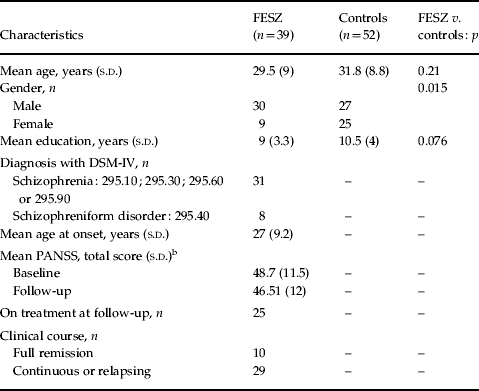
In the final sample of the longitudinal study, there were no differences between patients (n=39) and controls (n=52) regarding their mean interscanning interval [patients: 16.6 (s.d.=6.7) months, controls: 15.6 (s.d.=5.3) months, t=0.85, p=0.4] or number of participants examined in each scanner (χ2=4.6, p=0.09). Sociodemographic and clinical characteristics of the sample are shown in Table 1.
Sociodemographic and clinical characteristics of subjects who completed the follow-up magnetic resonance imaging studyFootnote a

FESZ, First-episode schizophrenia or schizophreniform disorder; s.d., standard deviation; DSM-IV, Diagnostic and Statistical Manual of Mental Disorders; PANSS, Positive and Negative Syndrome Scale.
a The analysis of continuous variables was carried out using the t test for independent samples, paired t test and analysis of variance; the analysis of categorical variables was carried out with the χ2 test.
b Paired t test for within-group analysis of PANSS for patients with FESZ: p=0.38.
The remitted FESZ subsample (comprising two patients with a diagnosis of schizophrenia but who were fully remitted plus eight patients who retained a diagnosis of schizophreniform disorder at follow-up) was not significantly different from the non-remitted subgroup (n=29) in terms of sociodemographic and clinical variables, with the exception of lower total PANSS scores at follow-up [35.7 (s.d.= 4.3) versus 50.2 (s.d.=11.7), respectively, t=5.6, p<0.001].
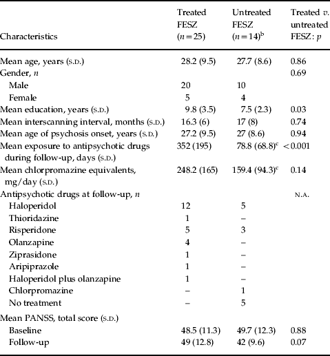
In the FESZ subgroup, 25 patients were on antipsychotic treatment at follow-up, while 14 had not been exposed to treatment or had stopped their medication during the follow-up interval (as shown in Table 2).
FESZ according to antipsychotic drug exposure during follow-upFootnote a

FESZ, First-episode schizophrenia or schizophrenia disorder; s.d., standard deviation; n.a., not applicable; PANSS, Positive and Negative Syndrome Scale.
a Continuous variables were analysed with the t test for independent samples and categorical variables were analysed with the χ2 test or Fisher's exact test.
b The untreated group comprised five patients with no exposure to antipsychotic drugs during follow-up and nine patients who interrupted the treatment during follow-up.
c Excluding five patients who had not been exposed to any treatment during follow-up.
Between-group VBM comparisons
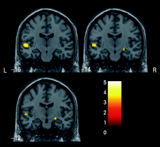
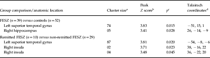
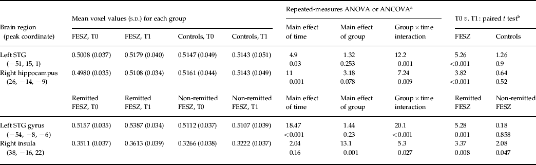
Significant ANCOVA group×time interactions (with gender as covariate) were found when FESZ subjects (n=39) were compared with controls (n=52) in the left STG and right hippocampus (Table 3; Fig. 1). The distribution of GM peak values (voxel values extracted from the coordinate of maximal statistical significance, corrected for the total GM in the brain) of each subject in these clusters revealed that the significant interaction was due to a GM increase in FESZ patients, with no change in the controls in these brain regions (Table 4).
Clusters of significant longitudinal differences in grey matter volumes between patients with first-episode schizophrenia/schizophreniform disorder (n=39) and healthy controls (n=52), obtained from voxel-based morphometry analyses (repeated-measures analysis of covariance, group×time interaction, p<0.05, family-wise error-corrected for multiple comparisons). The left superior temporal gyrus and the right hippocampus are highlighted in yellow, overlaid on coronal slices spatially normalized into an approximation to the Talairach & Tournoux (Reference Talairach and Tournoux1988) stereotactic atlas. L, Left; R, right.

Voxel-based morphometry: longitudinal between-group analyses

FESZ, First-episode schizophrenia or schizophreniform disorder.
a Total number of contiguous voxels in each region above an initial cut-off of Z>3.09.
b Z scores for the voxels of maximal statistical significance.
c Repeated-measures analysis of covariance, group×time interaction. Statistical significance after correction for multiple comparisons (voxel level).
d Talairach & Tournoux (Reference Talairach and Tournoux1988) coordinates of the voxel of maximal significance within each region.
Grey matter clusters of significant between-group differences: mean voxel values

s.d., Standard deviation; ANOVA, analysis of variance; ANCOVA, analysis of covariance; T0, baseline; T1, follow-up; FESZ, first-episode schizophrenia or schizophreniform disorder; STG, superior temporal gyrus.
a F values and p values.
b t values and p values.
Between-group comparisons of VBR measurements
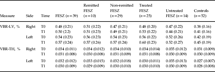
VBR values for each group are shown in Table 5.
Patients with first-episode psychosis and controls: VBRsFootnote a

Data are given as mean (standard deviation).
VBR, Ventricle:brain ratio; FESZ, first-episode schizophrenia or schizophreniform disorder; LV, lateral ventricle (encompassing the anterior, body and posterior portions of the lateral ventricles); T0, baseline; T1, follow-up; TH, temporal horns.
a VBR values are percentage of the region of interest in the brain volume.
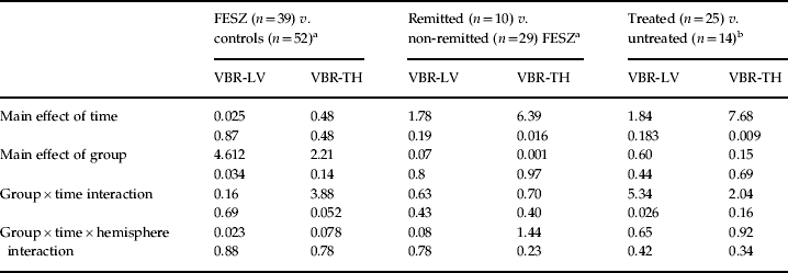
For FESZ subjects versus controls, a significant main effect of group indicated larger ventricle size in FESZ patients relative to controls in respect of the VBR-LV (F=4.61, p=0.034), but there was no statistical significance regarding the main effect of time or group×time interaction (Table 6).
Longitudinal analyses of VBRs in first-episode psychosis patients and controls

VBR, Ventricle:brain ratio, expressed as a percentage of the brain volume that is occupied by the region of interest; FESZ, patients with first-episode schizophrenia or schizophreniform psychosis; VBR-LV, the region of interest includes the anterior, body and posterior portion of the lateral ventricle; VBR-TH, the region of interest includes the temporal horns of the lateral ventricle.
a Repeated-measures analysis of covariance with time and hemisphere as within-subject factors, group as between-subject factor and gender as covariate. Analysis repeated with interscanning interval or scanner did not alter the results. F and p values.
b Repeated-measures analysis of variance with time and hemisphere as within-subject factors and group as between-subject factor. Analyses repeated with interscanning interval or scanner did not alter the results. F and p values.
However, the VBR comparison of the temporal horns (VBR-TH) between FESZ subjects and controls showed a trend towards significance for the diagnosis×time interaction (F=3.8, p=0.052, with gender included as a covariate) (Table 6). Paired t tests showed a significant reduction in VBR-TH over time in patients (significant at right, t=2.7, p=0.01; trend level at left, t=1.9, p=0.06), but not in controls.
Effects of clinical course in the FESZ subgroup
The VBM comparison between remitted and non-remitted FESZ subjects revealed significant ANCOVA group×time interactions in the left STG and right insula (Table 3). Inspection of GM peak values in these clusters revealed that GM increased over time in the remitted FESZ subjects (Table 4). There were no significant interactions between non-remitted FESZ subjects and controls.
In the VBR comparisons between remitted and non-remitted FESZ subjects, there was a significant main effect of time for the VBR-TH (baseline>follow-up), but there were no significant group×time interactions regarding either the VBR-LV or VBR-TH indices (Table 6). Also, the non-remitted FESZ subgroup showed no significant progression of ventricle size compared with controls.
Effects of treatment in the FESZ subgroup
The VBM comparison between treated and untreated FESZ subjects revealed no significant ANCOVA group×time interactions. Separate comparisons of each of these subgroups against controls showed no significant differences in GM change.
However, in the VBR comparisons between treated and untreated FESZ patients, there was a significant group×time interaction for the VBR-LV index (F=5.34, p=0.026) (Table 6). This was due to trends in opposite directions in the treated and untreated FESZ subgroups: VBR values increased over time in medicated subjects (paired t test; right, t=2.4, p=0.020; left, t=2.84, p=0.009), while there was a non-significant VBR decrease in unmedicated subjects. In regard to VBR-TH, there was a main effect of time (baseline >follow-up). Separate comparisons of treated and untreated FESZ patients against controls showed no significant group×time interactions in VBR.
Inclusion of interscanning interval and scanner as confounding covariates in each analysis did not alter the VBM or VBR results.
Discussion
To the best of our knowledge, this is the first longitudinal morphometric MRI evaluation of a group of first-episode schizophrenia spectrum disorder (comprising schizophrenia and schizophreniform disorder patients, FESZ) subjects drawn from a population-based sample of incident cases of psychosis recruited consecutively from a circumscribed geographical region, using epidemiological methods. Our strategy of recruiting healthy controls from the same neighbourhood was not employed in any previous longitudinal MRI studies of psychosis. While our baseline assessment had revealed regional GM decrements in fronto-temporal and hippocampal areas in FESZ subjects relative to controls (Schaufelberger et al. Reference Schaufelberger, Duran, Lappin, Scazufca, Amaro, Leite, de Castro, Murray, McGuire, Menezes and Busatto2007), the present follow-up MRI evaluation found no evidence of accelerated volumetric GM reductions in patients compared with controls. These findings indicate that, in psychosis, brain volume abnormalities are already present at the onset of illness but are not necessarily progressive.
Contrary to our a priori hypothesis, FESZ subjects exhibited GM volume changes, indicating a reversible course in the left STG and right hippocampus, regions that we previously found to show volumetric reductions in the FESZ group compared with controls at the baseline investigation (Schaufelberger et al. Reference Schaufelberger, Duran, Lappin, Scazufca, Amaro, Leite, de Castro, Murray, McGuire, Menezes and Busatto2007). Consistent with such longitudinal VBM results is the lack of increment in lateral ventricular size in patients relative to controls over time. Indeed, we found a reduction in lateral temporal horn volumes over time in FESZ patients, but not in controls, resulting in a group×time interaction at trend level. Such results are not in agreement with a number of previous longitudinal morphometric MRI investigations of FESZ which have reported progression of brain abnormalities in schizophrenia (Cahn et al. Reference Cahn, Hulshoff Pol, Lems, van Haren, Schnack, van Der Linden, Schothorst, van Engeland and Kahn2002; Kasai et al. Reference Kasai, Shenton, Salisbury, Hirayasu, Lee, Ciszewski, Yurgelun-Todd, Kikinis, Jolesz and McCarley2003a; Nakamura et al. Reference Nakamura, Salisbury, Hirayasu, Bouix, Pohl, Yoshida, Koo, Shenton and McCarley2007; Théberge et al. Reference Théberge, Williamson, Ayoama, Drost, Manchanda, Malla, Northcott, Menon, Neufeld, Rajakumar, Pavlosky, Densmore, Schaefer and Williamson2007; Mané et al. Reference Mané, Falcon, Mateos, Fernandez-Egea, Horga, Lomeña, Bargalló, Prats-Galino, Bernardo and Parellada2009), but are in accordance with other studies which failed to find this progression or even showed reversal of STG deficits (Keshavan et al. Reference Keshavan, Haas, Kahn, Aguilar, Dick, Schooler, Sweeney and Pettegrew1998) or lesser reduction of temporal lobe GM volume in patients than in controls (Gur et al. Reference Gur, Cowell, Turetsky, Gallacher, Cannon, Bilker and Gur1998).
One potential explanation for our findings is that any progressive GM changes associated with schizophrenia might have already occurred maximally by the time of the baseline MRI scan, for instance in prodromal phases of the disorder (Pantelis et al. Reference Pantelis, Yucel, Wood, Velakoulis, Sun, Berger, Stuart, Yung, Phillips and McGorry2005; Lappin et al. Reference Lappin, Dazzan, Morgan, Morgan, Chitnis, Suckling, Fearon, Jones, Leff, Murray and McGuire2007; Wood et al. Reference Wood, Pantelis, Velakoulis, Yücel, Fornito and McGorry2008). If substantial changes had already occurred, further structural brain changes would be minimal, and perhaps less readily demonstrable in the ensuing follow-up period. Meta-analytic evidence suggests that GM reductions in schizophrenia compared with age-matched controls are relatively minor in degree (Wright et al. Reference Wright, Rabe-Hesketh, Woodruff, David, Murray and Bullmore2000). It has been posited that these brain volume changes may be greatest in the early stages of illness, just as the greatest deterioration in social and occupational function in schizophrenia subjects often occurs in the first years following onset (McGlashan, Reference McGlashan2006).
One other potential explanation for the discrepancies between our findings and those of some previous longitudinal MRI studies of schizophrenia is the fact that there are important distinctions in the clinical characteristics of the samples included in each investigation. The present longitudinal MRI study of FEP is the first to use epidemiological methods to identify and assess cases directly from the community living in a circumscribed geographical region. Up to 25% of the FESZ group were in remission at the follow-up scan; thus, they are likely to be representative of the full range of possible outcomes of FEP (Menezes et al. Reference Menezes, Arenovich and Zipursky2006). In contrast, previous MRI studies evaluated patients recruited in in-patient hospital settings (Lieberman et al. Reference Lieberman, Chakos, Wu, Alvir, Hoffman, Robinson and Bilder2001; Ho et al. Reference Ho, Andreasen, Nopoulos, Arndt, Magnotta and Flaum2003; Kasai et al. Reference Kasai, Shenton, Salisbury, Hirayasu, Lee, Ciszewski, Yurgelun-Todd, Kikinis, Jolesz and McCarley2003a, Reference Kasai, Shenton, Salisbury, Hirayasu, Onitsuka, Spencer, Yurgelun-Todd, Kikinis, Jolesz and McCarleyb) and/or specialized psychiatric services (Cahn et al. Reference Cahn, Hulshoff Pol, Lems, van Haren, Schnack, van Der Linden, Schothorst, van Engeland and Kahn2002; van Haren et al. Reference van Haren, Hulshoff Pol, Schnack, Cahn, Brans, Carati, Rais and Kahn2008). In such previous studies, subjects diagnosed as suffering from schizophreniform psychosis at the outset invariably fulfilled DSM-IV criteria for schizophrenia at 1-year follow-up (Hulshoff Pol et al. Reference Hulshoff Pol, Schnack, Mandl, van Haren, Koning, Collins, Evans and Kahn2001; Cahn et al. Reference Cahn, Rais, Stigter, van Haren, Caspers, Hulshoff Pol, Xu, Schnack and Kahn2009). Thus the design of these previous investigations privileged the inclusion of more severe cases of schizophrenia, and they are highly likely to have missed remitting cases.
Consistent with this possibility, the subset of FESZ subjects who presented with a remitting course in the current study displayed a significantly different pattern of longitudinal GM volumes in the STG and insula compared with the FESZ subjects with a continuous or relapsing course of illness. Indications of a direct association between favourable outcome of schizophrenia and lesser brain volume deficits at follow-up were described in a number of previous MRI studies (Lieberman et al. Reference Lieberman, Chakos, Wu, Alvir, Hoffman, Robinson and Bilder2001; Cahn et al. Reference Cahn, Hulshoff Pol, Lems, van Haren, Schnack, van Der Linden, Schothorst, van Engeland and Kahn2002; Ho et al. Reference Ho, Andreasen, Nopoulos, Arndt, Magnotta and Flaum2003; Nakamura et al. Reference Nakamura, Salisbury, Hirayasu, Bouix, Pohl, Yoshida, Koo, Shenton and McCarley2007; van Haren et al. Reference van Haren, Hulshoff Pol, Schnack, Cahn, Brans, Carati, Rais and Kahn2008). In accordance with our study, the work by Kasparek et al. (Reference Kasparek, Prikyl, Scharwz, Kucerova, Marecek, Mikl, Vanicek and Ceskova2009) failed to find a progression of GM deficits in FESZ subjects over 1 year and the authors also could show differences between patients according to outcome, with the poor functional patients exhibiting greater longitudinal prefrontal GM decrements than the good outcome subgroup.
Our results showing absence of a progressive course in brain volume abnormalities in the FESZ group suggest that the notion of schizophrenia as a progressive brain disorder (Lieberman, Reference Lieberman1999; DeLisi, Reference DeLisi2008) may not be applicable to all types of the schizophreniform syndrome. Rather, our MRI findings of reversible GM deficits in key brain structures in subjects with remitting schizophrenia/schizophreniform psychosis possibly indicate neuroplastic changes occurring in the brains of those patients. Several lines of research have supported theories suggesting disturbances in brain plasticity in schizophrenia. These include evidence that genes implicated in neuronal plasticity are involved schizophrenia and that adult neurogenesis is disturbed in this disorder (Toro & Deakin, Reference Toro and Deakin2007). There is also evidence indicating decreased synaptic markers and altered brain apoptotic regulatory proteins in schizophrenia (Glantz et al. Reference Glantz, Gilmore, Lieberman and Jarskog2006) and findings that glutamatergic and dopaminergic systems are involved in the mediation of neuroplastic processes in the brain (Scott & Aperia, Reference Scott and Aperia2009). Findings that brain structural abnormalities in schizophrenia may be reversible, such as those generated by the present study, may further encourage the pursuit of therapeutic strategies for schizophrenia based on regenerative plasticity changes.
One important issue in longitudinal MRI studies of FEP is whether the use of antipsychotic drugs may influence any progression of brain volume abnormalities (Navari & Dazzan, Reference Navari and Dazzan2009; Smieskova et al. Reference Smieskova, Fusar-Poli, Allen, Bendfeldt, Stieglitz, Drewe, Radue, McGuire, Riecher-Rösller and Borgwardt2009). In all previous longitudinal studies that reported progressive brain abnormalities in schizophrenia subjects, the vast majority of patients remained treated with antipsychotic drugs over the follow-up period (Cahn et al. Reference Cahn, Hulshoff Pol, Lems, van Haren, Schnack, van Der Linden, Schothorst, van Engeland and Kahn2002; Kasai et al. Reference Kasai, Shenton, Salisbury, Hirayasu, Lee, Ciszewski, Yurgelun-Todd, Kikinis, Jolesz and McCarley2003a, Reference Kasai, Shenton, Salisbury, Hirayasu, Onitsuka, Spencer, Yurgelun-Todd, Kikinis, Jolesz and McCarleyb; Nakamura et al. Reference Nakamura, Salisbury, Hirayasu, Bouix, Pohl, Yoshida, Koo, Shenton and McCarley2007; Théberge et al. Reference Théberge, Williamson, Ayoama, Drost, Manchanda, Malla, Northcott, Menon, Neufeld, Rajakumar, Pavlosky, Densmore, Schaefer and Williamson2007; van Haren et al. Reference van Haren, Hulshoff Pol, Schnack, Cahn, Brans, Carati, Rais and Kahn2008; Mané et al. Reference Mané, Falcon, Mateos, Fernandez-Egea, Horga, Lomeña, Bargalló, Prats-Galino, Bernardo and Parellada2009). Some of these studies failed to demonstrate an influence of antipsychotic drug exposure on the progression of volumetric brain abnormalities in schizophrenia (Kasai et al. Reference Kasai, Shenton, Salisbury, Hirayasu, Lee, Ciszewski, Yurgelun-Todd, Kikinis, Jolesz and McCarley2003a; Nakamura et al. Reference Nakamura, Salisbury, Hirayasu, Bouix, Pohl, Yoshida, Koo, Shenton and McCarley2007); however, other studies showed an association between antipsychotic drug use and longitudinal GM decrements (Cahn et al. Reference Cahn, Hulshoff Pol, Lems, van Haren, Schnack, van Der Linden, Schothorst, van Engeland and Kahn2002; Lieberman et al. Reference Lieberman, Tollefson, Charles, Zipursky, Sharma, Khan, Keefe, Green, Gur, McEvoy, Perkins, Hamer, Gu and Tohen2005). It has been proposed that drug effects on brain volumes may vary depending on whether typical or atypical antipsychotic drugs are used, with the use of atypical antipsychotic drugs posited to attenuate progression of brain volume deficits over the course of the disorder (Lieberman et al. Reference Lieberman, Tollefson, Charles, Zipursky, Sharma, Khan, Keefe, Green, Gur, McEvoy, Perkins, Hamer, Gu and Tohen2005; van Haren et al. Reference van Haren, Hulshoff Pol, Schnack, Cahn, Brans, Carati, Rais and Kahn2008). Based on such assumptions, one could argue that the results of the present study could have been determined by protective effects of antipsychotic drug treatment. This is, however, unlikely, as there was not a predominance of use of atypical antipsychotic drugs in our sample and, also, ours is the first MRI follow-up study in which a substantial proportion of FEP patients remained largely untreated over several months during the follow-up period. Moreover, we found no difference in GM volume changes over time between subjects who spent the majority of the follow-up period on treatment and those who did not. Unexpectedly, we did find a difference between treated and untreated patients on VBR measures, suggesting that antipsychotic drugs may have influenced the enlargement of LVs. However, such interpretation has to be made with caution, as it reflects both a ventricular enlargement in medicated FESZ subjects and a trend-level ventricular volume decrement in unmedicated patients. Of note, there was no difference in VBRs between medicated FESZ subjects and controls.
Our findings suggest that brain volume abnormalities, evident at the onset of psychotic disorders, may not necessarily have a progressive course over the first year. Rather, we found evidence that FEP-related GM abnormalities are reversible in a proportion of individuals with schizophrenia/schizophreniform psychoses, and this reversal may be directly associated with a better illness course. These results indicate that schizophrenia should not be invariably seen as a progressive brain disorder and warrant further investigation of hypotheses implicating potentially reversible brain plasticity changes in psychotic disorders.
Limitations
In our follow-up investigation, we were not able to acquire MRI scans in a proportion of the FESZ subjects (37%) who had taken part in the baseline assessment. However, those did not differ from those who participated in both assessments in regard to any baseline clinical, sociodemographic or functional characteristics and in regard to their outcomes (data not shown).
As the magnitude of volumetric brain abnormalities in schizophrenia is subtle, the FESZ sample size must be considered modest, although the samples recruited in previous longitudinal MRI studies that did detect progression of brain volume abnormalities in FEP subjects are comparable in size with, or smaller than, those examined here (Pantelis et al. Reference Pantelis, Yucel, Wood, Velakoulis, Sun, Berger, Stuart, Yung, Phillips and McGorry2005; DeLisi, Reference DeLisi2008).
It is also possible that the use of high levels of significance with correction for multiple comparisons in the current study might increase the risk of not detecting a between-group difference that is truly present in the population. Indeed, it must be recognize that, although this study had a major a priori hypothesis based on the literature (progression of brain volumetric differences in FESZ compared with controls) it must be noted that secondary analyses, regarding outcome and medication status, were conducted. However, the significance levels employed herein are similar to those used in several VBM studies of psychosis (Honea et al. Reference Honea, Crow, Passingham and Mackay2005), thus improving the comparability of our findings with those reported in the previous literature. Furthermore, the use of the SVC approach for inspection of findings in frontal and temporal brain portions drastically decreases the risk of β-errors in such hypothesized regions (Kubicki et al. Reference Kubicki, Shenton, Salisbury, Hirayasu, Kasai, Kikinis, Jolesz and McCarley2002; Honea et al. Reference Honea, Crow, Passingham and Mackay2005).
It should also be noted that our range of inter-scanning intervals was considerably wide. Nevertheless, the repeated analyses including inter-scanning interval as a covariate produced the same results, and did not uncover findings of progressive brain volume deficits in patients.
One other important limitation of our study is that we combined imaging data acquired using two different MRI scanners. However, the two scanners and acquisition protocols were identical, and we obtained very high inter-equipment reliability indices for the neocortical and limbic regions that were the main focus of the investigation. Additionally, the inclusion of scanner as a covariate did not alter the results.
Finally, limitations of the VBM methodology should be highlighted, including the risk of systematic registration errors during spatial normalization (Bookstein, Reference Bookstein2001) and segmentation biases in brain areas where tissue contrast is poorly defined in MRI scans (Kennedy et al. Reference Kennedy, Erickson, Rodrigue, Voss, Colcombe, Kramer, Acker and Raz2009).
Acknowledgements
This study was supported with funding from the Wellcome Trust, UK and Fundação de Amparo à Pesquisa do Estado de São Paulo (FAPESP), Brazil (no. 2003/13627-0). G.F.B., P.R.M. and M.S. are partly funded by Conselho Nacional de Desenvolvimento Científico e Tecnológico (CNPq), Brazil. M.S.S. is funded by Coordenação de Aperfeiçoamento de Pessoal de Nível Superior (CAPES), Brazil (PRODOC Post-Doctorate Fellowship).
Declaration of Interest
None