INTRODUCTION
Onchocerciasis is a major public health problem in many developing countries in the tropics. In endemic areas, it is estimated that 40 million people are infected with Onchocerca volvulus (see Basañez et al. Reference Basañez, Pion, Churcher, Breitling, Little and Boussinesq2006; WHO, 2007). Current control regimes aim to interrupt transmission by reducing the levels of microfilariae in infected individuals. Since adult worms can survive up to 15 years, chemotherapy to control microfilariae requires multiple years of treatment with anti-microfilarial drugs. A goal of the World Health Organization is the development of new therapeutics which (i) kill microfilaria in a shorter time, (ii) sterilize or kill adult worms, (iii) can replace ivermectin for treating patients with onchoceriasis/loaiasis and/or (iv) be used if ivermectin resistance is present in worms (WHO, 2007).
Endosymbionts of the genus Wolbachia within a filarioid nematode were first described in the 1970s (Kozek and Figueroa Marroquin, Reference Kozek and Figueroa Marroquin1977). These endosymbionts have been found in a range of species, including the major causative agents of human filariasis (Bandi et al. Reference Bandi, Anderson, Genchi and Blaxter1998; Taylor and Hoerauf, Reference Taylor and Hoerauf1999). In O. volvulus, the bacteria are located in the hypodermis of the adult worms (both sexes), in the oocytes, developing embryos and all larval stages. The muscles, the epithelia of the digestive and genital tracts, and the sperm are devoid of Wolbachia (Kozek and Figueroa Marroquin, Reference Kozek and Figueroa Marroquin1977; Franz, Reference Franz1988; Büttner et al. Reference Büttner, Wanji, Bazzocchi, Bain and Fischer2003). The presence of Wolbachia within filariae led to the innovation of using tetracycline therapy to deplete the endosymbionts from worms (Molyneux et al. Reference Molyneux, Bradley, Hoerauf, Kyelem and Taylor2003; Taylor et al. Reference Taylor, Bandi and Hoerauf2005a; Hoerauf, Reference Hoerauf2006). This depletion of the endobacteria causes the degeneration of embryos, the sterilization of female worms and eventually leads to the death of adult worms (Hoerauf et al. Reference Hoerauf, Nissen-Pähle, Schmetz, Henkle-Dührsen, Blaxter, Büttner, Gallin, Al-Qaoud, Lucius and Fleischer1999; Taylor et al. Reference Taylor, Makunde, Mcgarry, Turner, Mand and Hoerauf2005b).
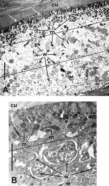
Although the hypodermis of filarial worms is syncytial, Wolbachia endobacteria are found in distinct zones (Franz and Büttner, Reference Franz and Büttner1983; Franz, Reference Franz1988). An examination of an electron microscopic section of the hypodermis (with the cuticle oriented upward) reveals that there is an outer zone under the cuticle that is usually free from Wolbachia but filled with mitochondria that are densely packed together (Supplemental Fig. 1, Online version only), followed by a zone containing a low-density mix of Wolbachia and mitochondria. Finally, there is an inner zone with many Wolbachia. The ultrastructure of Wolbachia is morphologically distinct from that of mitochondria, being both larger (0·3–0·6 μm), surrounded by a membrane, and not as heavily stained as mitochondria. The internal structures of the endobacteria are also different from those of mitochondria, i.e. lacking the characteristic cristae of mitochondria.
Wolbachia endobacteria and mitochondria localize to different zones of the hypodermis and are morphologically different. (A) The hypodermis can be divided into 3 zones based on the presence or absence of Wolbachia and mitochondria. Beginning from the cuticle (top right) there is a zone composed mostly of mitochondria (lines), in some of which the cristae can be seen. Also seen are folds in the hypodermal membrane (white arrows). Next is a zone with both mitochondria and Wolbachia (black arrows). Finally, there is a third region mostly free from mitochondria, but filled with Wolbachia. (B) A higher magnification of the outer and middle zones showing the morphological differences between Wolbachia endobacteria and mitochondria. The endobacteria are larger and contained within a vesicle of host-cell origin. Cristae are readily seen in some of the mitochondria under the hypodermal folds (white lines). cu=cuticle, m=mitochondria, w= Wolbachia.

Although Wolbachia have been detected using specific antiserum against Wolbachia surface protein (WSP) (Büttner et al. Reference Büttner, Wanji, Bazzocchi, Bain and Fischer2003; Kramer et al. Reference Kramer, Passeri, Corona, Simoncini and Casiraghi2003; Brattig et al. Reference Brattig, Bazzocchi, Kirschning, Reiling, Büttner, Ceciliani, Geisinger, Hochrein, Ernst, Wagner, Bandi and Hoerauf2004), much of the published immuno-histochemistry (both light and electron microscopic) has used various antisera against the heat shock-protein 60 (HSP60) (Hoerauf et al. Reference Hoerauf, Nissen-Pähle, Schmetz, Henkle-Dührsen, Blaxter, Büttner, Gallin, Al-Qaoud, Lucius and Fleischer1999; Taylor and Hoerauf, Reference Taylor and Hoerauf1999; Hoerauf, Reference Hoerauf2000; Büttner et al. Reference Büttner, Wanji, Bazzocchi, Bain and Fischer2003). In all cases, Wolbachia are seen as distinct spots in the middle and inner zones of the hypodermis and in the oocytes and embryos, but not in other tissues.
Examination by immunohistochemistry has proven invaluable for the monitoring of the efficacy of tetracyclines in depleting Wolbachia (Hoerauf et al. Reference Hoerauf, Nissen-Pähle, Schmetz, Henkle-Dührsen, Blaxter, Büttner, Gallin, Al-Qaoud, Lucius and Fleischer1999; Hoerauf, Reference Hoerauf2000). In some instances, diffuse staining in the outer zone of the hypodermis or in the embryos in the uterus was sometimes seen following the depletion of the Wolbachia. This has been attributed to mitochondrial HSP60, but has not been examined. Therefore, in the present study, we examined whether mitochondrial HSP60 was specifically labelled by anti-Yersinia HSP60 antiserum and whether this related to the up-regulation of mitochondrial HSP60 expression in O. volvulus.
MATERIALS AND METHODS
Worms from human patients
Onchocercomas (=subcutaneous nodules that develop around O. volvulus) were collected from patients treated 6 weeks with 100 mg/day doxycycline alone or 100 mg/day doxycycline plus once with 0·15 mg/kg/day ivermectin, once with 0·15 mg/kg/day ivermectin alone or untreated, as published previously (Hoerauf et al. Reference Hoerauf, Mand, Volkmann, Büttner, Marfo-Debrekyei, Taylor, Adjei and Büttner2003). The study design was approved by both the ethics committees of the Medical Board of Hamburg, Germany as well as the School of Medical Sciences of the Kwame Nkrumah University of Science and Technology, Kumasi, Ghana. The procedures used were in accordance with the Declaration of Helsinki (1975 and its revisions in 1983 and 2000). Onchocercomas were extirpated 5, 12 or 18 months following the commencement of doxycycline treatment.
Worms from animals
To support the hypothesis that tetracycline has no direct effect on the embryogenesis of filariae that do not contain Wolbachia, Acanthocheilonema viteae in Mastomys coucha were treated as described previously (Hoerauf et al. Reference Hoerauf, Nissen-Pähle, Schmetz, Henkle-Dührsen, Blaxter, Büttner, Gallin, Al-Qaoud, Lucius and Fleischer1999). Also, onchocercomas containing Onchocerca flexuosa (another filarial nematode without endobacteria) were collected from red deer (Cervus elaphus) in northern Germany (Plenge-Bönig et al. Reference Plenge-Bönig, Kromer and Büttner1995).
Immunohistochemistry
Acanthocheilonema viteae worms and onchocercomas extirpated from patients or red deer were fixed in 4% buffered formaldehyde and embedded in paraffin. Blocks were cut in 5 μm sections and blocked with 10% bovine serum albumin (BSA) in Tris-buffered saline (TBS). Wolbachia were probed with antiserum raised in rabbits against HSP60 from Yersinia enterocolitica (anti-Y-HSP60; diluted 1:1000 in TBS, 0·1% BSA) or the Wolbachia surface protein (WSP, diluted 1:4000) from Dirofilaria immitis as described in previous studies (Büttner et al. Reference Büttner, Wanji, Bazzocchi, Bain and Fischer2003; Hoerauf et al. Reference Hoerauf, Mand, Volkmann, Büttner, Marfo-Debrekyei, Taylor, Adjei and Büttner2003; Kramer et al. Reference Kramer, Passeri, Corona, Simoncini and Casiraghi2003; Brattig et al. Reference Brattig, Bazzocchi, Kirschning, Reiling, Büttner, Ceciliani, Geisinger, Hochrein, Ernst, Wagner, Bandi and Hoerauf2004). After washing with TBS containing 0·1% Tween-20, anti-rabbit polyclonal antibody (DakoCytomation, Hamburg, Germany; D0651) was used as secondary antibody according to manufacturer's recommendations for the Alkaline Phosphatase Anti-Alkaline Phosphatase kit (APAAP) (DakoCytomation). Fast red TR salt (Sigma, Deisenhofen, Germany) was used as the chromogen and haematoxylin (Merck, Darmstadt, Germany) served as the counter-stain. The specificity of the primary antibodies was verified using a different specific antiserum or buffer instead of antiserum on consecutive sections of the respective paraffin block.
Immunogold electron microscopy
For immunogold staining, onchocercomas were fixed in 2% paraformaldehyde/0·025% glutaraldehyde and embedded in LR-White (Merck, Darmstadt, Germany). Ultra-thin sections were cut with diamond knives using a Reichert-Jung Ultra Cut E (Leica, Bensheim, Germany). Sections were then blocked for 30 min in 10% BSA in phosphate-buffered saline (PBS). The sections were incubated with rabbit anti-Y-HSP60 serum diluted 1:500 in PBS with 1% BSA, followed by incubation with protein A-gold (10 nm in size) diluted 1:50 in PBS, 1% BSA. Sections were washed after each step with PBS, 1% BSA. Staining of subcellular structures was performed in 1% uranyl acetate and lead-citrate and then examined with a Philips CM 10 electron microscope (Philips, Eindhoven, The Netherlands). The specificity of the staining was verified using a specific antiserum (raised in rabbits) against glutathione S-transferase of O. volvulus (Ov-GST) (unpublished data) and diluted 1:500 in PBS with 1% BSA as a negative control.
Quantitative PCR
Total RNA was isolated from onchocercomas (16 untreated controls and 23 doxycycline treated) extirpated prior to ivermectin treatment, 5 months after the commencement of doxycycline treatment using the RNeasy Maxi Kit (Qiagen, Hilden, Germany). The collagenase protocol for heart and muscle tissue was followed. DNase treatment of the RNA was performed on the column. One microgram of RNA was reverse transcribed with oligo dT primers (Invitrogen, Karlsruhe, Germany) using the OmniScript kit (Qiagen). PCR primers specific to the mitochondrial gene hsp60 of O. volvulus (GenBank Accession number AF121264) were designed using Primer 3 (http://frodo.wi.mit.edu/cgi-bin/primer3/primer3_www.cgi). Briefly, the PCR was performed in 20 μl, containing a standard buffer (Qiagen), 4·5 mm of MgCl2, 50 μm of each dNTP, 900 nm of the forward primer (CCG GTA TGG GAG GAA TGT ACT), 300 nm of the reverse primer (TAA GAC TCC CTG TTC GCT CAA), 0·2 μl of SYBR Green (1:1000 diluted in DMSO, Roche, Mannheim, Germany), 0·8 μg bovine serum albumin (BSA) (Roche), 2·5 U of HotStar Taq polymerase (Qiagen) and 2 μl of cDNA or RNA sample in the cDNA buffer without reverse transcriptase as a control for genomic DNA contamination. Cycling was performed in a Light Cycler 1.0 (Roche) using the following protocol: 95°C for 15 min, followed by 40 cycles of 94°C for 20 sec, 58°C for 20 sec, 72°C for 20 sec, and 75°C for 10 sec, the fluorescence being acquired on the FAM channel. The additional temperature step at 75°C ensured that the signal recorded related to hsp60 rather than primer dimers, which are sometimes detected when cDNA amounts are limiting.
The level of transcription (copies per μl) from the hsp60 gene was estimated by comparing crossing point data for the test samples to a standard curve determined using a plasmid containing the mitochondrial hsp60 fragment produced by PCR using the hsp60 primers, as described previously for other genes (Heider et al. Reference Heider, Blaxter, Hoerauf and Pfarr2006). Transcription levels for hsp60 were normalized against actin, as described recently (Gilbert et al. Reference Gilbert, Nfon, Makepeace, Njongmeta, Hastings, Pfarr, Renz, Tanya and Trees2005).
Statistics
Significant differences in hsp60 transcription were tested using the Mann-Whitney non-parametric test employing GraphPad Prism v.4.00 for Windows, GraphPad Software, San Diego, California, USA, www.graphpad.com. Significance was set at P⩽0·05.
RESULTS
Localization of Wolbachia using immunogold electron microscopy
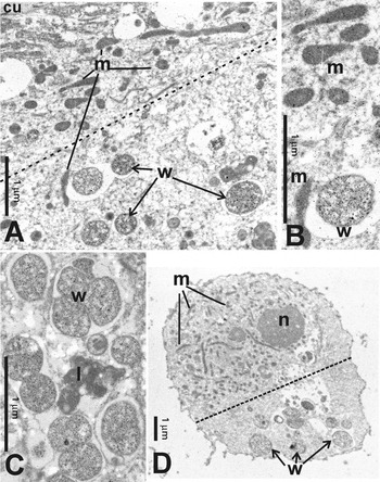
Previously, the depletion of Wolbachia has been monitored by immunohistochemistry using specific antisera against bacterial HSP60 and WSP (Hoerauf et al. Reference Hoerauf, Nissen-Pähle, Schmetz, Henkle-Dührsen, Blaxter, Büttner, Gallin, Al-Qaoud, Lucius and Fleischer1999). To confirm the specificity of anti-Y-HSP60 serum for the endobacteria and mitochondria, the antiserum was used for immunogold staining of ultra-thin sections of worms in onchocercomas from patients not treated with doxycycline. It should be noted that 1 patient had been treated with ivermectin 3 days before the excision of the nodule. Using the anti-Y-HSP60 serum, the typical localization pattern of Wolbachia and mitochondria was detected, in accordance with previous results (Kozek and Figueroa Marroquin, Reference Kozek and Figueroa Marroquin1977; Franz and Büttner, Reference Franz and Büttner1983; Franz, Reference Franz1988; Büttner et al. Reference Büttner, Wanji, Bazzocchi, Bain and Fischer2003) (Fig. 1A). In the hypodermis, mitochondria are concentrated under the cuticle and become sparser in the middle zone where Wolbachia are also found (Fig. 1A). The anti-Y-HSP60 serum appeared to detect Wolbachia more intensely than the mitochondria (Fig. 1B).
Immunogold, electron microscopy of female Onchocerca volvulus worms in nodules from untreated patients were incubated with anti-Y-HSP60 serum to specifically localize HSP60. (A) Mitochondria (lines) are located in the outer zone of the hypodermis and Wolbachia (arrows) are located in the middle zone. There is also weak labelling of the outer folds of the hypodermal membrane (near to the cuticle in the upper left corner). (B) Detail of (A) showing the weaker labelling of the mitochondria and the stronger labelling of the Wolbachia indicative of more HSP 60 in the endobacteria. (C) In another section of the hypodermis the Wolbachia are strongly labelled but the lysosomes in the same zone, and often of a similar size, are not labelled. (D) Wolbachia are also localized to a single area of an oocyte. Dashed lines represent the division between regions where mitochondria and the endobacteria are found. cu, Cuticle; l, lysosome; m, mitochondria; n, nucleus; u, uterus; w, Wolbachia.

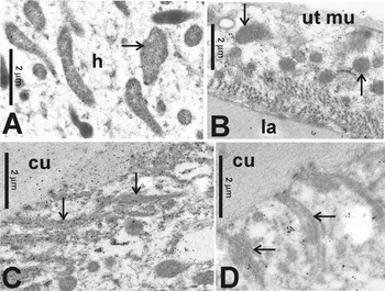
From the immunochemical staining, it was apparent that the antiserum did not react with other organelles, such as lysosomes, granular structures also located in the middle layer of the hypodermis, or the cytoplasm of the hypodermis and the epithelia of O. volvulus (Fig. 1C). There was weak labelling of the folded outer cell membrane of the hypodermis and the cuticle (Fig. 1A and 2C). The specific localization of both Wolbachia and mitochondria was even more pronounced when oocytes were examined. Wolbachia were detected near one or both ends of the oocytes, whereas the mitochondria were localized to the rest of the Wolbachia-free cytoplasm surrounding the chromosomes (Fig. 1D). Mitochondria were detected using HSP60 antiserum but not with the antiserum Ov-GST on sections from the same block (cf. Fig. 2A and B). Also, the cuticle and the outer membrane of the hypodermis were stained for HSP60 but not for glutathione S-transferase (Fig. 2C and D), although both antisera were detected with the same secondary antibody, showing the specificity of the anti-Y-HSP60 antibody for Wolbachia.
Rabbit anti-HSP60 serum and Alkaline Phosphatase Anti-Alkaline Phosphatase (APAAP) reagents are specific. Labelling of mitochondria in the hypodermis (A, arrow), the outer folds of the hypodermal membrane (C, arrows) and the cuticle is seen in some worms and can be misjudged as non-specific staining. However, the mitochondria of a muscle cell of the uterus (B, arrows) and the lamina incubated with another rabbit antiserum against an Ov-GST are not stained. (D) Labelling of the cuticle and hypodermal membrane is also not seen with the Ov-GST antiserum (arrows). Patient was treated with ivermectin 3 days before the excision of the nodule. cu, Cuticle; hy, hypodermis; la, lamina of the uterus epithelium, ut mu, uterus muscle cell.

Anti-Y-HSP60 labelling of O. volvulus mitochondrial HSP60 increases when Wolbachia are depleted
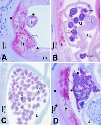
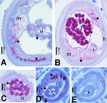
Using the specific anti-Y-HSP60 serum, granular staining of Wolbachia was seen in the hypodermis, oocytes, as well as in embryos of filariae not exposed to doxycycline (Fig. 3A–D, open arrows). Weak staining, representing mitochondrial HSP60, was also seen in worms not exposed or exposed to ivermectin. This staining was diffuse in the outer zone of the hypodermis (Fig. 3, panels A and D, closed arrows), the epithelia of the intestine (Fig. 3A) and genital tracts, muscles of the body wall (Fig. 3A) and uterus, oocytes (Fig. 3C), embryos (Fig. 3B), and the developing spermatozoa, but not in elongated, mature spermatozoa. The intensity of staining varied greatly, being strongest in portions of the filaria in which no bacteria are present and often in moribund worms, such as those with neoplastic cells in the pseudocoeloma (Fig. 3D; cf.Duke et al. Reference Duke, Marty, Peett, Gardo, Pion, Kamgno and Boussinesq2002). Also, ivermectin-exposed worms more frequently showed an increase of mitochondrial HSP60 (intestine in Fig. 3A) compared with untreated worms (Fig. 3, panels C and D).
Immunohistochemical labelling of HSP60 in Wolbachia endobacteria and in the tissues of female Onchocerca volvulus after treatment with ivermectin (A, 20 months; B, 5 months) or untreated. Granular endobacteria in the hypodermis (open arrows in A, B and D), in morulae (B) and in oocytes in the uterus (C) are strongly labelled. The mitochondria of the filaria are weakly labelled in the outer zone of the hypodermis (closed arrows in A), in the intestine (A), and in neoplastic cells in the pseudocoeloma (D). Stronger labelling of the intestinal mitochondria after ivermectin (A) is often seen. h, Hypodermis; i, intestine; m, muscle; u, uterus.

The examination of sections from onchocerciasis patients treated for 6 weeks with doxycycline showed that the hypodermis, oocytes and embryos were devoid of Wolbachia (Fig. 4, panels A–E). These worms showed a noticeable increase in mitochondrial HSP60 staining in the outer zone of the hypodermis, the oocytes and embryos. The labelled mitochondria appeared as small dots or as a diffuse staining that were distinct from the granular or ring-like structures of labelled Wolbachia. The diffuse staining was probably due to the labelled outer membrane folding of the hypodermis (lamellae). Tissues that were unstained or only lightly stained in untreated worms also had increased mitochondrial HSP60 expression (Fig. 4A–D, closed arrows). The staining seen in doxycycline-treated worms was not due to Wolbachia. Anti-Y-HSP60 serum specifically labelled the muscles in worms from patients who had received doxycycline (Fig. 4, panels A, B and D). However, serial sections labelled with anti-WSP serum, specific for bacteria, showed clearly that the Wolbachia had been depleted from these worms (Fig. 4E), indicating that the staining detected using the anti-Y-HSP60 serum represented the filarial mitochondria. The comparison of mitochondria in consecutive sections labelled using the anti-Y-HSP60 but not the WSP antiserum demonstrated the specificity of the antibody probes (cf. Fig. 4, panels D and E).
Female and male Onchocerca volvulus collected 5 (A, C) or 18 months (B, D, E) after doxycycline treatment showing increased labelling of filarial mitochondrial HSP60 in the hypodermis (closed arrows in A and D), muscles of the body wall (A, B, D), muscle cells of uterus (A) or vagina (D), the entire oocytes (C, in contrast to the granular bacteria in Fig. 3C), and developing spermatozoa in the testis (B). (D+E) Serial sections of a female worm demonstrated that endobacteria were absent by staining with antiserum against Wolbachia surface protein (E), and is also a negative control for the specificity of the HSP60 antiserum in (D). h, Hypodermis; i, intestine; m, muscles; t, testis; u, uterus, v, vagina.

We hypothesize that the increase in mitochondrial HSP60 was not a direct effect of the antibiotic on the filariae but due to the depletion of the Wolbachia from the nematodes. After the administration of doxycycline for 2 weeks, a treatment that does not deplete Wolbachia, increased staining of the mitochondria was not detected at any point of time up to 30 months after treatment. In contrast, after 6 weeks of doxycycline administration, a regimen that depletes Wolbachia, labelling increased at 18 and 24 months. We cannot exclude damage to the worms from the 2 weeks of doxycycline treatment, but the worms did not show more damage than that seen in untreated worms (data not shown).
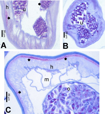
Acanthocheilonemea viteae (subfamily Onchocercinae) does not contain endobacteria (Hoerauf et al. Reference Hoerauf, Nissen-Pähle, Schmetz, Henkle-Dührsen, Blaxter, Büttner, Gallin, Al-Qaoud, Lucius and Fleischer1999). Comparing sections from untreated (Fig. 5A) and tetracycline-treated A. viteae (Fig. 5B), there was no increase in HSP60 staining in worms. The staining in the oocytes, muscles and gut was the same between both groups.
(A and B) No difference is seen in the labelling of filarial HSP60 in untreated (closed arrow, A) and treated (B) female Acanthocheilonemea viteae, a species without Wolbachia (after 70 days treatment with 25 mg/kg/day tetracycline). (C) In an untreated female Onchocerca flexuosa, another species without endobacteria, only the outer zone of the hypodermis (closed arrow) and the entire cytoplasm of oocytes in the ovary are labelled for filarial HSP60. h, Hypodermis; m, muscles; o, ovary; u, uterus.

Onchocerca flexuosa, is another species that does not harbour Wolbachia (see Plenge-Bönig et al. Reference Plenge-Bönig, Kromer and Büttner1995). Untreated female O. flexuosa often showed strong labelling in the entire cell of the oocytes and some staining in the outer hypodermis using anti-Y-HSP60 serum, but no labelling in regions of the hypodermis where Wolbachia are known to be found (Fig. 5C, compare with Fig. 2). The experiments using A. viteae and O. flexuosa support the hypothesis that the increased labelling of HSP60 in the mitochondria of O. volvulus is seen after depletion of Wolbachia.
Quantification of hsp60 transcription following Wolbachia depletion
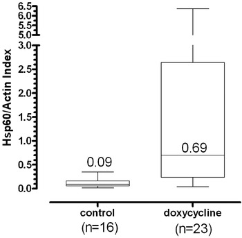
One argument for the increase in mitochondrial HSP60-staining could be that the specific antiserum has a higher affinity for the bacterial HSP60 and thus only stains weakly the mitochondrial HSP60 until the Wolbachia are depleted. To address this, we measured hsp60 transcription from onchocercomas extirpated from untreated and doxycycline-treated patients 5 months after the commencement of doxycycline treatment to demonstrate that the increase in mitochondrial HSP60 is also reflected at the level of transcription. The primers to the gene hsp60 had no similarity to the sequence of the hsp60 gene of Wolbachia, as determined by BLAST analysis (http://www.ncbi.nlm.nih.gov/BLAST/). The actin primers were tested on human cDNA and did not amplify the human gene (data not shown). The hsp60 index generated by quantitative PCR clearly showed a significant increase (7·7 fold; P=0·0002) in hsp60 transcription after Wolbachia had been depleted by doxycycline treatment (Fig. 6).
Filarial mitochondrial hsp60 transcription is up-regulated after depletion of Wolbachia endosymbionts from O. volvulus. Up-regulation of mitochondrial hsp60 at the mRNA level was demonstrated by quantitative PCR using the ratio of filarial hsp60 to actin. The line in the boxes represents the median value (the exact value represented by the number in each column); the bottom and top of the boxes represent the 25th and 75th percentiles, respectively; the lower and upper lines represent the minimum and maximum values, respectively. P=0·0002, as determined by the Mann-Whitney test.

DISCUSSION
Using the specific anti-Y-HSP60 serum, we were able to show that mainly HSP60 from Wolbachia, the filarial mitochondria, the outer folding of the hypodermis, and the cuticle are labelled, but not other organelles. Using electron microscopy, we confirmed that the mitochondria and Wolbachia localized to discrete regions of the hypodermis, the oocytes, zygotes, and embryos, thus allowing the differentiation of Wolbachia from mitochondria based on their location in the worm (cf. Franz and Büttner, Reference Franz and Büttner1983; Franz, Reference Franz1988).
Strong mitochondrial HSP60-labelling was also detected in some worms from untreated persons. These latter worms were classified moribund, as the worms contained neoplastic cells in the pseudocoeloma (cf. Duke et al. Reference Duke, Marty, Peett, Gardo, Pion, Kamgno and Boussinesq2002). Increased HSP60 expression in such worms is probably related to a normal stress response to the uncontrolled growth of the neoplastic cells. Increased HSP60 labelling was also detected in developing (but not mature) spermatozoa within the testis of male worms depleted of Wolbachia. This result was unexpected because Wolbachia in filariae had not been reported to occur in the male genital tract (Franz, Reference Franz1988; Wu et al. Reference Wu, Egerton, Ball, Tanguay and Bianco2000; Hoerauf et al. Reference Hoerauf, Mand, Volkmann, Büttner, Marfo-Debrekyei, Taylor, Adjei and Büttner2003). The higher levels of HSP60 in the testis may reflect ‘general stress’ in the male worms after Wolbachia have been depleted.
In the present study, we demonstrated that the up-regulation of mitochondrial HSP60 in worms that had been depleted of Wolbachia was also reflected in transcription levels, with 7·7-fold higher levels in worms from nodules from patients treated with doxycycline but not ivermectin compared with those from untreated patients. The quantitative PCR used to measure mitochondrial hsp60 was specific for filarial mitochondria. This was inferred from in silico analysis of the primer sequences. This was also inferred empirically as human hsp60 transcription would be higher than mitochondrial hsp60 due to the larger amount of human tissue in the onchocercomas, making detection of the mitochondrial signal impossible.
The broad range of the hsp60/actin ratio in the onchocercomas from doxycycline treated patients, while undesirable, is difficult to reduce because of the nature of the samples. Frozen onchocercomas were used in this study rather than worms from collagenase-treated onchocercomas to avoid inducing HSP60 during overnight incubation with an enzyme that is also active against the worms. Thus, the exact nature and status of the worms is unknown. Since whole onchocercomas were used for the RNA extraction, we were unable to undertake histological examinations to establish the relative age and status of individual worms in each nodule, as we have done before (see Hoerauf et al. Reference Hoerauf, Specht, Büttner, Pfarr, Mand, Fimmers, Marfo-Debrekyei, Konadu, Debrah, Bandi, Brattig, Albers, Larbi, Batsa, Adjei and Büttner2007). This limitation could affect the hsp60/actin ratio in 2 ways. (1) These patients live in a hyperendemic region and are therefore constantly exposed to new infections. Newly acquired worms in the same onchocercomas with doxycycline treated worms would result in a lower ratio. (2) Another possibility is that different aged worms may regulate HSP60 differently, resulting in onchocercomas with worms that only have up-regulated HSP60, onchocercomas with worms that are too old to react to the loss of their endosymbionts or onchocercomas with a mixture of the previous worms.
We believe that the up-regulation of filarial mitochondrial HSP60 is not merely a reaction to the stress of accumulated dead bacteria, but due to a long-term disruption of the homeostasis in the worms following the depletion of their essential endobacteria. This is reflected clearly in Fig. 4, panels A, B, and D, in which HSP60 staining is greater in musculature and spermatozoa usually lacking Wolbachia. Since Wolbachia and mitochondria are located in different tissues or zones of the hypodermis and oocytes of the worms and stain differently by immunohistochemistry (granular/ring-like vs diffuse), the increased staining of mitochondrial HSP60 infers increased protein expression.
The up-regulation of the mitochondrial protein HSP60 may indicate efforts by the filarial cells to compensate for an interruption in some cell processes requiring Wolbachia. This interruption may lead to an accumulation of proteins which require HSP60 for proper folding, as has been described in the free-living nematode Caenorhabditis elegans (see Yoneda et al. Reference Yoneda, Benedetti, Urano, Clark, Harding and Ron2004). In the latter nematode, the accumulation of unfolded proteins leads to an up-regulation of HSP60 specifically in the mitochondria and is associated with proteins imported from the cytoplasm into the mitochondria which subsequently form multi-protein complexes. HSP60 is not up-regulated in the mitochondria due to other stresses, such as reactive oxygen species (Zhao et al. Reference Zhao, Wang, Levichkin, Stasinopoulos, Ryan and Hoogenraad2002; Yoneda et al. Reference Yoneda, Benedetti, Urano, Clark, Harding and Ron2004). Whether the up-regulation of the expression of mitochondrial HSP60 is due to a direct interaction between Wolbachia and mitochondria has not yet been shown. However, it is more likely to be the result of a long-term stress induced by the disruption of the homeostasis of metabolic pathway(s) for which Wolbachia may be needed by the nematode (Foster et al. Reference Foster, Ganatra, Kamal, Ware, Makarova, Ivanova, Bhattacharyya, Kapatral, Kumar, Posfai, Vincze, Ingram, Moran, Lapidus, Omelchenko, Kyripide, Ghedin, Wang, Goltsman, Joukov, Ostravskaya, Tsukerman, Mazur, Comb, Koonin and Slatko2005; Pfarr and Hoerauf, Reference Pfarr and Hoerauf2005).
In conclusion, the depletion of Wolbachia from the filarial worms examined herein leads to both an increase in transcription of the hsp60 gene and the translated protein. This increase is probably not a direct effect of the antibiotic treatment, but a response to the loss of the essential Wolbachia. Since this up-regulation lasts for up to 18 months or longer, doxycycline treatment has a long-lasting effect on the metabolism of O. volvulus, and eventually results in detrimental effects on the adult worm (Taylor et al. Reference Taylor, Makunde, Mcgarry, Turner, Mand and Hoerauf2005b; Hoerauf et al. Reference Hoerauf, Specht, Büttner, Pfarr, Mand, Fimmers, Marfo-Debrekyei, Konadu, Debrah, Bandi, Brattig, Albers, Larbi, Batsa, Adjei and Büttner2007). It would be interesting to establish whether HSP60 is up-regulated in a similar manner in arthropods that are cured of their Wolbachia infections. If results are specific to filarial Wolbachia, it may provide insight into proteins involved in this mutualistic symbiosis.
The authors would like to thank Mrs Ingeborg Albrecht for her technical assistance. We are particularly grateful to Professor I. Authenrieth, University of Tübingen, for kindly providing the Yersinia HSP60 antiserum; Dr C. Bandi, University of Milan, for the Dirofilaria WSP antiserum; Professor Dr E. Liebau, University Münster, for the Ov-GST antiserum, Professor R. Lucius for providing A. viteae worms; and Dr A. Plenge-Bönig for the O. flexuosa nodules. This work was supported by the German Research Foundation (DFG grant numbers: HO 2009/5-1 and PF 673/1-1).