Implications
The data show that return to testicular function is highly variable and is reflected by the increase of testosterone after second vaccination, as shown earlier. Return to a physiological level of spermatogenesis that is comparable to untreated boars of the same age requires an additional period of 13 weeks.
Introduction
Traditionally, male piglets are surgically castrated without anaesthesia during the first week of life. This practice is performed to prevent later synthesis of the sex odour of androstenone in testicular Leydig cells and its accumulation in adipose tissue (Claus et al., Reference Claus, Weiler and Herzog1994).
Surgical castration, however, also eliminates sex-specific fattening advantages (Walstra, Reference Walstra1974) due to the anabolic function of testicular androgens and estrogens (Claus and Hoffmann, Reference Claus and Hoffmann1980; Metz et al., Reference Metz, Hohl, Waidelich, Drochner and Claus2002).
More recently, the aspect of animal welfare became an urgent issue so that alternatives to surgical castration without anaesthesia could be discussed. Such attempts include the use of anaesthesia combined with analgesia and genetic selection for low tissue concentrations of androstenone (Willeke et al., Reference Willeke, Claus, Pircher and Alsing1980; Robic et al., Reference Robic, Larzul and Bonneau2008), but they inevitably lead to a loss of anabolic advantages. In addition, genetic abolishment of androstenone, for example, by marker assisted genetic selection (Robic et al., Reference Robic, Larzul and Bonneau2008), would have other disadvantages, because this pheromone is a component of fertility regulation in the pig and thus closely linked to the other components of testicular function and in consequence, reproduction. Immunological castration is another alternative which at least maintains the main part of anabolic properties (Metz and Claus, Reference Metz and Claus2003). It is based on vaccination against gonadotropin-releasing hormone (GnRH) which in turn leads to inhibition of LH and thus its effect on steroid synthesis (Nieschlag, Reference Nieschlag1975), including the sex odour of androstenone, in Leydig cells of pigs.
This principle, so far, was applied not only for the pig (e.g. Awoniyi et al., Reference Awoniyi, Chandrashekar, Arthur, Schanbacher, Amador and Falvo1988a; Bonneau et al., Reference Bonneau, Dufour, Chouvet, Roulet, Meadus and Squires1994; Meloen et al., Reference Meloen, Turkstra, Lankhof, Puijk, Schaaper, Dijkstra, Wensing and Oonk1994), but also to a variety of farm animal (e.g. Robertson et al., Reference Robertson, Fraser, Innes and Jones1982; Schanbacher and Pratt, Reference Schanbacher and Pratt1985; Godfrey et al., Reference Godfrey, Walkden-Brown, Martin and Speijers1996) and pet species (Ladd et al., Reference Ladd, Tsong, Walfield and Thau1994) to eliminate hormone-dependent aggressive behaviour for a limited time.
For the pig, the vaccine IMPROVAC® (CSL Animal Health, Victoria, Australia) is commercialized in several countries. It is applied two times, about four weeks apart, so that the second vaccination elicits the anamnestic effect which in turn leads to a cessation of Leydig cell steroidogenesis within one week (Claus et al., Reference Claus, Lacorn, Danowski, Pearce and Bauer2007). This second booster is applied about four weeks prior to slaughter to allow androstenone, which had been stored in adipose tissue already, to be eliminated from the body (Claus, Reference Claus1976). In consequence, the anabolic function of testicular hormones is still maintained until four weeks before slaughter.
It was not known, however, whether single animals’ testicular function returns within this four-week period. Moreover, it is well-known that time to reach final weight may vary considerably between the pigs so that final weight might only be reached after more than four weeks in some individuals. Therefore, the maintenance of the castration effect after the second vaccination is a relevant research object. First data were reported by Zamaratskaia et al. (Reference Zamaratskaia, Rydhmer, Andersson, Chen, Lowagie, Andersson and Lundstroem2008) who slaughtered pigs either 16 or 22 weeks after second vaccination. Systematic studies on the development of endocrine and spermatogenic activity after second vaccination are missing.
Therefore, in a recent study, we chronically cannulated six boars and determined blood plasma profiles of antibody titres and reproductive hormones, and found a highly variable return of Leydig cell function between 10 and 24 weeks after second vaccination (Claus et al., Reference Claus, Rottner and Rueckert2008). The variability of testicular histological parameters and recrudescence of spermatogenic functions was not investigated so far.
Therefore, in the present paper, data on individual variation of testes histology and morphology of the same boars are presented and compared to a group of four other boars of the same age (52 weeks), which were not immunized and served as untreated controls.
Material and methods
Experimental design, animals and feeding
The whole experiment had been approved by the animal welfare committee of the state of Baden-Württemberg.
First results from the study regarding the post-vaccination recurrence of hormone biosynthesis have previously been reported (Claus et al., Reference Claus, Rottner and Rueckert2008). The treatment group (German Landrace boars, n = 6; from two litters) was obtained from the University of Hohenheim swine herd. These animals received the first dose of IMPROVAC® at 19 weeks of age (mean weight 61.6 ± 5.8 kg) and were transferred to the experimental stables on the university campus at 23 weeks of age (mean weight 84.8 ± 7.6 kg). After a two-week adaptation period (25 weeks of age; mean weight 91.3 ± 6.2 kg), the pigs were surgically fitted with indwelling cephalic vein catheters (Claus et al., Reference Claus, Bingel, Hofäcker and Weiler1990). Booster vaccinations were given at 26 weeks of age (mean weight 98.4 ± 6.6 kg). The day of second vaccination is the time mark of the experiment and is further referred to as day 0. Blood was drawn till the end of the study, at an age of 52 weeks. The study ended when the last boar again exceeded 0.5 ng testosterone/ml of blood plasma.
Due to the duration till return to testicular function, slaughter weight was far above normal; thus, feeding had to be adapted to avoid excessive fat deposition. Till an age of 31 weeks (mean weight 122.3 ± 7.5 kg), the boars were fed 1.5 kg portions of a ration containing 13.5 MJ metabolizable energy/kg dry matter (MJ ME/kg DM) and 16.7% crude protein (CP), twice daily. Portions were increased to 1.75 kg for the following period of four weeks. From week 36 till week 41, boars received 1.5 kg portions of a ration with 11.2 MJ ME/kg DM and 13.6% CP. Thereafter, till the end of the experiment (52 weeks of age; mean weight 193.2 ± 14.1 kg), portions were reduced to 1.3 kg.
In addition, a control group (German Landrace boars, n = 4; from one litter, University of Hohenheim swine herd) was included for the investigation of normal histological criteria and spermatogenic function and therefore was not immunized. They were transferred to the experimental stables at 46 weeks of age (mean weight 172.5 ± 6.1 kg) and received 1.5 kg portions of a ration with 11.2 MJ ME/kg DM and 13.6% CP. These boars were euthanized at the same age as the treatment group (52 weeks of age; mean weight 196.9 ± 7.4 kg).
Blood and tissue sampling
Blood (10 ml) from vaccinated boars was drawn twice daily at 0800 and 1500 h into heparinized vials, and the plasma stored at −20°C till it was assayed. Catheters were rinsed with heparinized saline after each blood sample. Additionally, a single blood sample from all boars was taken immediately before euthanasia.
All boars were euthanized at 52 weeks of age by an intravenous injection of 0.1 ml pentobarbital/kg live weight (Narcodorm®; CP-pharma Handelsgesellschaft mbH, Burgdorf, Germany). Testes were removed and the weights of both testes including the epididymides and accessory sex glands (bulbourethral gland, vesicular gland and prostate) were determined. Testis tissue was rinsed with physiological saline and fixed in 4% paraformaldehyde within 10 min post mortem, for histomorphological evaluation.
Testosterone analysis
Measurements were performed every third day after extraction with tert-butyl methyl ether (Wagner and Claus, Reference Wagner and Claus2004) from blood plasma samples. The antiserum had been raised against testosterone-3-CMO-BSA as an antigen and was specific. Tritiated testosterone was used as a tracer. The coefficient of intra-assay variation was 4.2%, and 8.4% for the inter-assay variation at a concentration of 1 ng/ml. The lower limit of detection was 0.05 ng/ml and the recovery 95%.
Histological techniques
The paraformaldehyde-fixed testes samples (right testis was used for evaluation) were dehydrated through an ascending alcohol series (50% up to 100%) and embedded in paraffin (Romeis, Reference Romeis1989). With a sliding microtome (SM2000R; Leica, Nussbach, Germany) slices of 4 μm were obtained and mounted on microscope slides for staining.
After removing the paraffin (Roth, Karlsruhe, Germany) in a chloroform bath (performed twice for 15 min), slides were put into a descending alcohol series (100% to 70%) for 2 min.
Staining with haematoxylin (1.09249.2500; Merck, Darmstadt, Germany) was performed for 3 s followed by a hydrating step under flowing water for 10 min. Subsequently, slides were put into 0.2% eosin (1345; Merck) with 50 μl acetic acid (Merck) for 5 to 10 s and into an ascending ethanol series (70% up to 96%). Thereafter, an incubation step followed with 100% ethanol and xylol (Roth), each for 2 min. Slides were then embedded with depex (Serva, Heidelberg, Germany).
Slides were additionally stained immunohistochemically for GATA-4 and Ki-67.
GATA-4 is a marker which specifically stains Sertoli cells. It was used in this study to discriminate Sertoli cells from germ cells and thus allowing a more accurate characterization of the latter cell population.
The staining protocol has been reported earlier in detail (McCoard et al., Reference McCoard, Lunstra, Wise and Ford2001; Haeussler et al., Reference Haeussler, Wagner, Welter and Claus2007). In brief, slides were prepared as described for haematoxylin–eosin, and GATA-4 was detected with a purified commercial antibody (goat anti-mouse, C-20; Santa Cruz Biotechnology, Heidelberg, Germany). After blocking unspecific binding with normal rabbit serum, the primary and biotinylated secondary antibodies (rabbit anti-goat, Dako GmbH, Hamburg, Germany) were applied. The avidin–biotin–peroxidase system (Dako GmbH) then led to the brown colour that is specific for immunodetection of Sertoli cells. Counterstaining was done with haematoxylin.
Internal negative controls were treated identically except that the primary antibody was omitted to exclude unspecific binding of the secondary antibody.
Ki-67 is a cell proliferation antigen used to detect germ cells undergoing mitosis. Staining with the monoclonal antibody Ki-67 MIB-1 (LINARIS GmbH, Wertheim-Bettingen, Germany) was performed as described earlier, in detail (Mentschel et al., Reference Mentschel, Leiser, Mülling, Pfarrer and Claus2001). Secondary anti-mouse antibody had been raised in goats (Dako GmbH). Counterstaining again was performed with haematoxylin.
Because the primary antibody also stains meiotic stages of spermatocytes, only type A and type B spermatogonia were counted (magnification ×400).
To characterize the rate of total mitosis, one hundred tubules were evaluated for each boar.
Histomorphological evaluations
From each section, five areas were selected which were assigned as representatives for the whole testis. They were analysed at a magnification of ×50. The visual fields were recorded with a video camera (Donpisha, 3CCD color vision camera module, model XC-003P; Sony, LTF Labortechnik, Wasserburg, Germany) and printed (hp LaserJet 1200 series, Hewlett Packard GmbH, Böblingen, Germany) with a standardized magnification so that each area used for quantitative evaluation corresponded to 1.3 × 106 μm2 in situ. These prints were then evaluated by planimetry (OTT-Planimeter; A. Ott, Kempten, Bavaria, Germany).
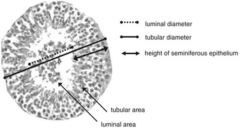
In addition to the relative areas occupied by tubuli or interstitial cells, the diameter of individual tubuli and the contribution and characterization of the spermatogenic epithelium were also determined. The principle of evaluation is outlined in Figure 1.
Principles of evaluation of individual tubuli.

Diameter and area of the whole seminiferous tubules, as well as their lumen, were determined in eight nearly-round tubules per boar at a magnification of ×100.
Leydig cell numbers were counted in five interstitial sections per boar at 400-fold magnification, each corresponding to an area of 106 μm2. For determination of cell and nuclei volumes, the method of Abercrombie (Abercrombie, Reference Abercrombie1946) was applied in ten well-identifiable Leydig cells.
The contribution of the eight stages of spermatogenesis (Swierstra, Reference Swierstra1968) was counted in one hundred tubules at 400-fold magnification. The spermatogenic cells were differentiated from Sertoli cells after GATA-4-staining. Sertoli cells were not further differentiated between boars because their number is regarded to be stable after puberty (Berndtson et al., Reference Berndtson, Igboeli and Parker1987).
Stages VI and VII were subsumed because they cannot be discriminated with certainty. Single stages were grouped as pre-meiotic (I, II, III), meiotic (IV, V) and post-meiotic (VI/VII, VIII) phases (Garcia-Gil et al., Reference Garcia-Gil, Pinart, Sancho, Badia, Bassols, Kadar, Briz and Bonet2002).
Statistical analysis
Because the study was performed to characterize individual reactions of boars, the relevant data in Tables 1–4 are given without standard errors. Data from control boars instead represent mean reactions of four boars and are given as means ± s.e.
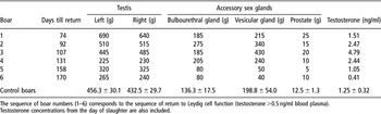
Weight of testes and accessory sex glands in individual vaccinated boars and in control boars (mean ± s.e.)

The sequence of boar numbers (1–6) corresponds to the sequence of return to Leydig cell function (testosterone > 0.5 ng/ml blood plasma).
Testosterone concentrations from the day of slaughter are also included.
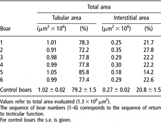
Absolute and relative total area of seminiferous tubules and interstitial tissue (mean ± s.e.)

Values refer to total area evaluated (1.3 × 106 μm2).
The sequence of boar numbers (1–6) corresponds to the sequence of return to testicular function.
For control boars the s.e. is given.
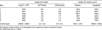
Characterization of interstitium: total area and relative contribution of Leydig cells and their nuclei and cytoplasm, respectively

The sequence of boar numbers (1–6) corresponds to the sequence of return to testicular function.
For control boars the s.e. is given.
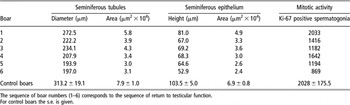
Characterization of seminiferous tubules, epithelial height and mitotic activity for the individual vaccinated boars and the controls

The sequence of boar numbers (1–6) corresponds to the sequence of return to testicular function.
For control boars the s.e. is given.
Data from spermatogenic epithelium were analysed for normal distribution using the Kolmogorov–Smirnov test. Then, they were evaluated statistically using the T-Test for independent random sampling, of the Statistical Package for the Social Sciences (version 15.0, 2006; SPSS, Chicago, IL, USA).
Results
Testis weight and gross morphology
Testis weight (Table 1) varied considerably; but, it is obvious that the mean testis weight of control boars was only reached by the three boars that were the first to return to testicular function. Boars 1 and 2 even exceeded testes weight of those of the controls; in contrast, boars 4 to 6 reached only about half the testes weight of those of the control animals. Boars 5 and 6 also had continued low testosterone concentrations.
The level of testosterone synthesis is also reflected by the weight of accessory sex glands, which were low in boars 5 and 6 both, compared to the other boars of the vaccinated group and to the controls. Boars 1 to 4 even exceeded the mean values for the bulbourethral and the vesicular gland of controls and tended to have higher weights of the prostate.
The relative contribution of tubules and interstitial tissue to testis cross-section is analysed in Table 2. Seminiferous tubule area was quite uniform, around 1.01 μm2 × 106 per total area evaluated, both within the group of vaccinated boars and when compared to the controls. In consequence, almost 80% of tissue area was represented by tubuli, whereas, interstitial tissue was responsible for the remaining 20%.
Thus, the relative contribution of these two compartments was not altered by increased testis weight.
Characterization of Leydig cells
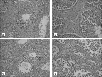
The change of Leydig cell characteristics with changing testis size is analysed in Table 3. An example is also shown in Figure 2.
Comparison of histological characteristics of tubuli (left side) and interstitium (right side) between boars which resumed testicular function early (boar 2; top) and late (boar 6; bottom).

The number of Leydig cells per evaluated area (106 μm2) (Abercrombie, Reference Abercrombie1946) is also referred to the total size of the testis.
The number of Leydig cells per testis (Table 3) was seen to be increased in all immunized boars, compared to the controls; but, there was no obvious relationship between the number of Leydig cells and testis size. Instead, the size of nuclei and cytoplasm reveals relationships to testicular size. Nuclei of the vaccinated boars revealed a minor dependency, because nuclear size differed maximally by a factor of 2 between boars 2 and 6. Control boars, however, had a mean nuclear volume of 154 μm3, which was clearly higher than in boar 2 of the vaccinated group.
Even more pronounced were the differences of cytoplasm. In vaccinated boars, maximal difference (again boar 2 to 5 and 6) was nearly six-fold; but then again, cytoplasm volume was even between a factor of 9.5 (boars 5 and 6) and 1.7 (boar 2), when compared to the controls. Altogether, it appears that both the size of nuclei and cytoplasm in vaccinated boars are related to the degree of resumption of testicular function, and thus, its size; but control boars are still clearly superior.
Characterization of tubuli and mitotic activity
In contrast to Leydig cells, tubuli contributed less to testicular size. Minor differences in the diameter and area of tubuli (Table 4; Figure 2) were found in the vaccinated boars. Boars whose testicular function returned earlier revealed slightly elevated dimensions of the tubuli. Controls, however, were again superior. The improved tubular size apparently does not depend on differences of Sertoli cell number which were around 6 per tubule without much variation in the vaccinated boars v. 7.5 per tubule in the controls (Abercrombie, Reference Abercrombie1946). Thus, the spermatogenic activity is the main reason, as shown by the dimensions of the seminiferous epithelium, and even more pronounced by the mitotic activity of spermatogonia.
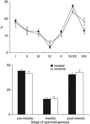
All boars had epithelia which represented all stages of spermatogenesis and all types of spermatogenic cells (Figure 3). We saw a tendency, though, that more advanced stages of spermatogenesis, like stage IV and VIII, were underrepresented in the immunized boars compared to control boars. A further differentiation within the group of treated boars did not lead to an obvious significant difference. Pre-meiotic (I, II, III) stages of spermatogenesis in treated boars tended to exceed the control boars while they approached boar level in terms of meiotic (IV, V) and post-meiotic (VI/VII, VIII) stages.
Frequency of different stages of spermatogenesis in vaccinated and control boars (top: individual stages; bottom: contribution to mitotic activities).

Discussion
Two doses of a GnRH vaccine given about four weeks apart to boars effectively led to antibody formation and a drop of LH, and consequently to the drop of testicular steroids. It was shown that the second dose of vaccine leads to maximal GnRH antibody titre and the concurrent drop of LH within one week (Claus et al., Reference Claus, Lacorn, Danowski, Pearce and Bauer2007).
Surprisingly, FSH is not altered by vaccination of boars (Awoniyi et al., Reference Awoniyi, Chandrashekar, Arthur, Schanbacher, Amador and Falvo1988a and Reference Awoniyi, Chandrashekar, Arthur, Schanbacher, Amador and Falvo1988b; Wagner and Claus, Reference Wagner and Claus2004) and it was concluded that it is regulated either by a separate releasing hormone, or by the hypothalamic secretion of activin (Padmanabhan and McNeilly, Reference Padmanabhan and McNeilly2001).
At the tissue level, vaccination – and in consequence the loss of reproductive hormones – may lead to a loss of Leydig cell cytoplasm by 90%, while the number of Leydig cells remained fairly constant. In addition, seminiferous tubule diameter was reduced by 30%. Mitosis was reduced, but except for type A spermatogonia, the spermatogenic cells were reduced by about 60% (Wagner and Claus, Reference Wagner and Claus2004).
From other species, it is well known that the effect of vaccination against GnRH is reversible; but the duration until resumption of reproductive functions varies considerably between species (e.g. Schanbacher and Pratt, Reference Schanbacher and Pratt1985; Ladd et al., Reference Ladd, Tsong, Walfield and Thau1994; Godfrey et al., Reference Godfrey, Walkden-Brown, Martin and Speijers1996).
In a preceding paper, therefore, we studied in detail the time course of return to testicular function and we found that it is apparently characterized by a drop of antibody titre, establishment of LH receptors in Leydig cells, loss of refractoriness of the hypothalamus and an increase of LH concentrations. In consequence, Leydig cell function is resumed and steroid production increases (Claus et al., Reference Claus, Rottner and Rueckert2008).
In addition, it was described that boars have a high potential to return to normal testicular and thus reproductive function even after repeated booster vaccinations up to 60 weeks after the fifth vaccination (Hilbe et al., Reference Hilbe, Jaros, Ehrensperger, Zlinszky, Janett, Hässig and Thun2006).
Detailed analyses on return of testicular function at tissue level were missing until now. The present study gives first data on testis and accessory sex glands morphology and the histological changes of Leydig cells and tubuli.
Based on a testosterone value of 0.5 ng/ml plasma, we found a high individual variation between 10 and 24 weeks until reestablishment of steroid synthesis by Leydig cells, after repeated GnRH immunization. This variation was only partly dependent on the GnRH antibody titre so we concluded that the individual sensitivity of the pituitary to react to minimal values of GnRH, and of the testis to react to LH, are more important in inducing resumption of synthesis of steroids by the Leydig cells (Christensen, Reference Christensen1975).
In the present study, we also found that histological criteria reveal a considerable variation, which can only be partly explained by the anti GnRH titre or by peripheral steroid concentrations. Therefore, further experiments are needed to clarify the mechanisms. Status of testis morphology showed that the weight of the testis is related to testosterone concentrations, but only a part of those boars, which started early to resume hormone production, had reached the weight of testis in controls, while others even exceeded testis weights of controls. The latter finding might be due to a rebound phenomenon as it was also shown during pubertal development, where testicular function is optimized in a wave-like pattern until reaching the mature status (Allrich et al., Reference Allrich, Christenson, Ford and Zimmerman1983; Wagner and Claus, Reference Wagner and Claus2008).
In contrast, weight of accessory sex glands is well known to be controlled by gonadal steroids, which include both testosterone and estradiol-17β in boars (Joshi and Raeside, Reference Joshi and Raeside1973; Claus and Hoffmann, Reference Claus and Hoffmann1980).
The main determinant for testis size is the functional status of the Leydig cells. It is generally accepted that in mature males, the number of Leydig cells is kept constant and, thus, does not explain changes of the testis size as also found in the present study (Mendis-Handagama and Ariyaratne, Reference Mendis-Handagama and Ariyaratne2001).
Instead, the size of the nuclei and the amount of cytoplasm partly determine the size of the testis. The increase in the size and, consequently the functional status of Leydig cells, is initiated by LH when decrease of GnRH antibodies allows the secretion of LH by the pituitary. It was shown earlier that LH receptors in the testis are found exclusively in the Leydig cells of boars (Peyrat et al., Reference Peyrat, Meusy-Dessolle and Garnier1981).
In consequence, LH acts a stimulus for Leydig cell enlargement, leading to increase in testis size and an increase in testosterone and estradiol concentrations.
Our data additionally show that tubular diameter contributes to testicular morphology.
Studies in strains of mice which differ in the testis size lead to the conclusion that testis size is under the control of at least two autosomal genes which exert their effect by regulating the number of Sertoli cells (Chubb, Reference Chubb1992).
In our study with genetically uniform pigs, Sertoli cells did not play a role inregulating the testis size, as also described earlier (Lunstra et al., Reference Lunstra, Wise and Ford2003; Wagner and Claus, Reference Wagner and Claus2009).
Rather, the effect of hormonal regulation of spermatogenesis and Leydig cell function explain the differences in testis size.
The height of the seminiferous epithelium showed a slight dependency on testosterone secretion and was lower in late returning boars of the vaccinated group than in early returners.
In addition, the height of the epithelium in control boars exceeded that of vaccinated boars, probably due to differences in the mitotic activity. It was demonstrated that mitotic activity is improved both by the stimulating effects of estradiol via its α receptor – present in the type A and type B spermatogonia up to mid pachytene primary spermatocytes – and via its oestrogen receptor which is found in Sertoli cells (Lekhkota et al., Reference Lekhkota, Brehm, Claus, Wagner, Bohle and Bergmann2006); thus, oestrogen substitution of GnRH vaccinated boars led to an increased mitotic activity (Wagner et al., Reference Wagner, Messe, Bergmann, Lekhkota and Claus2006).
Altogether, our data show that normalization of testicular functions after the onset of hormone synthesis (testosterone >0.5 ng/ml) still requires another 13 weeks until testis function is similar to untreated controls.
Acknowledgements
We thank Dr Ulrike Weiler for participating in the animal surgery, and Claudia Fischinger, William Dunne and Mohamed Mecellem for excellent care and handling of the animals. We also thank Helga Hägele and Anna Wagner for their assistance in the histological performances.