Introduction
Chitin is the second most abundant polymer on earth after cellulose and is found in many organisms, including arthropods (Merzendorfer, Reference Merzendorfer2006; Hegedus et al., Reference Hegedus, Erlandson, Gillott and Toprak2009), fungi (Ruiz-Herrera and Ortiz-Castellanos, Reference Ruiz-Herrera and Ortiz-Castellanos2010; Roncero, Reference Roncero2002), protozoa (Das et al., Reference Das, Van Dellen, Bulik, Magnelli, Cui, Head, Robbins and Samuelson2006), protobacteria (Esseling and Emons, Reference Esseling and Emons2004), protists (Jeuniaux and Voss-Foucart, Reference Jeuniaux and Voss-Foucart1991), sponges (Ehrlich et al., Reference Ehrlich, Krautter, Hanke, Simon, Knieb, Heinemann and Worch2007a,Reference Ehrlich, Maldonado, Spindler, Eckert, Hanke, Born, Goebel, Simon, Heinemann and Worchb), nematodes (Zhang et al., Reference Zhang, Foster, Nelson, Ma and Carlow2005), molluscs (Weiss et al., Reference Weiss, Schönitzer, Eichner and Sumper2006) and cuttlefish (Hunt and El Sherief, Reference Hunt and El Sherief1990). It is structurally the simplest of the glycosaminoglycans, being a β (1-4) linked linear homopolymer of N-acetylglucosamine (GlcNAc).
Chitin-degrading enzymes play an important role during insect larval moulting and pupation. Different types of enzymes that include the chitinases {poly [1,4-(N-acetyl- β-d-glucosaminide)] glycanohydrolase; EC 3.2.1.14} and β-N-acetylglucosaminidases (β-N-acetyl-β-d-hexosaminide N-acetylhexosaminohydrolase; EC 3.2.1.52) biodegrade chitin. These enzymes catalyse the hydrolysis of β-(1-4)-glycosidic bonds of chitin polymers and oligomers. Insect chitinases additionally catalyse transglycosylation reactions (Fukamizo Reference Fukamizo2000; Kondo et al. Reference Kondo, Matsumoto, Kojo and Maeda2002). A vast variety of organisms (including bacteria, fungi, invertebrates and vertebrates) produce chitinases (Bhattacharya et al., Reference Bhattacharya, Nagpure and Gupta2007; Merzendorfer, Reference Merzendorfer2013). Chitinase and chitinase-like proteins are among a group of proteins that insects use to digest the structural polysaccharide in their exoskeletons and peritrophic membrane during growth and development (Fukamizo, Reference Fukamizo2000). During moulting, chitinases are synthesized by epidermal cells and have important roles in the degradation of inner parts of the chitinous exoskeleton (Samuels and Paterson, Reference Samuels and Paterson1995; Reynolds and Samuels, Reference Reynolds and Samuels1996). In the intestinal tract, chitinases assist in digestion of chitin-containing food. In addition, chitinase-like proteins have important roles in the innate immune system (Merzendorfer, Reference Merzendorfer2013). Based on amino acid sequence, structure and catalytic mechanism similarity of chitinases, chitinases have been grouped into two families of glycoside hydrolases, GH18 and GH19 (Cantarel et al., Reference Cantarel, Coutinho, Rancurel, Bernard, Lombard and Henrissat2009). Most insect chitinases isolated to date exhibit endosplitting activity and belong exclusively to family GH18. Presumably, all insect orders produce chitinases, and particularly, chitinases diversify during evolution. Based on the primary structures and domain architectures of chitinases, Arakane and Muthukrishnan (Reference Arakane and Muthukrishnan2010) classified insect chitinases into eight different phylogenetic groups. In the literature, the functions of the chitinases belonging to different groups have been determined in detail. With respect to chitinolytic activities and the inhibitory effects on the growth and development of fungi and insects, chitinases may be considered for various applications in crop protection. From earlier reports, Kabir et al. (Reference Kabir, Sugimoto, Tado, Endo, Yamanaka, Tanaka and Koga2006b) evaluated the biocontrol potential of purified chitinase from the silkworm, Bombyx mori to control the Japanese pine sawyer, Monochamus alternatus. Zhang et al. (Reference Zhang, Liu, Tian and Hu2011) observed the antifungal potential of the 75 kDa chitinase from the cotton bollworm, Helicoverpa armigera against Saccharomyces cerevisiae and Penicillium.
Extensive studies are required to identify chitinases from insect species, because of their distinctive roles in the physiological processes (including moulting, digestion, cell proliferation and immune responses) within different insect tissues. Besides, chitinases are interesting targets, as they may prove useful in insect pest control. For instance, chitinases can interrupt insect growth and can be used in development of biotechnological strategies. The present study is the first attempt to report on the purification and biochemical characterization of a chitinase from the Mediterranean flour moth (an important pest of agricultural products) integument. Moreover, we also investigate the antimicrobial activity of the purified chitinase against some pathogenic bacteria.
Materials and methods
Chemicals
Chitin from shrimp, bovine serum albumin (BSA) and 3,5-dinitrosalycilic acid (DNS) were purchased from Sigma Aldrich Chemicals (Sigma Aldrich, Germany). Carboxymethyl–Chitin–Remazol Brilliant Violet (CM–Chitin–RBV) was purchased from Loewe Biochemica GmbH (Sauerlach, Germany) and SP-Sepharose was purchased from Pharmacia (Uppsala, Sweden). Unless otherwise noted, all other chemicals used were of analytical grade or the best grade commercially available.
Insect rearing and tissue preparation
A laboratory strain of Ephestia kuehniella used for this study was reared on a diet of wheat flour. The colonies were kept under laboratory conditions, in a climatic chamber with 27 ± 2 °C, 65 ± 10% relative humidity and a 12 h photoperiod (12 h light/12 h dark). Integuments were collected by dissection from fifth-instar larvae that were late in the making of the cocoon. Shortly after dissection, the integuments were rinsed twice with 30 mM Na phosphate buffer (pH 7.3) containing 20% (w/v) sucrose, 1 mM diisopropylphosphofluoridate (DFP, an inhibitor of serine proteases) and 10 μM phenylthiourea (PTU, an inhibitor of tyrosinase) in an ice bath. Unless otherwise noted, all operations were done at 4 °C.
Extraction and purification of chitinase from E. kuehniella larvae
Integuments from about 100 larvae were ground in dry ice using a mortar and pestle, and homogenized in 2 volumes of ice-cold 30 mM Na phosphate buffer (pH 7.3). The homogenate was centrifuged at 10,000 g for 25 min. The supernatant recovered constituted the crude enzyme extract. To purify enzyme, 6 ml of the supernatant was dialyzed against sodium acetate (20 mM, pH 4.2) overnight. Then, the sample was subjected to cation-chromatography on SP-Sepharose column (1.5 × 5 cm) previously equilibrated with 20 mM sodium acetate pH 4.2. After loading, the bound proteins were eluted by the use of stepwise gradient of 20 mM sodium acetate pH 4.2 containing 0.1 to 1 M NaCl at a flow rate of 2 ml/min. Fractions were monitored at 280 nm, and assayed for chitinase activity. The resulting active fractions were pooled and stored for subsequent studies.
Protein determination
The protein concentration was determined by measurement of absorbance at 280 nm with BSA as the standard protein following the Bradford method (Reference Bradford1976).
Preparation of colloidal chitin
Colloidal chitin was prepared using the method of Khan et al. (Reference Khan, Hamid, Ahmad, Abdin and Javed2010). The practical grade chitin powder was used to prepare the colloidal chitin. Chitin powder (5 g) was dissolved in 100 ml of concentrated hydrochloric acid (10 N) and continuously stirred at room temperature for 2 h. After stirring, the hydrolyzed chitin was washed several times with distilled water to remove the acid completely and adjust the pH to 7.0. Then, the colloidal chitin was filtered through Whatman filter paper No.1. The sieved colloidal chitin was subsequently collected and stored at 4 °C.
Enzyme assay
Chitinase activity was measured colourimetrically by detecting the amount of GlcNAc released from colloidal chitin substrate. The enzyme solution (0.02–0.03 ml) was added to 0.5 ml, 30 mM Na phosphate buffer (pH 7.0) containing 10 mg/ml, colloidal chitin, and this was incubated for 2 h at 37 °C. The mixture was boiled for 10 min, chilled and centrifuged for 20 min at 7000 × g to remove insoluble chitin. After centrifugation, the amount of reducing sugar produced in the supernatant was determined by the dinitrosalicylic acid (DNS) method (Miller, Reference Miller1959) with GlcNAc as a reference compound. One unit of chitinase activity was defined as the amount of enzyme required to produce 1 μmol of GlcNAc from colloidal chitin per min under the standard conditions of this study.
Protein quantification and electrophoresis
Sodium dodecyl sulfate polyacrylamide gel electrophoresis (SDS-PAGE) was carried out to determine the purity and molecular weight of chitinase. SDS-PAGE was performed on a 10% separating gel and 5% stacking gel by the method of Laemmli (Reference Laemmli1970). The protein samples were prepared by mixing with a 2×gel loading buffer (0.1 M Tris-HC1, pH 6.8, 4% SDS 0.2% Bromophenol Blue, 20% glycerol) and β-mercaptoethanol (0.2 M), boiled for 5 min, centrifuged at 10,000 g for 2 min and then loaded on the gel. As a standard molecular weight marker (Cat No., A101261, Pars Tous), Phosphorylase b (97 kDa), serum albumin (66 kDa), ovalbumin (45 kDa), carbonic anhydrase (30kDa), trypsin inhibitor (20.1 kDa) and lysozyme (14.4 kDa) were used. After electrophoresis, the gel was stained with Coomassie Brilliant Blue R-250 and the protein bands visualized by destaining gel in methanol and acetic acid.
pH profile
The effects of pH were determined using colloidal chitin as substrate. Chitinase activity was assayed over a pH range of 3.0–11.0 at 37 °C for 30 min, and the concentration of all buffers was 30 mM. Buffer compositions were sodium citrate for pH 3.0–4.5, sodium acetate for pH 4.5–6.0, sodium phosphate for pH 6.0–8.0, Tris-HCl for pH 8.0–9.0 and glycine–NaOH for pH 9.0–11.0. This experiment was replicated three times.
Temperature profile
The effects of temperature on enzyme activity were studied over temperatures ranging from 10 to 60 °C using colloidal chitin as substrate in 30 mM sodium phosphate buffer (pH 7.0). The reaction mixture consisted of 0.4 ml of buffer containing 10 mg/ml, colloidal chitin and 0.4 ml enzyme (0.4 μg). The absorbance at 550 nm was recorded. This experiment was done in triplicate.
Effects of heavy metal ions on the enzyme activity
The effects of several metallic ions assayed at two different concentrations of 5 and 10 mM were investigated by adding [Mn2+ (MnCl₂), Co2+ (CoSo₄), Mg2+ (MgCl₂), Ca2+ (CaCl₂), Cu2+ (CuSO₄), Zn2+ (ZnSO4), Fe2+ (FeSO₄), Ag+ (AgNO₃), Hg2+ (HgCl₂), and K+ (KCl)] metallic ions to the reaction mixture. The solution mixture having no metals was treated as control. Each metal ion was added into the reaction mixture, and then the chitinolytic activity determined immediately following the method described above. The activity was expressed as a percentage of the activity level in the absence of metallic ions. The experiments were replicated three times.
Kinetic measurements
Kinetic parameters were calculated from the initial rate activities of the purified enzymes using colloidal chitin as an insoluble polymeric substrate and CM–Chitin–RBV as a soluble polymeric substrate at concentrations ranging from 0.4 to 20 mg/ml at 37 °C for 1 h in Na phosphate buffer (pH 7.0). Assays were carried out in triplicate and kinetic parameters estimated by Lineweaver–Burk double reciprocal plots.
Antibacterial activity
Our method was based on the observation of the inhibition of growth of microorganisms in plates. Microbial growth inhibition was assayed with the agar diffusion method. The Gram-positive bacteria, Bacillus subtilis (Ehrenberg) subspecies spizizenii ATCC 6633 and Bacillus thuringiensis (Berliner) subspecies kurstaki EG 2348, and the Gram-negative bacterium, Escherichia coli (Migula) ATCC 8739, were used as the bacterial indicators. Bacillus subtilis and B. thuringiensis were chosen because they have long been used as biological control agents against plant bacterial diseases and pests, respectively. Petri dishes were plated with Nutrient Agar Medium (Merck, Darmstadt, Germany). Petri dishes with Nutrient Agar Medium were overlaid with 2 ml of Nutrient Broth (Merck, Darmstadt, Germany) inoculated with 1 × 10⁸ B. subtilis, B. thuringiensis and E. coli spores, separately. Paper discs were placed on the surface of the medium, and the enzyme solution (50 µg) to be assayed pipetted onto each disc. As negative control, an equal amount of sterile buffer was added on the paper discs. Each experiment was assayed in triplicate. After 24 h of incubation at 28 or 37 °C, the susceptible cells grew uniformly in the medium, except for the area where enzyme had diffused into the medium. It was considered positive inhibition when the diameter of the inhibited zone was more than 5 mm.
Results and Discussion
Purification and determination of molecular weight of E. kuehniella chitinase
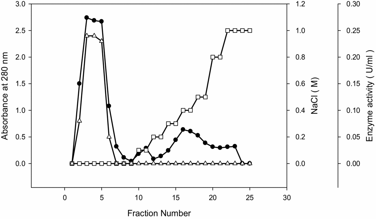
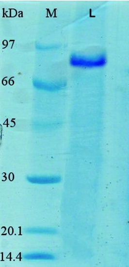
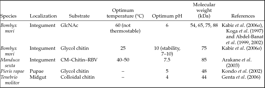
The elution profile obtained using the optimized conditions for cation exchange chromatography on SP-Sepharose described in Methods is shown in Fig. 1. Cation exchange chromatography of the sample resulted in elution of three main peaks of proteins. Chitinase activity assay revealed that the most enzyme activity was found to be in the unbound fraction in fractions of 2 to 6. The second peak (fractions of 10 and 11) was eluted at 0.1 NaCl and the third peak (fractions of 15 to 19) showing enzyme activity was eluted at 0.3 to 0.5 NaCl. A shoulder in the third peak was observed by eluting at 0.8 NaCl. Altogether, three peaks possessed enzyme activity. Under conditions applied on the SP-Sepharose column, the most enzymatic contents have taken the same charge of the column, so that they have eluted as unbound samples, due to repulsion of the same charge on resin and sample. Owing to higher activity of the first peak, fractions 3–5 were selected and combined for further characterization. At the one-step procedure, the chitinase from E. kuehniella was purified approximately 11-fold with a recovery of 41.45%. When colloidal chitin was used as the substrate, the enzyme showed a specific activity of 1.86 μmol/min/mg. Results of the purification procedure are summarized in Table 1. SDS-PAGE analysis of chitinase fraction revealed a single protein bond with 79.7 kDa (Fig. 2). As represented in Table 2, different molecular masses that ranged from 44 to 88 kDa have been reported for insect chitinases. Workers have reported production of several chitinases with molecular weights of 54, 65, 75 and 88 kDa by B. mori (Koga et al., Reference Koga, Sasaki, Uchiumi, Hirai, Arakene and Nagamatsu1997; Abdel-Banat et al., Reference Abdel-Banat, Kameyama, Yoshioka and Koga1999, Reference Abdel-Banat, Zhou, Karasuda and Koga2002; Kabir et al., Reference Kabir, Hirowatari, Watanabe and Koga2006a). Koga et al. (Reference Koga, Sasaki, Uchiumi, Hirai, Arakene and Nagamatsu1997) isolated the 88 kDa chitinase from the silkworm, B. mori larval integument at the early stage of 1 day before ecdysis. They also suggested that the 88 kDa chitinase may be successively processed to a 65 kDa chitinase at a later stage. Therefore, we tried to purify the chitinase from E. kuehniella larval integument 1 day before ecdysis. The cation chromatography, SP-Sepharose column chromatography purified the 79.7 kDa chitinase in earlier fractions. In comparison with the characteristics of other insect chitinases (Table 2), E. kuehniella chitinase is similar to other insect chitinases with respect to the molecular mass. The moulting fluid of Manduca sexta has three chitinases with three sizes (85, 62 and 50 kDa) and B. mori has four chitinases (88, 75, 65 and 54 kDa).
Purification of Ephestia kuehniella chitinase on SP-Sepharose chromatography: The column was equilibrated with 20 mM sodium acetate pH 4.2. The bound proteins were eluted by use of stepwise gradient of 20 mM sodium acetate pH 4.2 containing 0.1 to 1 M NaCl at a flow rate of 2 ml/min. The highest chitinase activity was observed in fraction numbers 3 to 5. Absorbance at 280 nm (●), chitinase activity (∆), step-gradient of salt (□).

Summary of the purification of chitinase from Ephestia kuehniella

* One unit of chitinase activity was defined as the amount of enzyme required to produce 1 μmol of GlcNAc from colloidal chitin per min under the standard conditions of this study. The values are mean ±SD, (n = 3).
SDS-PAGE of purified Ephestia kuehniella chitinase: The proteins were stained with Coomassie Brilliant Blue R 250. Lane L, purified chitinase enzyme band obtained from SP-Sepharose chromatography. Lane M, represents broad range protein molecular mass markers, from the top, Phosphorylase b (97.2 kDa), bovine serum albumin (66.4 kDa), ovalbumin (45 kDa), carbonic anhydrase (30 KDa), trypsin inhibitor (20.1 KDa) and lysozyme (14.4 kDa).

Biochemical characterization of some of the previously reported chitinases from insects

Effect of pH on the activity and stability of E. kuehniella chitinase
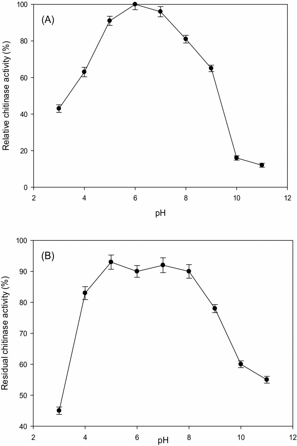
We examined the optimal pH values for chitinase activity and stability of the chitinase. The enzyme was most active between pH 5.5 and 6.5 (Fig. 3A). Some chitinases, including E. kuehniella chitinase, showed an optimum pH requirement of the acidic range. Studies of insect chitinases have shown that the optimum pH of the 54 kDa chitinase from B. mori depends on the substrate being used, about pH 6.0 towards a short substrate, GlcNAc and pH 10.0 (high activity) together with pH 4.0 (low activity) towards a long substrate, glycol chitin (Abdel-Banat et al., Reference Abdel-Banat, Kameyama, Yoshioka and Koga1999). This pH activity profile is similar to that of the 65 and 88 kDa chitinases from B. mori (Koga et al., Reference Koga, Sasaki, Uchiumi, Hirai, Arakene and Nagamatsu1997) and that of M. sexta chitinase (Koga et al., Reference Koga, Jilka and Kramer1983). Finally, we could not compare the optimal pH requirement of the colloidal chitin to other insect chitinases, because we did not measure their optimum pHs. Determination of pH stability of the chitinase indicated that the enzyme was stable between pH 4.5 and 8.0 and it maintained 90% of its original activity in this range after incubation for 4 h at 4 °C (Fig. 3B). The pH stability of the chitinase varies depending on the species.
Effect of pH on enzyme activity (A) and stability (B): The enzyme activity was assayed at 37 °C for 30 min and the stability measured after 4 h pre-incubation of the enzyme at 4 °C in various buffers (pH 3.0–11.0). The values are presented in percentages of the maximum activity under standard assay conditions. The points represent the mean values of three independent measurements. The ranges are shown by the vertical bars.

Effect of temperature on the activity of E. kuehniella chitinase
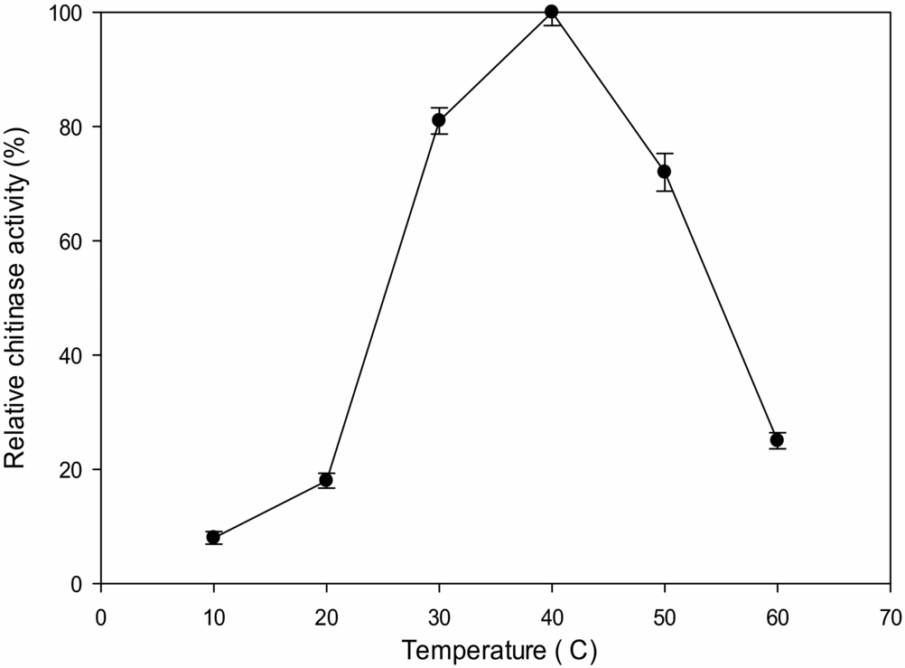
Using colloidal chitin as the substrate, we investigated the optimum temperature of E. kuehniella chitinase. The maximal activity for the enzyme was 40 °C (Fig. 4). Above 45 °C, the activity decreased. Compared to other insect chitinases, it is interesting to note that the optimum temperature of the enzyme differed according to substrate. As in the integument of B. mori, all of the 88, 75, 65 and 54 kDa chitinase isozymes showed an optimum temperature of 60 °C towards GlcNAc, but the 75-kDa chitinase isozymes showed an optimum temperature of 25 °C towards glycolchitin (Abdel-Banat et al., Reference Abdel-Banat, Kameyama, Yoshioka and Koga1999; Kabir et al., Reference Kabir, Hirowatari, Watanabe and Koga2006a; Koga et al., Reference Koga, Sasaki, Uchiumi, Hirai, Arakene and Nagamatsu1997).
Effect of temperature on the activity of Ephestia kuehniella chitinase: The chitinase activity was measured over temperatures ranging from 10 to 60 °C using colloidal chitin as substrate in 30 mM sodium phosphate buffer (pH 7). The points represent the mean values of three independent measurements. The ranges are shown by the vertical bars.

Effects of heavy metal ions on the enzyme activity
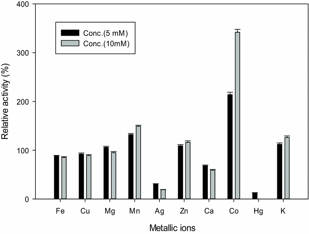
The effects of various metal ions on the chitinase activity were investigated in an assay system, where 5 or 10 mM of each ion was present. From Fig. 5, it is evident that the chitinase activity was inhibited by Ag+ and Hg2+, and was relatively moderately inhibited by Ca2+. The chitinase activity was increased with the addition of Mn2+ and Co2+. The presence of Fe²+ ions showed slight stimulation in enzyme activity, even after increasing the metal ion concentration. Mg2+ ions showed an increase in the chitinase activity at low concentration (5 Mm), but a slight decrease at high concentrations (10 mM). The enzyme was activated by Zn2+ and K+. The particular sensitivity of the E. kuehniella chitinase to Co2+ and the stimulation of enzyme by Fe2+ and Cu2+ could be mainly related to the importance of aspartic and glutamic acid residues in chitinases, as these carboxylic groups have been found to bind certain divalent cations with the active sites and to enhance the efficiency of an enzyme (Milewski et al., Reference Milewski, O'Donnell and Gooday1992; Lee et al., Reference Lee, Park, Yoo, Chung, Lee, Cho, Ahn, Kim and Choi2007).
Effect of metal ions on chitinase activity. Values represent means of three replicates. The ranges are shown by the vertical bars.

Determination of the kinetic properties of E. kuehniella chitinase
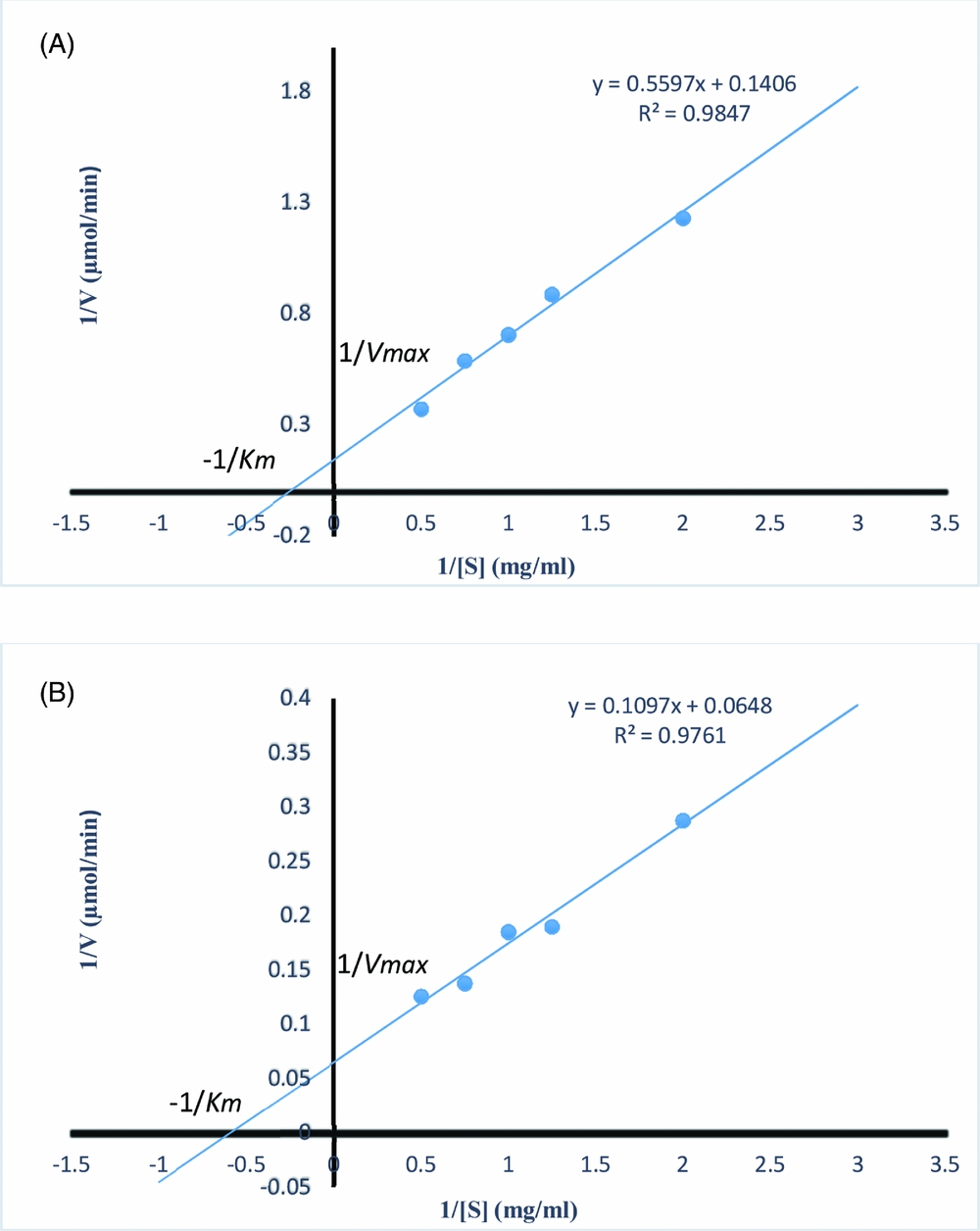
To investigate the enzymatic action of this chitinase, the kinetic parameters of purified chitinase from E. kuehniella using an insoluble substrate (colloidal chitin) and a soluble substrate (CM–Chitin–RBV), were studied. Kinetic parameters were obtained from Lineweaver–Burk double reciprocal plots (Fig. 6A and B). The enzyme was active towards each of the substrates. As shown in Table 3, the K m and V max values were 3.98 mg/ml and 7.11 μmol/min/mg using colloidal chitin as substrate, respectively. With CM–Chitin–RBV as a soluble substrate, the purified chitinase showed Km and V max values of 1.69 mg/ml and 15.4 μmol/min/mg, respectively. Conclusively, when comparing the substrate affinity (1/K m) obtained with colloidal chitin and CM–Chitin–RBV (Table 3), the E. kuehniella chitinase has a 2.36-fold higher affinity for CM–Chitin–RBV. It has been observed in polysaccharide-degrading enzymes that one or more domains are responsible for hydrolysis and one or more domains are responsible for associating with the solid polysaccharide substrate. Arakane et al. (Reference Arakane, Zhu, Matsumiya, Muthukrishnan and Kramer2003) showed that the domain structure of insect chitinase evolves for efficient degradation of the insoluble polysaccharide to soluble oligosaccharides during the moulting process. One study suggested that chitin-binding domains help localize the enzyme on the insoluble substrate to enhance the efficiency of degradation (Linder and Teeri, Reference Linder and Teeri1997). The chitin-binding domain of insect chitinase facilitates hydrolysis of insoluble but not soluble substrates, and also influences the size of the oligosaccharide products generated. With respect to our results, the kinetic behaviour of E. kuehniella chitinase could be related to the domain structure of this enzyme.
Lineweaver–Burk plots of the purified chitinase with colloidal chitin (A) and CM-Chitin-RBV (B) as substrate.

Kinetic parameters of purified chitinase from Ephestia kuehniella toward colloidal chitin substrate and CM-Chitin-RBV

Antibacterial action of the enzyme
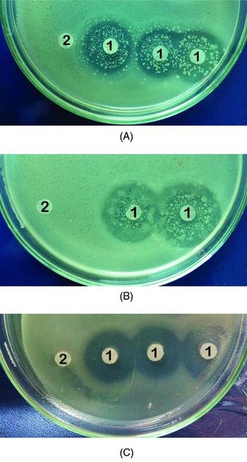
The chitinase was examined for its potential antibacterial activity by paper disc diffusion bioassay against both Gram-positive and Gram-negative bacterial strains. The present study reveals the antibacterial potential of the chitinase against all the tested bacteria (Fig. 7A, B and C). This was shown by the formation of clear zones of inhibition. While the enzyme inhibited the growth of tested bacteria, zones of microbial inhibition were visible. The purified chitinase showed inhibitory activity against B. subtilis, B. thuringiensis and E. coli where the average diameter of inhibition zone reached 20, 23 and 19 mm, respectively. The antibacterial activity of purified chitinase was investigated to evaluate antagonistic activity of the chitinase against some bacterial pathogens of insects and phytopathogens. The antibacterial properties found in this study are similar to the pattern for chitinases from other organisms, such as Monascus purpureus CCRC31499, Streptomyces sp. and Bacillus sp. (Jagadeeswari and Panneer Selvam, Reference Jagadeeswari and Panneer Selvam2012; Mathur et al., Reference Mathur, Rawat, Bhatt, Baweja, Ahmad, Grover, Madhav, Dhand, Mathur, Verma, Singh and Dua2011; Wang et al., Reference Wang, Hsiao and Chang2002).
Antibacterial activity of Ephestia kuehniella chitinase: Antibacterial activity on the growth of Bacillus subtilis (A), Bacillus thuringiensis (B) and Eschershia coli (C) by agar diffusion assay; (1) inhibitor activity, (2) the control (sterile buffer).

This study describes the characterization of the E. kuehniella chitinase and its antibacterial activity. This would suggest the possibility of using the chitinase as a biocontrol agent, which is an attractive and environmentally safe strategy. Recently, Kabir et al. (Reference Kabir, Sugimoto, Tado, Endo, Yamanaka, Tanaka and Koga2006b), while studying the 75 kDa chitinase from B. mori for control of Japanese pine sawyer M. alternatus, found that oral ingestion of purified chitinase caused high mortality in the beetle. Zhang et al. (Reference Zhang, Liu, Tian and Hu2011) compared the antifungal potential of the 88 kDa chitinase from B. mori and the 75 kDa chitinase from H. armigera on the growth of S. cerevisiae and Penicillium. The chitinase from H. armigera exhibited inhibitory effect on spore germination of the tested organisms. Moreover, chitinases have the ability to control insects. Degradation of chitin by chitinase perforates the peritrophic membrane and exoskeleton, and makes insects vulnerable to attack by different pathogens and to lose water. It has been shown that the chitinase from Aeromonas hydrophila SBK1 lyses the musculatures and internal canals of Culex quinquefasciatus larvae and can be used as an insecticide (Halder et al., Reference Halder, Maity, Jana, Pati and Keshab2012). Consequently, identification and characterization of various types of insect chitinases based on the structure and function is important.
Conclusion
Insect chitinases play important roles in moulting and digestion, and defensive roles against their own parasites. Apart from the physiological role of chitinases, chitinases can be used as inhibitors and biopesticides. Further work on characterization of isozymes of E. kuehniella chitinase will provide us with the tools to make better use of them.
Acknowledgements
We thank Dr Ahmad Reza Bahrami for technical assistance during this work.