I. INTRODUCTION
Lanthanide complexes with organic ligands have long been the subject of extensive research for their special luminescence properties with high color purity and long lifetimes as well as high quantum efficiency, which can be expected to have potential applications in a variety of fields such as luminescent sensors, lasers, optical amplification, organic-light emitting diodes, and light-conversion molecular devices. Reference Koeppen, Yamada, Jiang, Garito and Dalton1–Reference Kido and Okamoto4 However, due to their poor thermal stabilities and low mechanical strength, the lanthanide complexes have been excluded from practical applications. One solution is to incorporate the lanthanide complexes into stable host matrices such as inorganic gel or organic polymer to construct organic–inorganic hybrid materials, which not only improved the photo and thermal stabilities of the complexes but also avoided the self-quenching resulting from the concentration effect. Reference Li, Lin, Zhang, Fu, Meng and Wang5–Reference Zhang, Wang, Qiao, Tang, Liu and Huo7 The mesoporous molecular sieves used as a support for rare earth complexes have attracted considerate attention because they possess properties such as photostability, rigidity, and well-defined hydrophilic/hydrophobic properties, which allow for sophisticated tuning of the rare earth complex microenvironment. Reference Guo, Guo, Fu, Deng, Chen, Feng, Dang and Zhang8 Among them, SBA-15 has certainly become one of the most attractive hosts due to its large pore size, thick walls, adjustable pore size from 3 to 30 nm, high hydrothermal and thermal stability. Reference Zhao, Sun, Li and Stucky9,Reference Madhugiri, Dalton, Gutierrez, Ferraris and Balkus10 Until recently, Zhang Reference Peng, Zhang, Yu, Meng, Fu, Li, Sun and Guo11–Reference Peng, Zhang, Meng, Li, Yu, Guo and Sun13 and Yan Reference Li, Yan and Yang14–Reference Li, Yan and Li18 group reported the synthesis and luminescent properties of lanthanide complexes chemically bonded to mesoporous silica SBA-15. It is shown that excellent visible-luminescent properties can be obtained via linking the lanthanide complexes to mesoporous silica materials.
In this paper, hexafluoroacetylacetone (HFAA)-functionalized SBA-15 mesoporous hybrid (HFAASi-SBA-15) was prepared, in which HFAA was covalently bonded to the framework SBA-15 by co-condensation of modified HFAA (denoted as HFAASi) and tetraethoxysilane (TEOS) in the presence of Pluronic P123 surfactant as a template. The highly luminescent ternary lanthanide (Eu3+, Tb3+) complex on functionalized SBA-15, denoted as Ln(HFAASi-SBA-15)3phen (Ln = Eu, Tb), was obtained by introducing Ln3+ and 1,10-phenanthroline (phen) into the hybrid material HFAASi-SBA-15 through chemically bond assembling method. Full characterization and detail studies of luminescent properties of all these synthesized hybrid materials were investigated.
II. EXPERIMENTAL SECTION
A. Chemicals
Europium and terbium nitrates were obtained by dissolving their respective oxides (Eu2O3 and Tb4O7) in concentrated nitric acid. Hexafluoroacetylacetone (HFAA), 3-(triethoxysilyl)-propyl isocyanate (TESPIC), and Pluronic P123 (EO20PO70EO20) were purchased from the Lancaster company (Shanghai, China). Tetraethoxysilane (TEOS, Aldrich, Shanghai, China) was distilled and stored under nitrogen atmosphere. The solvent tetrahydrofuran (THF) was introduced after desiccation with anhydrous calcium chloride. All the other reagents are analytically pure.
B. Synthesis
1. Synthesis of HFAA-functionalized SBA-15 mesoporous material (HFAASi-SBA-15)
The modified precursor HFAASi was synthesized according to the procedure reported in the literature, Reference Wang and Yan19 described as follows: first, HFAA (1 mmol, 0.21 g) was dissolved in 20 mL of dehydrate THF with stirring, and NaH (2 mmol, 0.048 g) was added to the solution. Two hours later, 2.0 mmol (0.49 g) of TESPIC was then added to the above mixture solution by drops for half an hour. The whole mixture was refluxing at 70°C under an atmosphere of argon for 12 h. A yellow oil liquid sample, i.e., the precursor HFAASi was obtained after isolation and purification. The mesoporous material HFAASi-SBA-15 was prepared from acidic mixture with the following molar composition: 0.0172 P123: 0.96 TEOS: 0.04 HFAASi: 6 HCl: 208.33 H2O. 1.0 g of P123 was dissolved in 7.5 g of deionized water and 30 g of 2 M HCl solution at 35 °C under vigorous stirring. A mixture of TEOS and HFAASi was added to the above solution, which was stirred at 35 °C for 24 h and transferred into a Teflon bottle sealed in an autoclave, which was then heated at 100 °C for 48 h. The solid product was filtrated, washed thoroughly with deionized water, and air-dried at 60 °C for 10 h. Removal of copolymer surfactant P123 was conducted through Soxhlet extraction with ethanol under reflux for 48 h to give the sample denoted as HFAASi-SBA-15.
2. Synthesis of SBA-15 mesoporous material chemically bonded with the ternary Ln3+ complexes [denoted as Ln(HFAASi-SBA-15)3phen (Ln = Eu, Tb)]
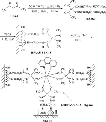
The sol–gel-derived hybrid mesoporous material Ln(HFAASi-SBA-15)3phen was prepared as follows: while being stirred, HFAASi-SBA-15 was soaked in an appropriate amount of Ln(NO3)3 and 1,10-phenanthroline (phen) ethanol solution with the molar ratio of Ln3+:HFAASi-SBA-15:phen = 1:3:1. The mixture was stirred at room temperature for about 12 h followed by filtration and extensive washing with EtOH. The resulting materials Ln(HFAASi-SBA-15)3phen were dried at 60 °C under vacuum overnight. The predicted structure of Ln(HFAASi-SBA-15)3phen was obtained as outlined in Fig. 1.
Synthesis process and predicted structure of the ternary mesoporous hybrid material Ln(HFAASi-SBA-15)3phen (Ln = Eu, Tb).

C. Characterization
Fourier transform infrared (FTIR) spectra were measured within the 4000–400 cm−1 region on a Nicolet 6700 spectrophotometer (Thermo Electron Corporation, MA) with the KBr pellet technique. Nitrogen (N2) adsorption/desorption isotherms were measured by using a Nova 2000 analyzer (Quantachrome, Beijing, China) at the liquid nitrogen temperature. Before the measurements, the sample was outgassed for 3 h at 423 K. Surface areas were calculated by the Brunauer–Emmett–Teller (BET) method and pore size distributions were evaluated from the desorption branches of the nitrogen isotherms using the Barrett–Joyner–Halenda (BJH) model. X-ray powder diffraction patterns were recorded on a Rigaku D/max2500VB+/PC diffractometer (Hitachi Limited, Tokyo, Japan) equipped with a Cu anode in a 2θ range from 0.6° to 6°. Transmission electron microscope (TEM) observation was made on a JEOL2011 microscope (Hitachi Limited, Tokyo, Japan) operated at 200 kV and on a JEM-4000EX microscope (Hitachi Limited, Tokyo, Japan) operated at 400 kV. Reflectivity spectra were recorded on a UV-2501 spectrometer (Shimadzu, Tokyo, Japan) equipped with a diffuse reflectance accessory. The fluorescence excitation and emission spectra were obtained on a Hitachi F-4500 spectrophotometer (Hitachi Limited, Tokyo, Japan). The luminescence lifetime measurements were measured with an Edinburgh FLS920 phosphorimeter (Edinburgh Instruments Ltd., UK) using a 450 W xenon lamp as the excitation source.
III. RESULTS AND DISCUSSION
A. FTIR spectra
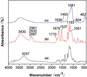
The presence of the organic ligand HFAA covalently bonded to the mesoporous SBA-15 was characterized by FTIR spectra. The FTIR spectra for HFAA (a), the precursor HFAASi (b), and HFAA-functionalized mesoporous hybrid material HFAASi-SBA-15 (c) are shown in Fig. 2. From a to b, it can be observed that the stretching of –CH2– at 3257 cm−1 (a) was replaced by the peaks at 2981, 2930, and 2887 cm−1 (b), which originated from three methylene groups of TESPIC. In addition, the spectrum of HFAASi is dominated by ν (C–Si, 1193 cm−1) and ν (Si–O, 1081 cm−1) absorption bands, characteristic of trialkoxylsilyl functions, which suggests formation of the siloxane bonds. The band centered at 3430 cm−1 corresponds to the stretching vibration of grafted –NH– groups. Besides, the bending vibration (δNH, 1537 cm−1) further proves the formation of amide groups. New bands at 1770 and 1679 cm−1 in curve b were due to the absorptions of the C=O of TESPIC, proving that TESPIC was successfully grafted onto the ligand HFAA. In Fig. 2(c), the formation of Si–O–Si framework is evidenced by the broad bands located at 1081 cm−1 (νas, Si–O), 804 cm−1 (νs, Si–O), and 462 cm−1 (δ, Si–O–Si), which is ascribed to the success of hydrolysis and copolycondensation (where ν represents stretching; δ, plane bending; s, symmetric; and as, asymmetric vibrations). Moreover, the bands at 1636 and 1493 cm−1 originating from –CONH– group of HFAASi, can also be observed in mesoporous hybrid material HFAASi-SBA-15 (c), which is consistent with the fact that the HFAA group in the framework remains intact after both the hydrolysis–condensation reaction and the surfactant extraction procedure. Reference Peng, Zhang, Yu, Meng, Fu, Li, Sun and Guo11
FTIR spectra of HFAA (a), HFAASi (b), and HFAA-functionalized mesoporous hybrid HFAASi-SBA-15 (c).

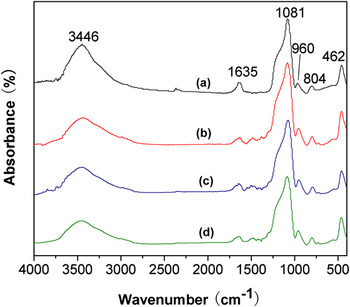
Figure 3 gives the FTIR spectra of SBA-15 (a), HFAASi-SBA-15 (b), Eu(HFAASi-SBA-15)3phen (c), and Tb(HFAASi-SBA-15)3phen (d). In the spectrum of the SBA-15 host material (a), the obvious absorption bands from –OH– (3446 cm−1), Si–O–Si (νas, 1081 cm−1; νs, 804 cm−1), Si–OH (νs, 960 cm−1), Si–O (δ, 462 cm−1), and H2O (1635 cm−1) are present. Reference Yu, Lin and Fang20,Reference Yu, Lin, Wang, Fu, Wang, Zhang and Han21 Compared with SBA-15, the mesoporous hybrid materials HFAASi-SBA-15 and Ln(HFAASi-SBA-15)3phen (Ln = Eu, Tb) not only show the similar absorption bands as the silica framework, but also the bands at the range of 2850–3000 cm−1 attributed to the vibrations of methylene –(CH2)3– and the bands at 1380–1630 cm−1 range originating from the –CONH– group of HFAASi, indicates that HFAASi has been grafted onto the wall of SBA-15.
FTIR spectra of SBA-15 (a), HFAASi-SBA-15 (b), Eu(HFAASi-SBA-15)3phen (c), and Tb(HFAASi-SBA-15)3phen (d).

B. X-ray diffraction
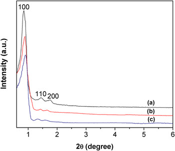
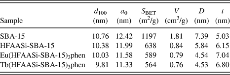
The small-angle powder x-ray diffraction (SAXRD) measurement is a very popular and efficient method to characterize the nanostructured host-guest hybrid materials. Figure 4 shows the SAXRD patterns of HFAASi-SBA-15 (a), Eu(HFAASi-SBA-15)3 (b), and Tb(HFAASi-SBA-15)3phen (c). These patterns exhibit three well-resolved diffraction peaks in the 2θ range of 0.6–2°, which can be indexed as (100), (110), and (200) reflections of a two-dimensional hexagonal structure (p6mm) of SBA-15 material. Compared with the SAXRD pattern of HFAASi-SBA-15 material, the d 100 spacing values of Eu(HFAASi-SBA-15)3phen and Tb(HFAASi-SBA-15)3phen are nearly unchanged (see Table I), suggesting that their framework ordering has been retained well after the introduction of Ln3+. However, it is worth noting that Ln3+ complex functionalized materials Eu(HFAASi-SBA-15)3phen and Tb(HFAASi-SBA-15)3phen appear slight decrease in diffraction intensity as compared with the parent HFAASi-SBA-15 material, which is probably because of the steric constraints introduced by the linking of ligands via complexation with the lanthanide ions, resulting in a larger heterogeneity of the pores. Reference Li, Yan and Li22
SAXRD patterns of HFAASi-SBA-15 (a), Eu(HFAASi-SBA-15)3phen (b) and Tb(HFAASi-SBA-15)3phen (c).

Textural data of SBA-15, HFAASi-SBA-15, Eu(HFAASi-SBA-15)3phen and Tb(HFAASi-SBA-15)3phen. a

a d 100 is the d(100) spacing, a 0—the cell parameter (a 0 = 2 d 100/√3), S BET—the BET surface area, V—the total pore volume, D—the average pore diameter, and t—the wall thickness, calculated by a 0 − D.
C. Nitrogen sorption measurement
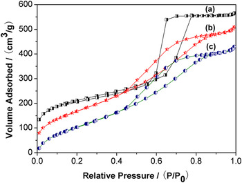
The measurements of nitrogen adsorption/desorption isotherms further provide proof for the preservation of the ordered mesoporous structure upon the introduction of the lanthanide ion. The nitrogen adsorption/desorption isotherms of HFAASi-SBA-15 (a), Eu(HFAASi-SBA-15)3phen (b), and Eu(HFAASi-SBA-15)3phen (c) are presented in Fig. 5. All these samples exhibit type IV isotherm curves with a clear H1 type hysteresis at high relative pressure, characteristic of highly ordered mesoporous materials according to the IUPAC classification. Reference Yan, Wang and Li23–Reference Zhang, Lu, Xiu, Hua, Zhang, Robertson, Shi, Yan and Holmes25 By using BET and BJH methods, the specific area and the pore size have been calculated. And the structural data of all these mesoporous hybrid materials (BET surface area, total pore volume, BJH average pore size, etc.) are summarized in Table I. It was observed that HFAASi-SBA-15 exhibits a smaller specific area, and a slightly smaller pore size and pore volume compared with those typically reported for pure SBA-15 mesoporous silica, Reference Peng, Zhang, Yu, Meng, Fu, Li, Sun and Guo11 which might be due to the presence of both organic ligand HFAA on the pore surface and the cosurfactant effect of HFAASi, which interacts with surfactant and reduces diameter of the micelles. Reference Kong, Yan and Li26 Furthermore, upon introduction of Ln3+ into the HFAASi-SBA-15, the specific area, pore size, and pore volume of the hybrid materials containing Ln3+ were less than those of HFAASi-SBA-15. This further confirmed the incorporation of the Ln3+ complexes in the channels of SBA-15.
N2 adsorption–desorption isotherms of HFAASi-SBA-15 (a), Eu(HFAASi-SBA-15)3phen (b) and Tb(HFAASi-SBA-15)3phen (c).

D. Transmission electron microscope
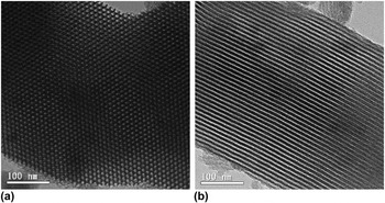
The hexagonal mesostructures of Eu(HFAASi-SBA-15)3phen are further confirmed by TEM images (see Fig. 6). As presented in Fig. 6, the Eu3+ complex-functionalized mesoporous material Eu(HFAASi-SBA-15)3phen presents a P6mm symmetry and regular hexagonal structure, which suggests that the mesostructure of Eu(HFAASi-SBA-15)3phen can be substantially conserved after the complexation process. In addition, the distance between the centers of the mesopores is estimated to be around 10 nm, which is in good agreement with the value determined from the corresponding SAXRD data (see Table I).
HRTEM images of Eu(HFAASi-SBA-15)3phen recorded along the [100] (a) and [110] (b) zone axes.

E. Ultraviolet-visible diffuse reflection absorption spectra
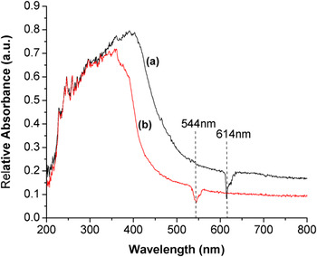
The ultraviolet-visible diffuse reflectance spectroscopy of the lanthanide mesoporous hybrids is given in Fig. 7 [(a) for Eu(HFAASi-SBA-15)3phen and (b) for Tb(HFAASi-SBA-15)3phen]. It is observed that both the spectra exhibit a broad absorption band in the UV-vis range (200–500 nm), which corresponds to transition from the ground state of the organic ligand to the first excited state (S0 → S1). Following Dexter's exchange energy transfer theory, Reference Fan, Feng, Song, Lei, Zheng and Zhang27 the luminescence intensity of hybrid materials depends on the matching degree between the triplet-state energy of ligand and excited-state energy of lanthanide ion. In terms of the above phenomena, it can be primarily predicted that the energy levels difference between HFAASi and lanthanide ions is suited and appropriate so that the organic ligand can absorb abundant energy in ultraviolet-visible extent to transfer the energy to the corresponding rare earth ions. Then the final hybrid materials can be expected to have excellent luminescence, which is proved by the emission spectra of these materials (see Figs. 8 and 9). In addition, the inverse peak at 614 nm for Eu(HFAASi-SBA-15)3phen and 545 nm for Tb(HFAASi-SBA-15)3phen, is the characteristic emission of europium and terbium ions, respectively.
The ultraviolet-visible diffuse reflection absorption spectra of Eu(HFAASi-SBA-15)3phen (a) and Tb(HFAASi-SBA-15)3phen (b).

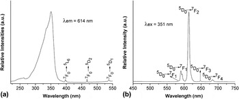
The excitation spectra (a) and emission spectra (b) of Eu(HFAASi-SBA-15)3phen.

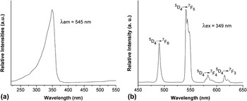
The excitation spectra (a) and emission spectra (b) of Tb(HFAASi-SBA-15)3phen.

F. Photoluminescence properties
The luminescence behavior of lanthanide mesoporous hybrid materials has been investigated at room temperature. The fluorescent excitation and emission spectra of europium mesoporous hybrid material are given in Fig. 8. The excitation spectrum of Eu(HFAASi-SBA-15)3phen was obtained by monitoring the strongest emission wave length of Eu3+ ion at 614 nm. The excitation spectrum displays a large broad band ascribed to the excited states of the ligands (220–370 nm, with one component at 351 nm) and a series of narrow lines attributed to transitions between the 7F0 levels and the 5L6, and 5D1,2 excited states. The higher intensity of the broad-band excitation component with respect to that of the intra-4f 6 lines indicates that the Eu3+ sensitization process is more efficient than the direct intra-4f 6 excitation. So the emission spectrum was measured under excitation wave length of 351 nm. As a result, five sharp emission peaks at 579, 591, 614, 648, and 684 nm were seen, which are corresponding to the 5D0 → 7F J (J = 0, 1, 2, 3, 4) transitions of Eu3+, respectively. Among these transitions, the 5D0 → 7F2 transition at about 614 nm shows the most prominent emission. It is well known that the 5D0 → 7F2 transition belongs to a typical electric dipole transition and strongly varies with the local symmetry of Eu3+, whereas the 5D0 → 7F1 transition corresponds to a parity-allowed magnetic dipole transition, which is independent of the host material. The intensity ratio of the red and orange is sensitive to the symmetry around Eu3+. When the ratio is higher, europium ion occupies a lower symmetry microenvironment. Reference Fan, Feng, Song, Lei, Zheng and Zhang27–Reference Guillet, Imbert, Scopelliti and Bünzli29 The intensity ratio of the two lines (5D0 → 7F2/5D0 → 7F1) is about 5.14, which indicates that the Eu3+ ion is not at the center of an asymmetric coordination field.
Figure 9 shows the excitation and emission spectra for terbium mesoporous hybrid Tb(HFAASi-SBA-15)3phen. The excitation spectrum of the material was obtained by monitoring the emission of the Tb3+ ions at 545 nm, which exhibits broad excitation band centered at 349 nm in the ultraviolet region, corresponding to the ligand-to-metal resonant energy transfer caused by interaction between the organic groups and the rare-earth ions. Reference Li, Yan and Wang30–Reference Malta, Brito, Menezes, Silva, Alves, Farias and DeAndrade32 No apparent f–f transitions could be observed in the spectra. This indicates that an effective energy transfer occurs from the ligands to the central Tb3+ ions. Excitation at 349 nm provides the typical luminescence of Tb3+ ion at 490, 541, 583, and 621 nm, corresponding to 5D4 → 7F J transitions (J = 6, 5, 4, and 3, respectively). As a result, the strong green luminescence was observed in the emission spectra. The mesoporous hybrid materials show relatively strong emissions due to the chemically covalently bonded molecular Si–O network structure between the complexes and the mesoporous silica.
G. Luminescence decay times (τ) and emission quantum efficiency (η)
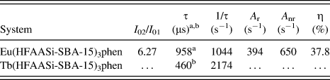
To further discuss the photophysical properties of the resulting mesoporous materials, the typical decay curves of the europium and terbium mesoporous hybrid materials are measured, and they can be described as a single exponential (ln(S(t)/S0) = −k 1 t = −t/τ), indicating that Eu3+ and Tb3+ ions occupy the same average coordination environment. The resulting lifetime data of rare earth mesoporous hybrid materials are given in Table II. It was found that the mesoporous hybrid material Eu(HFAASi-SBA-15)3phen presents longer luminescent lifetime than the hybrid material Tb(HFAASi-SBA-15)3phen, suggesting that HFAASi is more suitable for the luminescence of Eu3+ than that of Tb3+.
Luminescence data of the mesoporous hybrid materials.

a For the 5D0 → 7F2 transition of Eu3+.
b For the 5D4 → 7F5 transition of Tb3+.
Further, we selectively determined the emission quantum efficiencies of the 5D0 excited state of the europium ion on the basis of the emission spectra and lifetimes of the 5D0 emitting level. The detailed luminescent data are shown in Table II. Assuming that only nonradiative and radiative processes are essentially involved in the depopulation of the 5D0 state, η can be defined as follows Reference Malta, DosSantos, Thompson and Ito33–Reference Lima, Nobre, Freire, Junior, Mafra, Ferreira, Pischel, Malta and Carlos37 :
 $${\rm{\eta }} = {A_{\rm{r}}}/({A_{\rm{r}}} + {A_{{\rm{nr}}}})\quad ,$$
Here, A r and A nr are radiative and nonradiative transition rates, respectively. A r can also be obtained by summing over the radiative rates A 0J for each of the 5D0 → 7F J (J = 0–4) transitions. The branching ratio for the 5D0 → 7F5,6 transitions can be neglected as they are not detected experimentally, whose influence can be ignored in the depopulation of the 5D0 excited state. Since 5D0 → 7F1 belongs to the isolated magnetic dipole transition, it is practically independent of the chemical environments around Eu3+, and can be considered as an internal reference for the whole spectra, the experimental coefficients of spontaneous emission A 0J can be calculated according to the equation as follows:
 $${A_{0J}} = {A_{01}}({I_{0J}}/{I_{01}})({{\rm{\nu }}_{01}}/{{\rm{\nu }}_{0J}})\quad ,$$
Here, A 0J is the experimental coefficients of spontaneous emission. A 01 is the Einstein's coefficient of spontaneous emission between the 5D0 and 7F1 energy levels. In vacuum, A 01 could be used as a value of 14.65 s−1, whereas in the air atmosphere, the value of A 01 can be determined to be 50 s−1 approximately (A 01 = n 3 A 01(vac)), Reference Wang and Yan19 when an average index of refraction n equal to 1.506 was considered. I 01 and I 0J are the integrated intensities of the 5D0 → 7F1 and 5D0 → 7F J transitions (J = 0–4) with ν01 and ν0J (ν0J = 1/λ J ) energy centers, respectively. ν0J refers to the energy barrier and can be determined with the emission peaks of 5D0 → 7F J emission transitions of Eu3+. The emission intensity, I, is taken as the integrated intensity of the 5D0 → 7F0–4 emission curves.
On the basis of the above discussion, the quantum efficiencies of mesoporous hybrid material Eu(HFAASi-SBA-15)3phen can be determined as shown in Table II. In addition, compared with the hybrid Eu(HFAASi)3phen (34.8%) reported in the literature, Reference Wang and Yan19 the quantum efficiency of corresponding mesoporous hybrid material Eu(HFAASi-SBA-15)3phen (37.8%) is higher, which shows that SBA-15 is an excellent host for the lanthanide β-diketone complex.
IV. CONCLUSIONS
In summary, the organic–inorganic mesoporous luminescent hybrid materials Ln(HFAASi-SBA-15)3phen were obtained by linking the ternary lanthanide (Eu3+, Tb3+) complexes to the ordered SBA-15 mesoporous material by the modification of HFAA with TESPIC using a co-condensation method. The resulting materials Ln(HFAASi-SBA-15)3phen all have high surface area and preserve mesoscopically ordered structures. Further investigation into the luminescent properties of Ln(HFAASi-SBA-15)3phen mesoporous materials exhibits that the characteristic luminescence of the corresponding lanthanide ions (Ln3+) through the intramolecular energy transfers from the modified ligand (HFAASi) to the lanthanide ions. The excellent luminescent properties of these materials, together with the uniform tunable pore sizes and the ordered hexagonal channel structure of the organic group functionalized SBA-15 mesoporous hybrid materials will expand their applications in both optical and electronic areas.
ACKNOWLEDGMENTS
This work was supported by the Scientific Research Foundation of Hebei Province Education Department (No. Z2013015), the Doctoral Science Foundation of Hebei University of Science and Technology (No. QD201255), and the Xiaoli Foundation of Hebei University of Science and Technology (XL201257 and XL201250).