Introduction
Depressed individuals demonstrate a poorer ability to recognize the emotions of others (Gur et al. Reference Gur, Erwin, Gur, Zwil, Heimberg and Kraemer1992; Persad & Polivy, Reference Persad and Polivy1993), which may contribute to difficulties in interpersonal behaviour (Libet & Lewinsohn, Reference Libet and Lewinsohn1973; Youngren & Lewinsohn, Reference Youngren and Lewinsohn1980). Studies investigating the recognition of facial expressions suggest that depressed individuals are particularly impaired when they need to label emotions semantically, although they also have more subtle deficits in the perceptual categorization of emotions (Feinberg et al. Reference Feinberg, Rifkin, Schaffer and Walker1986; Rubinow & Post, Reference Rubinow and Post1992). Difficulties with emotion recognition and interpersonal behaviour improve with remission from depression, indicating that these abnormalities are dependent on the depressive state (Hinchliffe et al. Reference Hinchliffe, Lancashire and Roberts1971; Mikhailova et al. Reference Mikhailova, Vladimirova, Iznak, Tsusulkovskaya and Sushko1996).
Labelling of emotions requires the recruitment of a semantic network in addition to the neural network that is necessary for emotion recognition. The language processing network mainly exists of the left inferior frontal gyrus and left temporal cortex (Hagoort, Reference Hagoort2005), and the network involved in facial emotion recognition includes the amygdala, orbitofrontal cortex and occipitotemporal cortex (Adolphs, Reference Adolphs2002). Neuroimaging studies in depressed patients have demonstrated both structural and functional brain changes (Davidson et al. Reference Davidson, Pizzagalli, Nitschke and Putnam2002; Phillips et al. Reference Phillips, Drevets, Rauch and Lane2003) as well as altered neural responses to emotional faces (Fu et al. Reference Fu, Williams, Cleare, Brammer, Walsh, Kim, Andrew, Pich, Williams, Reed, Mitterschiffthaler, Suckling and Bullmore2004; Surguladze et al. Reference Surguladze, Brammer, Keedwell, Giampietro, Young, Travis, Williams and Phillips2005; Lee et al. Reference Lee, Seok, Lee, Cho, Yoon, Lee, Chae, Choi and Ham2008; Matthews et al. Reference Matthews, Strigo, Simmons, Yang and Paulus2008; Peluso et al. Reference Peluso, Glahn, Matsuo, Monkul, Najt, Zamarripa, Li, Lancaster, Fox, Gao and Soares2009) in parts of those networks. This suggests that these brain regions may also mediate emotion recognition deficits.
In the present study, we investigated the neural basis of emotion recognition deficits related to emotion labelling and emotion matching in major depression. We included a group of medication-naive patients with a first major depressive episode (MDE), a group of medication-free patients who had recovered from a first MDE and a group of matched healthy individuals. We investigated medication-free patients with a first depressive episode to control for acute medication effects (Murphy et al. Reference Murphy, Norbury, O'Sullivan, Cowen and Harmer2009) and structural brain changes associated with chronic disease (MacQueen et al. Reference MacQueen, Campbell, McEwen, Macdonald, Amano, Joffe, Nahmias and Young2003). The participants were requested to identify the emotion of angry and fearful face stimuli, either by matching them to other emotional faces on a perceptual basis or by labelling the emotion semantically, while their brain activity was measured with functional magnetic resonance (MR) imaging (MRI) (Hariri et al. Reference Hariri, Bookheimer and Mazziotta2000). This allowed us to investigate the neural basis of emotion recognition deficits under semantic categorization and perceptual matching response conditions, and to identify whether this is state or trait related. Based on the larger behavioural deficits when emotions are labelled semantically (Feinberg et al. Reference Feinberg, Rifkin, Schaffer and Walker1986), we hypothesized that neural differences in language processing and emotion recognition networks would particularly be present in the emotion-labelling condition. Furthermore, because the emotion recognition deficits appear to recover after remission (Mikhailova et al. Reference Mikhailova, Vladimirova, Iznak, Tsusulkovskaya and Sushko1996), we hypothesized that the neural differences would be state related.
Method
Participants
A total of 20 medication-naive patients diagnosed with a first MDE, 21 medication-free patients who had recovered from a first MDE and 30 healthy individuals were included in this study (van Wingen et al. Reference van Wingen, Van Eijndhoven, Cremers, Tendolkar, Verkes, Buitelaar and Fernández2010). The currently depressed patients were investigated just prior to the start of pharmacological or psychological treatment. The recovered patients had discontinued selective serotonin reuptake inhibitors or venlafaxine (four patients) treatment for at least 2 months [10.6 (s.d.=6.9) months], had received antidepressant treatment for 18.8 (s.d.=11.2) months, and were asymptomatic for 11.9 (s.d.=6.8; all ⩾6) months (Frank et al. Reference Frank, Prien, Jarrett, Keller, Kupfer, Lavori, Rush and Weissman1991). Major depressive disorder was diagnosed using the Structured Clinical Interview for the Diagnostic and Statistical Manual of Mental Disorders, 4th edition (DSM-IV) (First et al. Reference First, Spitzer, Gibbon and Williams1996) by an experienced psychiatrist (P.v.E.). The additional inclusion criterion for the depressed group was a moderate to severe depression (⩾17 on the 17-item Hamilton Rating Scale for Depression; HAMD), and for the recovered group the absence of clinically relevant symptoms defined as a HAMD score ⩽7 (Frank et al. Reference Frank, Prien, Jarrett, Keller, Kupfer, Lavori, Rush and Weissman1991). Patients with current or past bipolar, anxiety, substance abuse, psychotic and eating disorders as assessed with the Mini International Neuropsychiatric Interview (MINI) (Sheehan et al. Reference Sheehan, Lecrubier, Sheehan, Amorim, Janavs, Weiller, Hergueta, Baker and Dunbar1998) and medical history interview were excluded. Inclusion criteria for healthy individuals were no history of psychiatric disorders as confirmed by use of the MINI and medical history interview, and no history of psychiatric disorders in first-degree relatives. All participants were physically healthy, currently medication free and had no MRI contraindications.
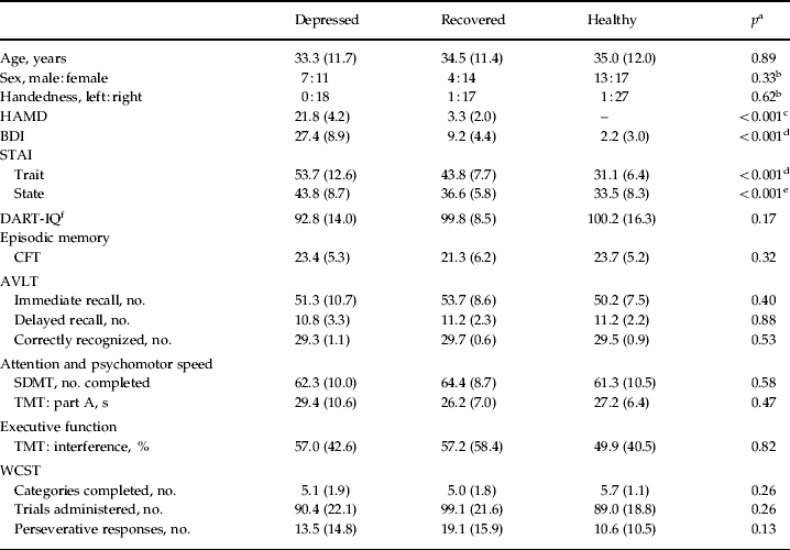
Of the participants, three were excluded from the analysis because of brain abnormalities (two depressed and one recovered patient), and two recovered patients were excluded because of technical difficulties. Therefore, the results are based on data of 18 depressed, 18 recovered and 30 healthy individuals. These groups did not differ significantly in age, sex ratio, general intelligence and handedness (see Table 1). The study was in accordance with the Declaration of Helsinki and approved by the local ethics committee (CMO Regio Arnhem-Nijmegen, The Netherlands). Volunteers were recruited by local newspaper advertisements and our out-patient clinic, and all participants gave written informed consent prior to participation.
Demographic, clinical and neuropsychological test data of currently depressed (n=18), recovered (n=18) and healthy individuals (n=30)

Data are given as mean (s.d.).
HAMD, Hamilton Rating Scale for Depression; BDI, Beck Depression Inventory; STAI, State Trait Anxiety Inventory; DART, Dutch version of the National Adult Reading Test (Schmand et al. Reference Schmand, Lindeboom and van Harskamp1992); IQ, intelligence quotient; CFT, Rey-Osterrieth Complex Figure Test (Osterrieth, Reference Osterrieth1944); AVLT, Dutch modified version of the Rey Auditory Verbal Learning Test (Saan & Deelman, Reference Saan and Deelman1986); SDMT, Symbol Digit Modalities Test (Smith, Reference Smith1982); TMT, Trail Making Test (Lezak, Reference Lezak1995); WCST, Wisconsin Card Sorting Test (Heaton et al. Reference Heaton, Chelune, Talley, Kay and Curtiss1993), s.d., standard deviation.
a One-way analysis of variance.
b Pearson χ2.
c Independent-samples t test.
d Depressed>recovered>healthy; independent-samples t tests.
e Depressed>recovered=healthy; independent-samples t tests.
f The DART-IQ estimate is about 0.5 s.d. lower than the Wechsler Adult Intelligence Scale estimate (Bouma et al. Reference Bouma, Mulder and Lindeboom1998).
Procedure
All individuals participated in the study on 2 days during the afternoon. Day 1 started with a clinical interview to confirm the assignment to the depressed, recovered or healthy control group (see above). Thereafter, participants completed the Beck Depression Inventory (BDI; Beck et al. Reference Beck, Ward, Mendelson, Mock and Erbaugh1961), the trait version of the State Trait Anxiety Inventory (STAI; Spielberger et al. Reference Spielberger, Gorsuch and Lushene1970) and the Edinburgh Handedness Inventory (Oldfield, Reference Oldfield1971). The scanning session took place on day 2, after which the participants completed the state version of the STAI. Finally, neuropsychological function was assessed using a neuropsychological test battery (see Table 1).
Behavioural paradigm
The experimental paradigm has previously been validated in healthy individuals (see Hariri et al. Reference Hariri, Bookheimer and Mazziotta2000) and consisted of a blocked design, including an emotion-labelling, emotion-matching and a baseline control task. Each trial consisted of three simultaneously presented stimuli, with one cue stimulus presented above one target and one distractor. In the emotion-labelling condition, an angry or fearful face (http://www.macbrain.org) was presented on top with the words ‘anger’ and ‘fear’ underneath. In the emotion-matching condition, again an angry or fearful face was presented on top, but now with one other angry and one other fearful face presented underneath. In the baseline control condition, a horizontally or vertically oriented ellipse was presented on top with one vertically and one horizontally oriented ellipse underneath. For each trial, the participant was requested to select with a button press the bottom stimulus that correctly described the emotional expression of the cue (labelling task), expressed the same emotion (matching task) or had an identical orientation (baseline control task). Half of the trials per block were from each target emotion or orientation. The 12 actors for the cue faces appeared once in the labelling and once in the matching task, once fearful and once angry, which was counterbalanced across participants. Two emotion-labelling blocks and two emotion-matching blocks were interleaved with five baseline control blocks. Each block consisted of six stimuli, resulting in a total of 12 labelling, 12 matching and 30 control stimuli, which were presented for 3.3 s each with a 0.2 s interstimulus interval, producing blocks of 21 s.
MRI data acquisition
MR data were acquired with a 1.5 T Siemens (Erlangen, Germany) Sonata MR scanner, equipped with a standard head coil. T2*-weighted blood-oxygenation level-dependent (BOLD) images were acquired using echo-planar images (EPI), with each image volume consisting of 33 axial slices (3 mm, 0.5 mm slice-gap, repetition time=2.290 s, echo time=30 ms, 64×64 matrix, field of view=224 mm, flip angle=90°). In addition, a high-resolution T1-weighted structural MR image was acquired for spatial normalization procedures [three dimensional magnetization-prepared rapid gradient-echo (3D MP-RAGE), 1×1×1 mm3 voxels].
Functional MRI data analysis
Image analysis was performed with SPM5 (University College London, UK; http://www.fil.ion.ucl.ac.uk/spm). The first five EPI-volumes were discarded and the remaining images were realigned to the first volume. Images were then co-registered to the anatomical scan, corrected for differences in slice acquisition time, spatially normalized to the Montreal Neurological Institute (MNI) T1 template, resampled into 2×2×2 mm3 voxels, and spatially smoothed (8 mm full width at half maximum).
Statistical analysis was performed within the framework of the general linear model. The emotion-labelling, emotion-matching and control tasks were modelled as separate box-car regressors, and convolved with the canonical haemodynamic response function of SPM5. In addition, the realignment parameters were included to model potential movement artifacts, as well as a high-pass filter (cut-off 1/128 Hz). The data were proportionally scaled to account for various global effects, and temporal autocorrelation was modelled with an AR(1) process. Parameter images contrasting the emotion-labelling and -matching conditions to the baseline control task were entered in a mixed-model analysis of variance (ANOVA) with non-sphericity correction for dependent measures. In addition, sex and age were entered as covariates of no interest. Statistical tests were family-wise error rate corrected for multiple comparisons. Because of our a priori hypothesis about the amygdala, we corrected the statistical tests for the amygdala on the voxel-level for the volume of the amygdala that was anatomically defined (Worsley et al. Reference Worsley, Marrett, Neelin, Vandal, Friston and Evans1996; Tzourio-Mazoyer et al. Reference Tzourio-Mazoyer, Landeau, Papathanassiou, Crivello, Etard, Delcroix, Mazoyer and Joliot2002; Maldjian et al. Reference Maldjian, Laurienti, Kraft and Burdette2003). For the rest of the brain, we corrected the statistical tests on the cluster-level (p<0.05) across the whole brain (Friston et al. Reference Friston, Worsley, Frackowiak, Mazziotta and Evans1994). Clusters were defined using an initial height threshold of p<0.005.
Behavioural data analysis
Reaction times were analysed using a group (3: depressed, recovered, healthy)×task (3: labelling, matching, control) mixed-model ANOVA. Decision accuracies were analysed using the non-parametric Kruskal–Wallis and Mann–Whitney tests, because the distributions were negatively skewed and data transformation did not result in normally distributed data. Because this required three separate group comparisons, a Bonferroni correction was applied to correct for multiple testing (α=0.017).
Results
Clinical and neuropsychological test data
The HAMD scores confirmed that the currently depressed patients had a moderate to severe depression, and that the currently asymptomatic patients had recovered from depression. As expected, the depressed patients scored higher on negative mood (BDI) and state and trait anxiety (STAI) on the self-report questionnaires than the recovered patients, and the recovered patients scored higher than the healthy individuals on the BDI and trait anxiety (all p<0.05; see Table 1) but not state anxiety (p>0.1). However, no significant differences between the groups were observed on any of the neuropsychological tests, including measures of intelligence quotient (IQ), visual and declarative memory, attention, psychomotor speed and executive function (all p>0.1; see Table 1). These results show that this medication-free population of first-episode depressed patients had no apparent impairment in neuropsychological functioning.
Behavioural results
Accuracy was significantly different between the task conditions [values are median±interquartile range (IQR)] [emotion labelling: 100.0±8.3; emotion matching: 91.7±16.7; control: 96.7±6.7; χ2(2)=19.4, p<0.001]. Emotion-matching accuracy was significantly lower than emotion-labelling (z=−4.4, p<0.001) and control task accuracy (z=−3.9, p<0.001), but the emotion-labelling and control conditions were not significantly different (z=−0.02). Importantly, emotion-labelling accuracy was significantly different between the groups [depressed: 91.7±22.9; recovered: 100.0±8.3; healthy: 100.0±8.3; H(2)=8.5, p<0.017], but emotion-matching and control task accuracies were not significantly different (medians, matching: depressed 91.7, recovered 91.7, healthy 91.7; control: depressed 96.7, recovered 96.7, healthy 96.7). Further testing showed that the currently depressed individuals performed worse on the emotion-labelling task than recovered (U=99.5, p=0.017) and healthy individuals (U=153.5, p<0.017). The performance of recovered and healthy individuals was not significantly different (U=253.5, p>0.3).
Reaction times were significantly different between the three tasks [F(2, 59)=171.5, p<0.001], but no significant group effects were observed (p>0.3). Reaction times were faster for the control [1236 (s.d.=366) ms] than the emotion-labelling [1697 (s.d.=284) ms] and emotion-matching task [1962 (s.d.=303) ms, all paired t tests p<0.001].
Imaging results
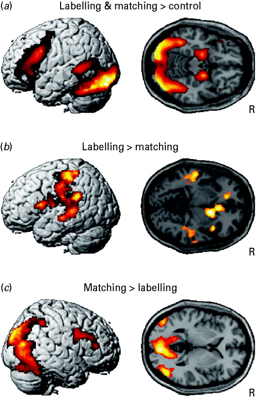
Across the groups, the emotion-labelling and -matching tasks elicited more activation than the baseline control task in clusters including the left and right amygdalae and hippocampi, left and right inferior frontal gyri, in the ventral visual stream ranging from the primary visual cortex to the fusiform gyrus, and supplementary motor area. The emotion-labelling task elicited more activity than the matching task in a cluster including the right putamen, caudate nucleus, insula and superior temporal gyrus, and a cluster including the left inferior frontal gyrus, posterior middle and superior temporal gyri, inferior parietal cortex, and motor, premotor and supplementary motor cortices. The emotion-matching task elicited more activity than the emotion-labelling task in a cluster including the ventral and dorsal visual stream, ranging from the primary visual cortex to the fusiform gyrus and superior parietal lobule, and the right inferior frontal gyrus (all p<0.05, corrected; see Fig. 1).
Task effects across depressed, recovered and healthy individuals (p cluster<0.05, corrected). (a) Emotion labelling and emotion matching>control. (b) Emotion labelling>emotion matching. (c) Emotion matching>emotion labelling (figures p<0.005; statistics p<0.05, corrected). R, Right.

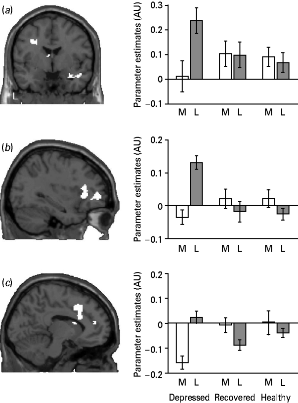
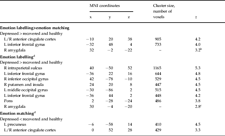
The between-group comparisons were restricted to differences between the emotion-labelling and emotion-matching tasks. To test for state-related effects, the currently depressed individuals were compared with the recovered and healthy individuals combined. The group×task condition interaction showed that the currently depressed individuals recruited clusters in the right amygdala, left inferior frontal gyrus and anterior cingulate cortex to a larger extent in the emotion-labelling than in the emotion-matching task than recovered and healthy individuals (p<0.05, corrected; see Fig. 2 and Table 2). These results remained significant after correcting for reaction times and accuracy for all conditions, suggesting that the results are unlikely to be explained by differences in attention, effort or accuracy. Testing for group differences within each task condition showed increased activity in the left inferior frontal gyrus and a statistical trend for the right amygdala in the emotion-labelling condition and decreased activity in the anterior cingulate cortex in the emotion-matching condition in the depressed individuals (p<0.05, corrected; see Table 2). In contrast, no significant clusters were observed that were recruited relatively more in the emotion-matching than in the emotion-labelling task in currently depressed than recovered and healthy individuals. To investigate trait-related effects, the currently depressed and recovered individuals were compared with the healthy individuals, but no significant group×task condition interactions were observed (p>0.05, corrected).
Currently depressed individuals recruit the right amygdala (a; y=−2), left inferior frontal gyrus (b; x=−32) and anterior cingulate cortex (c; x=−10) more than recovered and healthy individuals during semantic labelling of emotion in contrast to perceptual matching of emotion. The left panels show slices at peak activation (figures p<0.005; statistics p<0.05, corrected) and the right panels show the cluster means, with their standard errors, for the match (M) and label (L) conditions. AU, Arbitrary units.

Activation differences between the depressed, recovered and healthy individualsFootnote a

MNI, Montreal Neurological Institute; L, left; R, right.
a The local maxima of the significant clusters (p cluster<0.05) are reported in MNI coordinates.
b Corrected for multiple comparisons (p SVC<0.05).
c Statistical trend, corrected for multiple comparisons (p SVC=0.10).
d Statistics for brain regions that did not show a significant group×task condition interaction are also reported.
Discussion
These results show that deficits in semantic labelling of negative emotions are related to increased activation of the amygdala, left inferior frontal gyrus and anterior cingulate cortex. These abnormalities were present in medication-free depressed but not in recovered individuals, suggesting that the neural and behavioural deficits in emotion recognition are mood state-dependent. The depressed individuals performed worse when negative emotions were semantically categorized, but not when matching them on a perceptual basis. This suggests that accessing semantic knowledge about negative emotions stimulates task-unrelated processes in these brain regions, which subsequently impairs task-relevant behaviour.
The left inferior frontal gyrus is thought to integrate language with other information (Hagoort, Reference Hagoort2005; Willems et al. Reference Willems, Ozyurek and Hagoort2007). Therefore, we suggest that the inferior frontal gyrus may integrate the semantic knowledge about the concepts of anger and fear with the emotional information conveyed by the faces by interacting with the amygdala. The concurrent activation of these systems may subsequently trigger automatic negative thoughts and stimulate task-unrelated processes such as rumination (Siegle et al. Reference Siegle, Steinhauer, Thase, Stenger and Carter2002; Ray et al. Reference Ray, Ochsner, Cooper, Robertson, Gabrieli and Gross2005), and thereby hinder task-appropriate behaviour. This process does not appear to occur when emotion is categorized perceptually, indicating that the activation of negative concepts could be a crucial component for emotion-recognition deficits in depression. However, we did not include measures that may reflect task-unrelated processes such as rumination to test for a relationship with left inferior frontal gyrus and amygdala activation. Therefore, this interpretation is speculative and remains to be tested in future studies.
The increased recruitment of the left inferior frontal gyrus, anterior cingulate cortex and amygdala may also reflect an attempt to compensate for inadequate behaviour. A theory of anterior cingulate cortex function suggests that its enhanced recruitment indicates a conflict in response selection, which triggers strategic adjustments in cognitive control (Botvinick et al. Reference Botvinick, Cohen and Carter2004). Thus, signalling of the anterior cingulate cortex may have triggered additional involvement of the inferior frontal gyrus and amygdala, which were generally involved in task performance. However, differences in anterior cingulate cortex activation were mainly present during emotion matching, whereas differences in amygdala and left inferior frontal gyrus activation were mainly present during emotion labelling, indicating no direct relationship between those processes. Moreover, increased involvement of the inferior frontal gyrus and amygdala did not compensate for inadequate behaviour, indicating that increased involvement of these brain regions triggered worse performance rather than compensated for it.
Another possibility is that the increased recruitment of brain regions is related to altered coping with more demanding situations in depression. However, the behavioural results suggest that the emotion-matching task was the most demanding, because accuracy was lowest and response times were slowest during this condition, whereas the group differences in brain activity were observed in the emotion-labelling condition. Also the amount of attention and effort devoted to each of the different task conditions as reflected by the response times appears not different between the groups, and correcting the imaging data for response time and accuracy differences between conditions did not alter the pattern of results. Together, this suggests that the increased recruitment of the left inferior frontal gyrus, anterior cingulate cortex and amygdala during emotion labelling is unlikely to be explained by mere task demands or group differences in attention or effort.
The current study should be viewed in the light of some strengths and limitations. One potential limitation is the difference in state and trait anxiety scores between groups. However, increased anxiety is part of the symptomatology of depression, and elevated anxiety-related traits including neuroticism and trait anxiety are also observed pre-morbidly and in unaffected relatives. These personality traits are thought to contribute to the vulnerability for depression (Lauer et al. Reference Lauer, Bronisch, Kainz, Schreiber, Holsboer and Krieg1997; Ormel et al. Reference Ormel, Oldehinkel and Vollebergh2004). Importantly, none of the patients in our study fulfilled the criteria for an anxiety disorder. Thus, although differences in anxiety are inherently part of the observed group differences, the results remain specific for the diagnosis of major depression. A second limitation is the use of a cross-sectional design. Even though the groups did not differ significantly in demographic characteristics and neuropsychological test performance, longitudinal studies are required to confirm that the deficits observed in the present study are mood state-dependent. A third limitation is the use of only two negative emotions, namely anger and fear. Although emotion-labelling deficits do not appear to be specific for particular emotions, the more subtler emotion-matching deficits may be (Feinberg et al. Reference Feinberg, Rifkin, Schaffer and Walker1986; Rubinow & Post, Reference Rubinow and Post1992; Persad & Polivy, Reference Persad and Polivy1993). Therefore, additional studies are required to investigate whether neural abnormalities during emotion labelling are emotion specific. Obvious strengths of the study are the investigation of patients with a first depressive episode to control for structural brain changes associated with chronic disease (MacQueen et al. Reference MacQueen, Campbell, McEwen, Macdonald, Amano, Joffe, Nahmias and Young2003), the inclusion of medication-free patients to control for acute medication effects (Murphy et al. Reference Murphy, Norbury, O'Sullivan, Cowen and Harmer2009) and the inclusion of a group of recovered patients.
Cognitive theories of depression have postulated that adequate behavioural responses are hampered by activation of maladaptive, negative schemas that stimulate self-focused thinking, which subsequently reduce awareness and sensitivity to the concerns of others (Ingram, Reference Ingram1984; Beck & Clark, Reference Beck and Clark1988). In line with this theory, our results suggest that accessing semantic knowledge about negative emotions stimulates task-unrelated processes in the left inferior frontal gyrus and amygdala, which subsequently impairs emotion recognition. Therefore, we propose that accessing semantic knowledge about negative emotional information may trigger these negative schemas, and subsequently lead to further task-unrelated elaboration on negative content in the left inferior frontal gyrus and amygdala. This escalation could be a crucial component for emotion-recognition deficits during depressive episodes, which appear to subside after recovery.
Acknowledgements
The authors thank Sabine Kooijman and Sara Pieters for their assistance with data collection, and Paul Gaalman for his technical support.
Declaration of Interest
None.