Introduction
Lichens are stable symbiotic organisms in which only the fungal partner is able to carry out sexual reproduction. The development of special dispersal structures containing both symbionts, such as soredia or isidia, together with thallus fragmentation, allow the efficient propagation of the symbiotic dual organism. Soredia have been interpreted as being vegetative propagules selected during the evolutionary history of lichens (Poelt Reference Poelt1970, Reference Poelt1972) and have been accorded significant status as taxonomic characters. Earlier investigations defined ‘species pairs’ in lichens as two species, one fertile without vegetative propagules (primary species) and the other with soredia or isidia (secondary species); the latter being derived from the former. Hale (Reference Hale1965) presumed in his work on Parmelia (subgenus Amphigymnia) that sorediate species evolved from either isidiate or nonsorediate–nonisidiate ancestors, naming the two or three taxa involved ‘counterparts’.
The classic interpretation among lichenologists supports taxonomic separation at the species level for both members of a species pair. However, studies based on morphological and ecological information (Tehler Reference Tehler1982) or phylogenetic analysis (Lohtander et al. Reference Lohtander, Kallersjö and Tehler1998a, Reference Lohtander, Myllys, Sundin, Kallersjö and Tehlerb; Myllys et al. Reference Myllys, Lohtander, Källersjö and Tehler1999, Reference Myllys, Lohtander and Tehler2001; Articus et al. Reference Articus, Mattsson, Tibell, Grube and Wedin2002; Molina et al. Reference Molina, Crespo, Blanco, Hladun and Hawksworth2002; Cubero et al. Reference Cubero, Crespo, Esslinger and Lumbsch2004; Buschbom & Mueller Reference Buschbom and Mueller2006) show, at least for sorediate/nonsorediate species pairs, that these forms do not separate into monophyletic lineages. Consequently, pairs have been interpreted as representing two dispersal strategies and the specific status of each member of the pair has not been corroborated. Few cases of isidiate/ non-isidiate pairs have been studied, and recent results based on molecular techniques do not provide evidence of any clear ‘sister’ phylogenetic relationship for members of such ‘species pairs’ (Argüello et al. Reference Argüello, Del Prado, Cubas and Crespo2007).
It appears that the occurrence of isidia or soredia, particularly the latter, is a character that has been gained and lost in the phylogeny of lichens (Lohtander et al. Reference Lohtander, Kallersjö and Tehler1998a, Reference Lohtander, Myllys, Sundin, Kallersjö and Tehlerb; Myllys et al. Reference Myllys, Lohtander, Källersjö and Tehler1999, Reference Myllys, Lohtander and Tehler2001; Articus et al. Reference Articus, Mattsson, Tibell, Grube and Wedin2002; Molina et al. Reference Molina, Crespo, Blanco, Hladun and Hawksworth2002; Cubero et al. Reference Cubero, Crespo, Esslinger and Lumbsch2004; Buschbom & Mueller Reference Buschbom and Mueller2006). In many groups of lichens there are species bearing isidia or soredia but there are few, if any, examples of lichens bearing both kinds of structures at the same time. One vegetative dispersal strategy usually predominates in evolutionary monophyletic branches in the Parmeliaceae. Vegetative propagating members of the genera Xanthoparmelia (Hale Reference Hale1990), Melanelixia (Blanco et al. Reference Blanco, Crespo, Divakar, Esslinger, Hawksworth and Lumbsch2004), Parmelina (Hale Reference Hale1976) and Pseudevernia (Hale Reference Hale1968) mostly produce isidia, whereas the majority of commonly sterile species in Parmotrema and Flavoparmelia (Hale Reference Hale1965) disperse by means of soredia.
The genus Pseudevernia (a member of the family Parmeliaceae), which is thought to be closely related to Brodoa and Hypogymnia (Crespo et al. Reference Argüello, Del Prado, Cubas and Crespo2007), comprises only a small number of species. Depending on the taxonomic concepts applied, either six species (Hale Reference Hale1968) or only four species (recent concepts) are accepted. Three well defined species containing lecanoric acid [P. consocians (Vain.) Hale & W. Culb. with isidia, P. cladonia (Tuck.) Hale & W. Culb. and P. intensa (Nyl.) Hale & W. Culb. without isidia] occur in North America (see distribution map in Hale Reference Hale1968). Three other isidiate taxa, namely P. furfuracea (L.) Zopf (with physodic and oxyphysodic acids), P. olivetorina (Zopf) Zopf (with olivetoric acid) and P. soralifera (Bitt.) Zopf (with physodic and oxyphysodic acids; with soralia in addition to isidia), which were treated by Hale (Reference Hale1968) as separate species, are classified by several European lichenologists (e.g., Clerc Reference Clerc2004) within a single entity, P. furfuracea.
Pseudevernia furfuracea (L.) Zopf is a common and widespread species in Eurasia and North Africa; there are some very occasional reports from Mexico, Central America, Bolivia and Uganda (Hale Reference Hale1968). It is rarely infertile in several localities in Europe (Obermayer Reference Obermayer2008) and has abundant cylindrical, dark-brown-tipped, sometimes branched, isidia on the upper surface. In rare cases, in addition to isidia, capitate soralia have been found. Such sorediate specimens have been considered by Hale (Reference Hale1968) as a different species, P. soralifera. Recent studies tend to reject any systematic category for these specimens (Hafellner & Obermayer Reference Hafellner and Obermayer2004; Clerc Reference Clerc2004), but no phylogenetic studies have been performed until now to support such an opinion.
During the last 30 years, populations of P. furfuracea from different regions of Europe have been the subject of chemical analyses. Three chemical races or chemotypes have been described and their distribution in relation to their ecology has been widely discussed (Hawksworth & Chapman Reference Hawksworth and Chapman1971; Culberson et al. Reference Culberson, Culberson and Johnson1977; Halvorsen & Bendiksen Reference Halvorsen and Bendiksen1982; López & Manrique Reference López and Manrique1989; Rikkinen Reference Rikkinen, Tibell and Hedberg1997). In many European floras, the chemotypes are treated as varieties of such species: P. furfuracea var. furfuracea with physodic and oxyphysodic acids (corresponding to chemical race I) and P. furfuracea var. ceratea with olivetoric acid in addition,or not, to physodic acid (corresponding to chemical races II and III, respectively).
All specimens of P. furfuracea collected by us bearing soralia were infected by a filamentous fungus, growing both internally and externally. This infection was observed not only in sorediate/isidiate samples but also in exclusively isidiate samples.
The aim of our investigation was to study the morphology and phylogeny of sorediate specimens of P. furfuracea in order to evaluate the biological and taxonomic significance of propagules in this species.
Material and Methods
Taxon sampling
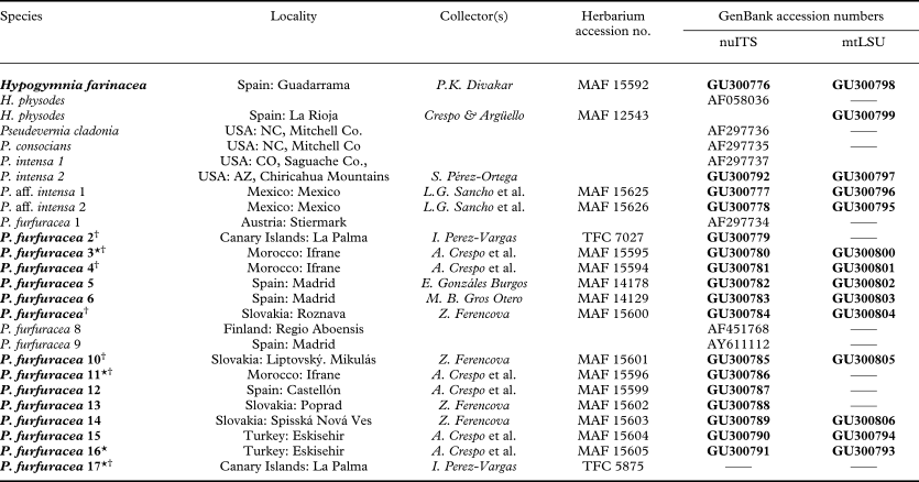
Sorediate samples of P. furfuracea were collected from the Canary Islands (North West Africa), the mountains of the north of Morocco and the west of Turkey. Specimens with soralia are rare and only four specimens were found as isolated sorediate thalli in populations of P. furfuracea; no separate populations of sorediate P. furfuracea were found. Non-sorediate samples of P. furfuracea were also collected both from the same or localities nearby and from several other regions of Europe. The details of the material, area of collection, location of voucher specimens and their GenBank accession number are presented in Table 1. The material is deposited in the herbarium at the Facultad de Farmacia of the Complutense University of Madrid, Spain (MAF) and in the University of La Laguna, Tenerife, Canary Islands, Spain (TFC).
Species and specimens used in the current study (taxa studied morphologically and newly obtained sequences are indicated in bold)

* sorediate specimens;
† indicates presence of olivetoric acid; all others contain physodic and oxyphysodic acids.
Morphological and anatomical studies
The morphology of the specimens of P. furfuracea (both non-sorediate and sorediate) listed in bold in Table 1 was studied under a dissecting microscope (Leica Wild M 8; Leica, Wetzlar, Germany).
Internal structure was studied in hand-cut transverse sections of thalli and vegetative propagules (isidia and soredia). Sections were stained with unheated lactophenol cotton blue (Panreac, Barcelona, Spain) and observed and photographed using a light microscope (Nikon Eclipse 80i; Nikon, Badhoevedrop, Netherlands).
Chemical analysis
Chemical analysis was carried out on the 16 samples of Pseudevernia listed in bold in Table 1. Chemical constituents were identified by thin-layer chromatography (TLC) using solvent systems A [benzene: dioxane : acetic acid, 180:45:5], B' [hexane : methyl t-butyl ether : formic acid, 140:72:18] and C [toluene : acetic acid, 85:15] (Culberson Reference Culberson1972; Culberson et al. Reference Culberson, Culberson and Johnson1981; Culberson & Johnson Reference Culberson and Johnson1982; Elix & Ernst-Russell Reference Elix and Ernst-Russell1993; Orange et al. Reference Orange, James and White2001).
DNA extraction, PCR and sequencing
Total DNA was extracted from freshly collected material (Table 1), using the DNeasy Plant Mini Kit (Qiagen, Hilden, Germany) following the manufacturer's instructions, with slight modifications described in Crespo et al. Reference Crespo, Blanco and Hawksworth2001. Primers for amplification were: (1) for the nu-ITS rDNA: ITS1F (Gardes & Bruns Reference Gardes and Bruns1993) and ITS4 (White et al. Reference White, Bruns, Lee, Taylor, Innis, Gelfand, Sninsky and White1990); (2) for the mt-LSU rDNA: ML3 and ML4 (Printzen Reference Printzen2002). Amplifications were performed in 50 μl volumes containing a reaction mixture of 5 μl 10 × DNA polymerase buffer (Biotools, Madrid, Spain; containing 2 mM MgCl2, 10 mM Tris-HCl, pH 8·0, 50 mM KCl, 1 mM EDTA, 0·1% Triton X-100), 1 μl deoxyribonucleotide triphosphate (dNTP) (Biotools), containing 10 mM of each base, 2·5 μl of each primer (10 μM) (Sigma Aldrich), 1·25 μl DNA polymerase (1 U/μl) (Biotools), and 27·75 μl distilled water. Finally, 40 μl of this mixture were added to 10 μl of DNA of each sample.
Amplifications were carried out in an automatic thermocycler (Techne Progene Jepson Bolton & Co. Ltd., Watford, Herts, UK) under the following conditions: initial denaturation at 94°C for 5 min, followed by 30 cycles of 94°C for 1 min, 54°C (nu ITS primers) or 47°C (mt LSU primers) for 1 min and 72°C for 1·5 min, and a final extension at 72°C for 5 min. Amplification products were viewed on 1% agarose gels stained with ethidium bromide and subsequently purified using the Bioclean Columns kit (Biotools, Madrid, Spain) according to the manufacturer's instructions. Fragments were sequenced using the Big Dye Terminator reaction kit (ABI PRISM, Applied Biosystems, Foster City, CA, USA). Sequencing and PCR amplifications were performed using the same sets of primers. Cycle sequencing was carried out as follows: initial denaturation at 94°C for 3 min, followed by 25 cycles of 96°C for 10 s, 50°C for 5 s and 60°C for 4 min. Sequencing reactions were electrophoresed on a 3730 DNA analyser (Applied Biosystems). Sequence fragments obtained were assembled with SeqMan 4.03 (DNAStar Madison) and adjusted manually.
Sequence alignments and phylogenetic analysis
We generated 17 new nu ITS and 14 new mt LSU rDNA sequences for this study. Details of the samples analysed are listed in Table 1. Seven nu ITS sequences were downloaded from GenBank (http://www.ncbi.nlm.nih.gov).
Two analyses were performed. 1) A single-gene analysis of the nu ITS sequences of Pseudevernia furfuracea (including three sequences of sorediate samples) in which 13 newly obtained sequences were aligned with three sequences obtained from GenBank (Table 1); one new sequence of P. cladonia and two new sequences of P. aff intensa were included as the outgroup. 2) A combined analysis of nu ITS and mt LSU rDNA sequences of P. furfuracea and species of Pseudevernia from Mexico and USA. Two data matrices were assembled for this analysis: 2a) 14 samples using the mt LSU rDNA region; 2b) 17 samples using those 14 sequences of nu ITS region for which sequences of the mt LSU rDNA region were available, and three sequences of P. cladonia, P. consocians and P. intensa whose mt LSU sequences failed; two taxa of Hypogymnia were included as outgroup since this genus is shown to be closely related (Crespo et al. Reference Argüello, Del Prado, Cubas and Crespo2007).
A single-gene analysis was also presented as mt LSU extraction failed in most cases of European P. furfuracea. Thus, the nu ITS rDNA data set allows us to study a larger taxon sampling of this species, and the combined analysis was performed to show the phylogenetic hypothesis for all species of the genus Pseudevernia.
Each data set was aligned separately using Clustal W (Thompson et al. Reference Thompson, Higgins and Gibson1994). Regions that could not be aligned with statistical confidence were excluded from the phylogenetic analysis. Ambiguously aligned regions were delimited manually (Lutzoni 2000).
The topology of the 95% majority rule consensus tree of the two Bayesian single-partition analyses for matrices (2a) and (2b) was congruent (data not shown), justifying their subsequent combined analysis. The alignment of the combined data set was analysed using maximum parsimony (MP), maximum likelihood (ML) and a Bayesian approach (B/MCMC). MP analyses were performed using the PAUP* program (Swofford Reference Swofford2003). A heuristic search with 2000 random taxon addition replicates, using the general time-reversible nucleotide substitution model (Rodriguez et al. Reference Rodríguez, Oliver, Martín and Medina1990) and assuming a gamma shape parameter of 0·5 was conducted with TBR branch swapping and the MulTrees option selected. Bootstrapping (Felsenstein Reference Felsenstein1985) was performed on the basis of 2000 pseudoreplicates with the same settings as for the heuristic search.
ML analyses were performed using PhyML on the program's online web server, http://atgc.lirmm.fr/phyml (Guindon et al. Reference Guindon, Lethiec, Duroux and Gascuel2005), assuming the general time-reversible model of nucleotide substitution (Rodriguez et al. Reference Rodríguez, Oliver, Martín and Medina1990) and a discrete gamma distribution with six rate categories for the combined data set. The bootstrap analysis was run with 500 pseudoreplicates.
The B/MCMC analyses were conducted using the MrBayes 3.1.2 program (Huelsenbeck & Ronquist Reference Huelsenbeck and Ronquist2001), adopting the general time-reversible model of nucleotide substitution (Rodríguez et al. Reference Rodríguez, Oliver, Martín and Medina1990) including estimation of invariant sites, assuming a discrete gamma distribution with six rate categories and allowing site-specific rates (GTR+I+G). The nucleotide substitution model was selected using jModelTest (Posada Reference Posada2008).
The combined data set was divided into the two parts (nu ITS and mt LSU), each of which was allowed to have its own parameters as suggested by Nylander et al. (Reference Nylander, Ronquist, Huelsenbeck and Nieves-Aldrey2004). No molecular clock was assumed. Parallel runs were made with 3 000 000 generations starting with a random tree and employing 12 simultaneous chains each. Trees were sampled every 200 generations for a total of 30 000 trees. The first 300 000 generations (i.e., the first 3000 trees) were discounted as burn-in for the chain.
The log-likelihood scores of sample points were plotted against generation time using TRACER 1.0 (http://evolve.zoo.ox.ac.uk/software.html/tracer/) to ensure that stationarity was achieved after the first 300 000 generations by checking whether the log-likelihood values of the sample points had reached a stable equilibrium (Huelsenbeck & Ronquist Reference Huelsenbeck and Ronquist2001). For the remaining 54 000 trees (27 000 from each parallel run) a majority-rule consensus tree with average branch lengths was calculated using the sumt option in MrBayes.
We used a Bayesian approach to examine the heterogeneity in phylogenetic signal among the two data partitions (DeQueiroz Reference DeQueiroz1993; Buckley et al. Reference Buckley, Arensburger, Simon and Chambers2002). Posterior probabilities (PPs) were approximated for each data set by sampling trees using an MCMC method. The PPs of each branch were calculated by counting their occurrence in trees that were visited during the course of the MCMC analysis. PPs were obtained for each clade. Only clades with PPs ≥ 95 % were considered as strongly supported. If no conflict was evident, it was assumed that the two data sets were congruent and could be combined.
In the combined data set, a conservative approach for interpreting support values was considered. Only clades that received bootstrap support ≥ 70% in MP and ML analyses and PPs ≥ 0·95 were considered as strongly supported.
The alignment of 19 samples of nu ITS (1) was analysed using a Bayesian approach (B/MCMC). Phylogenetic trees were drawn using TREEVIEW (Page Reference Page1996).
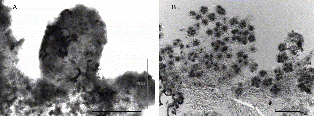
Pseudevernia furfuracea, transverse sections of the thallus and vegetative propagules of a sorediate specimen. A, parasitic fungal hyphae growing on and inside an isidium; B, uninfected soredia. Scales: A & B = 100 μm.

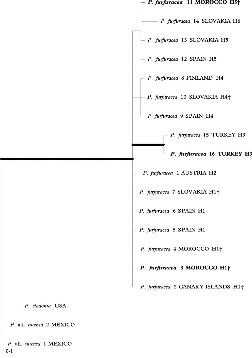
A 50% majority-rule consensus tree of nu ITS sequences of Pseudevernia furfuracea. This tree is based on 27 000 trees from a B/MCMC tree sampling procedure. Branches with posterior probabilities ≥ 0·95 are indicated in bold. Sorediate specimens are indicated in bold. Specimens with olivetoric acid are indicated with †, all others contain physodic and oxyphysodic acids. Different haplotypes are indicated by an ‘H’ followed by a number.

Results
Morphological observations
Sorediate thalli looked weaker, were shorter and showed reduced vitality compared with non-sorediate specimens. Isidia were morphologically similar in both kinds of thalli but, in the sorediate specimens, were less abundant and poorly developed. Soralia were regularly capitate, substipitate, whitish, laminar and marginal and were occasionally also present on the lower surface of the thalli.
All sorediate samples were infected by at least one filamentous fungus (Fig. 1A). Fungal hyphae grew on the surface of the lichen thallus including the isidia (cortical and medullar inner part). They also penetrated into the whole thalline cortex and reached the medulla. Interestingly soralia were not infected, at least not in the central zone where they are produced (Fig. 1B).
Chemical TLC analysis
All samples of the morphospecies Pseudevernia furfuracea contained atranorin. Seven samples, marked (†) in Table 1 and Fig. 2, contained olivetoric acid (hitherto treated in many European lichen floras as P. furfuracea var. ceratea). The other seven samples had oxyphysodic and physodic acids (named as P. furfuracea var. furfuracea). Specimens with mixed chemistry (containing both physodic and olivetoric acid), described from Spain by Culberson et al. (Reference Culberson, Culberson and Johnson1977) and López & Manrique (Reference López and Manrique1989), were not found. Samples with olivetoric acid did not form a monophyletic group and could not be treated as a separate valid taxon, P. olivetorina, as suggested by some authors (e.g., Hale Reference Hale1968).
Phylogenetic analysis
We generated 17 new nu ITS and 14 new mt LSU rDNA sequences for this study (Table 1). Two analyses were performed: 1) single gene analysis of the nu ITS sequences of Pseudevernia furfuracea, in which the data matrix had 525 unambiguously aligned nucleotide positions, 48 of which were variable; 2) a combined analysis of nu ITS and mt LSU rDNA sequences where the data matrix had 476 unambiguously aligned nucleotide positions in the nu ITS and 809 in the mt LSU rDNA partitions. Eighty-seven characters were variable in the nu ITS and 27 in the mt LSU data set. Since the topologies of the MP, ML and B/MCMC analyses did not show any supported conflicts, only the 50% majority-rule consensus tree of Bayesian tree sampling is shown (Fig. 3), the nodes in bold being those that received strong support in all three analyses (i.e., PP ≥ 0·95 in the B/MCMC analysis and ≥ 70% for the MP and ML bootstraps).
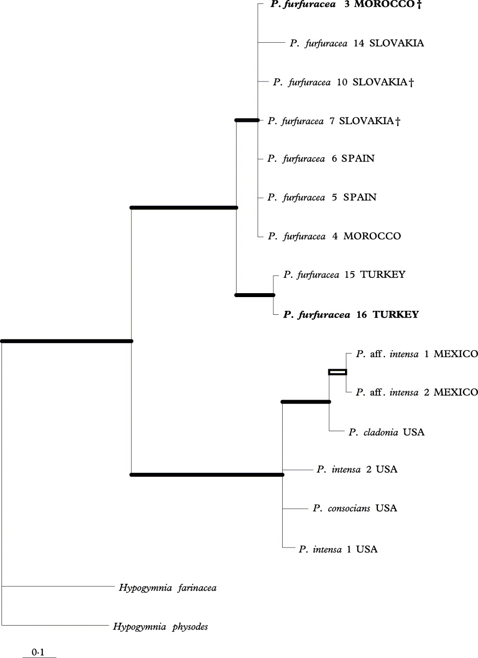
A 50% majority-rule consensus tree of the phylogenetic relationships in the genus Pseudevernia based on 54 000 trees from a B/MCMC tree-sampling procedure from a combined data set of nu ITS and mt LSU rDNA. Branches that received strong support in all three analyses (i.e., PP ≥ 0·95 in B/MCMC analysis and ≥ 70% in MP and ML bootstraps) are indicated in bold; the branch that received strong support in MP and PhyML is indicated by an empty rectangle. Specimens with olivetoric acid are indicated with †, all others contain physodic and oxyphysodic acids.

In the single-gene analysis (Fig. 2), the likelihood parameters of the sample had the following mean values (variance): LnL = −1076·7 (0·10), base frequencies π(A) =0·225 (0·0002), π (C) = 0·28 (0·0002), π (G) = 0·254 (0·0002), π (T) = 0·242 (0·0002), rate matrix r(AC) = 0·089 (0·0012), r(AG) = 0·242 (0·0018), r(AT) =0·143 (0·0014), r(CG) = 0·0109 (0·0011), r(CT) = 0·382 (0·0018), r(GT) = 0·035 (0·0008), gamma shape parameter alpha = 0·097 (0·0003) and proportion of invariable sites, p(invar) = 0·762 (0·0004).
In the two-gene analysis (Fig. 3), in the B/MCMC analysis, the likelihood parameters in the sample had the following mean values for the nu ITS partition (variance): LnL = −2381·69 (0·09), base frequencies π(A) = 0·214 (0·0002), π (C) = 0·283 (0·0002), π (G) = 0·263 (0·0002), π (T) =0·24 (0·0002), rate matrix r(AC) = 0·075 (0·0004), r(AG) = 0·228 (0·0008), r(AT) =0·09 (0·0006), r(CG) = 0·065 (0·0003), r(CT) = 0·495 (0·001), r(GT) = 0·046 (0·0004), gamma shape parameter alpha = 0·097 (0·0003) and the proportion of invariable site p(invar) = 0·55 (0·002). The following means were obtained for the mt LSU partition (variance): LnL = −2381·69 (0·09), base frequencies π(A) = 0·369 (0·0002), π (C) = 0·143 (0.0001), π (G) =0·189 (0·0002), π (T) = 0·3 (0·0002), rate matrix r(AC) = 0·071 (0·0014), r(AG) =0·174 (0·0017), r(AT) = 0·213 (0 ·0017), r(CG) = 0·186 (0·0025), r(CT) = 0·26 (0·0028), r(GT) = 0·095 (0·0013), gamma shape parameter alpha = 0·068 (0·0004) and p(invar) = 0·912 (0·0002).
The majority-rule consensus tree of the ITS data set included sequences from European and North African samples and is shown in Fig. 2. All the samples of P. furfuracea from Central and Western Europe, Turkey and North Africa (Morocco) nested in a monophyletic group resolved in a pectinated topology. One monophyletic clade that includes the two samples from Turkey is supported. Three sequences of sorediate specimens of P. furfuracea were obtained but the sorediate samples from the Canary Islands remained unamplified, most likely because of the ‘unhealthy’ state of the material and the heavy fungal infection. All specimens are monophyletic (Fig. 3) but six haplotypes were noted. Three of these haplotypes (H1, H3 and H5) included both sorediate and nonsorediate samples.
The phylogenetic relationships for all species of the genus (Fig. 3) shows that the New and Old World samples are separated into two sister monophyletic clades. The American species (P. intensa (Nyl.) Hale & W. Culb., P. cladonia (Tuck.) Hale & W. Culb., P. consocians (Vain.) Hale & W. Culb. and P. aff. intensa) form a sister group to the European P. furfuracea (including sorediate samples).
Discussion
Sorediate populations of Pseudevernia furfuracea are rare. Nevertheless, our collections are from distant localities around the Mediterranean basin and the Canary Islands, so it is notable that such a rare phenomenon is so widespread. A common origin for the sorediate specimens would be a straightforward hypothesis. However, no monophyletic separate lineage of sorediate specimens was found within the phylogenetic tree, and the same haplotypes were found in sorediate and non-sorediate samples. In other words, our phylogenetic analyses do not suggest that P. soralifera is a valid taxon (Hale Reference Hale1968), but rather that it should be included within P. furfuracea (as already has been done, for example, by Clerc Reference Clerc2004). A previous study based on biological observations of a population in the Austrian Alps also concluded that the sorediate collections do not merit recognition as a separate taxon (Hafellner & Obermayer Reference Hafellner and Obermayer2004).
Similarly, none of our studied samples containing olivetoric acid represents a phylogenetically supported lineage. Thus, the inclusion of the taxon P. olivetorina in P. furfuracea, as already proposed by López & Manrique (Reference López and Manrique1989) and Rikkinen (Reference Rikkinen, Tibell and Hedberg1997) and implemented by Clerc (Reference Clerc2004), is supported.
The presence of well-defined soralia characterized by a constant capitate form appears to be an important morphological feature for differentiating morphospecies (Taylor et al. Reference Taylor, Jacobson, Kroken, Kasuga, Geiser, Hibbett and Fisher2000), particularly when molecular data are not available. However, the status of P. soralifera had already been discussed by Hale (Reference Hale1968), who reported the rare simultaneous occurrence of isidia and soralia in his synopsis of the species. He suspected that this feature was “an abnormal condition of otherwise normal P. furfuracea” and was prompted to reduce the status of the species. Recently, Hafellner & Obermayer (Reference Hafellner and Obermayer2004) were more conclusive in their study of Austrian sorediate material, stating that “it is not convenient to apply a taxon name to sorediate Pseudevernia specimens.” Moreover, although a considerable number of sorediate ‘species’ persist in taxonomic index lists (www.indexfungorum.org), it has been demonstrated repeatedly in lichens that differentiation based on the presence of soralia does not merit acceptance of sorediate forms as valid species (Tehler Reference Tehler1982). In addition, all available data from molecular markers concur in demonstrating that sorediate counterparts in ‘species pairs’ do not appear as an isolated evolutionary monophyletic lineage that can be described as a single separate species (Lohtander et al. Reference Lohtander, Kallersjö and Tehler1998a, Reference Lohtander, Myllys, Sundin, Kallersjö and Tehlerb; Myllys et al. Reference Myllys, Lohtander, Källersjö and Tehler1999, Reference Myllys, Lohtander and Tehler2001; Articus et al. Reference Articus, Mattsson, Tibell, Grube and Wedin2002; Molina et al. Reference Molina, Crespo, Blanco, Hladun and Hawksworth2002; Cubero et al. Reference Cubero, Crespo, Esslinger and Lumbsch2004; Buschbom & Mueller Reference Buschbom and Mueller2006). There are examples analogous to these appealing capitate soralia, such as the profuse marginal soralia of Physconia perisidiosa, where the use of molecular markers did not indicate that the sorediate P. perisidiosa is a monophyletic species separate from the non-sorediate P. venusta (Cubero et al. Reference Cubero, Crespo, Esslinger and Lumbsch2004).
However, Pseudevernia soralifera is not a typical case of a ‘species pair’ but unusual in the occasional simultaneous presence of isidia and soredia. Pseudovernia furfuracea, probably propagating by means of isidia, is an extremely common species in most European regions and shows notable ecological plasticity. Therefore, isidia appear to be efficient propagules and the presence of capitate soralia in these populations of Pseudevernia may not be simply an additional reproductive strategy.
The three collections of sorediate specimens studied here all had regular capitate soralia, showed more or less reduced vitality and were infected or colonized by at least one fungal mycelium. The fungus was visible superficially but anatomical studies demonstrated that it also penetrated the upper cortex and that most of the isidia were also infected internally and externally. Interestingly, infection did not appear to penetrate the soralia. These morphological similarities among all sorediate samples from widely separated populations lead us to suggest that soralia arise as a consistent reaction to a similar agent, the lichenicolous or parasitic fungi, and that it may be a coevolved reproductive strategy.
The fungal infection in the lichen thallus covers the isidia, so their subsequent dispersal would also spread the fungal infection or possibly make the isidial propagules inviable. Interestingly, soralia are free of infection and their production could be an alternative reproductive strategy by P. furfuracea to ensure survival when the health of isidia is compromised by fungal infection.
Finally, the reconstruction of the phylogeny of Pseudevernia reveals two monophyletic sister branches of the genus. The two branches separate the American species (P. intensa, P. cladonia, P. consocians), from the Eurasiatic and North African P. furfuracea (including both sorediate samples and samples with olivetoric acid).
We thank Dr W. Obermayer and an anonymous reviewer for their suggestions, which have improved the quality of our work and Dr A. Green for his kind help in revising the English language of the manuscript. This project was funded by the Banco Bilbao Vizcaya Argentaria (BBVA) Foundation. We are grateful to the Spanish Ministerio de Educación y Ciencia for the FPU grant to ZF (CGL2007- 64652). Sequencing, for which we thank Maria Isabel García Saez, was carried out at the Unidad de Genómica (Parque Científico de Madrid).