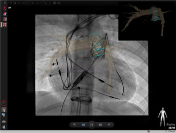
Novel commercially available software has enabled registration of both CT and MRI images to rapidly fuse with fluoroscopic imaging. This imaging overlay technology has enabled the 3D reconstruction of structures of interest, which has then provided guidance of transcatheter interventions in complex cardiac defects. Furthermore, it has facilitated more precise positioning of catheters, balloons, and devicesReference Fresse-Warin, Plessis and Benelhadj1 and has also allowed for careful planning of interventions prior to catheterisation. In turn, this has led to decreased need for diagnostic angiography which has reduced radiation and contrast exposure as well as procedure time.Reference Goreczny, Dryzek and Morgan2-Reference Nicole, Alkassar and Dittrich4 Given the improving life expectancy of patients with CHD defects, reducing radiation exposure is becoming an increasingly important consideration, especially as evidence has shown the correlation between radiation exposure and increased carcinoma risk in the adult CHD population.Reference Cohen, Liu and Gurvitz5
We describe our initial experience performing cardiac catheterisations with the guidance of 3D advanced imaging overlay using the VesselNavigator system (Philips Healthcare, Best, NL).
Materials and methods
This study was a retrospective chart review of all cardiac catheterisations (n = 33) guided with the VesselNavigator system performed at Children’s Medical Center in Dallas, TX, between 1 December, 2016 and 31 January, 2019. Institutional Review Board approval was obtained for retrospective analysis of patient data at UT Southwestern. Patient demographic information as well as pre-catheterisation imaging and catheterisation data was collected. Imaging was performed via CT (Siemens SOMATOM Flash) or MRI [Phillips Ingenia 1.5 Tesla (T)].
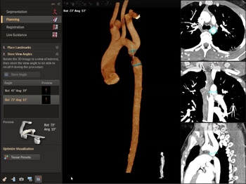
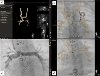
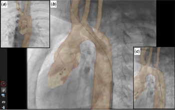
There were no strict inclusion or exclusion criteria for this descriptive study. Patients only needed to have had advanced imaging performed prior to catheterisation to be eligible for enrollment. No specific anatomy, interventions, or patient demographics were excluded from this study. Segmentation of the appropriate structures requiring intervention was performed with the VesselNavigator system, and ring landmarks were placed to locate vessel origin, stenosis, or estimated proximal/distal landing zone for stent placement (Fig 1). Pre-procedural planning included vascular access sites and camera angles for angiography. Guidewires and catheters were placed into great vessels before X-ray fluoroscopy to enable registration (Fig 2). Additionally, only two fluoroscopic images are required in comparison to previous 3D–3D registration with rotational angiography, which also reduces radiation and anaesthesia time. After co-registration, procedures were performed using biplane angiography alongside fused overlay images on the A plane (AP camera) to enhance guidance. All catheterisations were performed under general anaesthesia. Further analysis of radiation variables was performed on the coarctation of the aorta stent angioplasty overlay cohort (Fig 3) between 10 and 15 years of age. This cohort was compared to the same demographics undergoing coarctation of the aorta stent angioplasty without the use of overlay technology. These groups were then compared to recently published national radiation benchmarksReference Cevallos, Armstrong and Glatz6 for the same age group and procedure.
Segmentation and planning process is shown. CT images (right) are segmented (light blue) to create a 3D reconstruction. A preview is available for the interventionalist’s camera (bottom left) to ensure the image overlay image is feasible with the patient on the table.

VesselNavigator system for overlay in a Glenn anastomosis patient status post left pulmonary artery (LPA) stent placement. (a) Depiction of planning process after segmentation. (b ) Two light blue rings were digitally placed at the proximal and distal ends of a LPA stenosis to aid in precise stent placement. In addition, a small veno-venous collateral is filled with contrast from the left innominate to the left sided pulmonary veins. Image is post-LPA stent placement using the overlay guidance. (c) Angiogram post-LPA stent placement. (d) Overlay image with removal of left innominate vein depicting overlay image in panel C. Angiographic catheter aided overlay image fusion.

VesselNavigator system for 3D advanced imaging overlay with rapid registration to assist cardiac catheterisation in a patient with a discrete coarctation of the aorta. (a) AP view with overlay imaging. (b) Balloon stent angioplasty across discrete coarctation of the aorta. (c) Overlay guiding wire across jailed left subclavian.

Results
The VesselNavigator system was used in 33 patients undergoing cardiac catheterisation. Median age and weight were 8 years and 25 kg (ranging from 6 days to 21 years and 3.7 to 69.2 kg, respectively). Eleven overlay images were from prior CT reconstruction, 13 from MRI, and nine patients were direct transfers to the X-ray fluoroscopy lab from the Philips Ingenia 1.5 T magnet (XMR). Further demographic data are outlined in Table 1. Median time from image to procedure was 3 days [ranging from 0 (XMR patients) to 646 days].
Patient demographic and catheterisation data for 3D overlay procedures

BSA = body surface area; DAP = dose area product; XMR = X-ray MR.
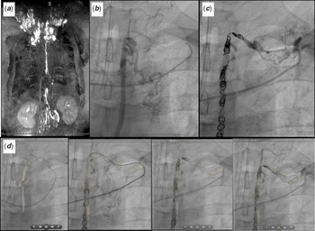
Of the 33 cases, three were diagnostic catheterisations; the remainder were interventional. Sixteen stents were placed – eight of which were for stenting of discrete coarctation of the aorta. Thirteen dilations were performed – seven of which were pulmonary artery angioplasties. Six patients underwent coiling of collateral vessels. Two procedures to manipulate the lymphatic system were performed [one thoracic duct closure (Fig 4) and one interruption with Chiba needle], two Melody valves were placed (Fig 5), and one occlusion of a right coronary artery fistula was performed.
Fontan patient with plastic bronchitis undergoing thoracic duct lymphatic embolisation with overlay guidance. (a) Dynamic contrast magnetic resonance lymphangiogram showing significant lymphatic insufficiency with a dilated, intact lymphatic duct draining to the left innominate vein. (b) X-ray fluoroscopic lymphangiogram. (c) Post-coil occlusion X-ray fluoroscopic lymphangiogram. (d) Series depicting thoracic duct lymphatic embolisation with overlay guidance.

VesselNavigator system for 3D overlay assisting with placement of a bare metal stent over the first covered stent for support before ultimately placing a 22 mm Melody valve placement in the RV to PA conduit (Tetralogy of Fallot patient).

Further analysis in Table 2 demonstrates our average fluoroscopy time for patients undergoing XMR was 35.8 minutes. Average fluoroscopic time for patients who had overlay registrations from previously obtained CT or MRI images was 39.7 minutes. Average total contrast given was 3.4 ml/kg for all patients. The number of total angiograms averaged 9.7 and ranged from 2 to 21. Total radiation exposure averaged 453.3 mGy, and average procedure time was 177.4 minutes (ranging from 53 to 378 minutes). Table 4 specifically looks at the overlay coarctation of the aorta cases (n = 7) for patients between 10 and 15 years of age. The average radiation exposure [2516 Dose Area Product (DAP, µGy.m2)] was markedly lower than national benchmarks (5052 µGy.m2) and other similar cases at our institution without overlay guidance (n = 17; 4422 µGy.m2).
Catheterisation data, imaging modalities, and interventions

A-P = aortopulmonary; BT = Blalock–Taussig; CoA = coarctation of aorta; LPA = left pulmonary artery; PDA = patent arterial duct; PPVI = percutaneous pulmonary valve implantation; PV = pulmonary valve; RIMA = right internal mammary artery; RPA = right pulmonary artery; RV-PA = right ventricle-to-pulmonary artery; V-V = veno-venous.
Overview of VesselNavigator system cases to assist cardiac catheterisation diagnostics and interventions

Comparison of radiation and contrast exposure during CoA stent angioplasty using 3D advanced overlay to guide the procedure. Overlay CoA radiation findings at Children’s Medical Center (CMC) are considerably lower than “Benchmark” and “No overlay” radiation exposure

CoA = coarctation of aorta.
Discussion
The study’s aim was to evaluate and describe our institution’s application of single-plane 3D image overlay technology in various cardiac transcatheter interventions and effects on radiation burden. This study adds to the growing body of literature which shows that 3D CT or MRI overlay for CHD transcatheter intervention with rapid registration is feasible and aids decisions regarding access and planned angiographic angles.Reference Fresse-Warin, Plessis and Benelhadj1–Reference Glöckler, Halbfaß and Koch3,Reference Goreczny, Dryzek, Moszura, Kühne, Berger and Schubert7–Reference Nordmeyer, Kramer, Berger and Schubert9 Novel applications of imaging overlay technology in this study include thoracic duct interventions (Fig 4) as well as a right coronary artery fistula occlusion. This information is further outlined in Tables 2 and 3. Average contrast use for all overlay patients was 3.4 ml/kg which is consistent with previously published studies.Reference Nicole, Alkassar and Dittrich4 In our anecdotal experience, operators found intraprocedural overlay fusion registration using placed vessel guidewires to be more accurate than using bony structures (Fig 5) especially in smaller children. CT images were registered with equal accuracy to MRI.
Furthermore, we investigated the effects of radiation reduction when using this technology in paediatric coarctation of the aorta stenting between 10 and 15 years of age. Our coarctation of the aorta stenting cohort showed a meaningful decrease in ionising radiation use when compared to similar cases at our institution without overlay technology as well as national benchmark radiation measurements for the same population (Table 4).Reference Cevallos, Armstrong and Glatz6 While the results are not statistically significant given the low number of coarctation of the aorta stenting patients, it is an important finding that is worth further investigation. Average contrast use for all overlay coarctation of the aorta stent angioplasty patients was 3.4 ml/kg (n = 7) compared to 3.8 ml/kg in our “no overlay” coarctation of the aorta cohort (n = 17) cohort.
Overall, we found overlay imaging useful in the guidance of complex cardiac procedures. The authors acknowledge that this study is limited due to the single centre nature in a specific patient population. Pre-procedural planning with the VesselNavigator system aids in planned access. Once fused, the image can be rotated without radiation leading to desired angiographic projection of the area of interest. Over the study period, the authors noted subjective improvement in image overlay when the advanced imaging was performed shortly before the planned catheterisation. Our most seamless overlay applications were performed for extracardiac structures due to the reduced cardiac motion. Static image overlay fusion was most challenging for interventions within the heart as the technology is limited by the dynamic nature of cardiac and respiratory motion. Further limitations include the lack of software compensation due to distortion of cardiac anatomy during catheterisation from arm positioning. In addition, placement of stiff wires and sheaths can distort the anatomy such that the overlay is not accurate. Future studies are necessary to establish additional benefits of this fusion software. Additional work is also needed in the use of overlay on both planes of the biplane system as biplane systems are commonly used for congenital procedures. The growing use and applications of this overlay fusion technology will allow interventionalists to perform procedures more efficiently with less radiation.
Acknowledgements
The authors would like to thank the cardiac catheterisation and advanced imaging staff at Children’s Medical Center Dallas.
Financial Support
Research reported in this publication was supported by The Pogue Family Distinguished Chair. This fund was awarded to Dr F. Gerald Greil in February, 2015. The purpose of the funds is to encourage research, personnel, and institution development.
Conflicts of Interest
None.
Ethical Standards
The authors assert that all procedures contributing to this work comply with the ethical standards of the U.S. Good Clinical Practice Guidelines and with the Helsinki Declaration of 1975, as revised in 2008, and have been approved by the institutional review board of UT Southwestern.