Introduction
The National Cancer Moonshot, announced in January 2016, has been compared with the more literal “moonshot” of the 1960s to send a man to the moon and guarantee his safe return within a decade. One important similarity between these two initiatives is that, much like models and simulations were critical to prepare the Apollo astronauts for different aspects of their mission, well-designed models will play an important role in the success of researchers in the Cancer Moonshot. Importantly, our scientific understanding of cancer has expanded in the past 35 years largely because of advancements in modeling cancer, and we now know that cancer is a complex disease encompassing a remarkable level of case-by-case and intratumor heterogeneity.[Reference Hanahan and Weinberg1] However, as our understanding of the tumor microenvironment (i.e., the mechanical, chemical, structural, and biological factors that surround the tumor and influence its progression and metastasis) has increased, it is clear that more sophisticated and reproducible tumor models are needed to interrogate cancer biology and improve the efficiency of the drug development pipeline. But how does one improve upon existing tumor models?
Traditionally, cancer biologists have utilized in vitro approaches in which cancer cells are grown on plastic dishes and in vivo methods in which tumors are grown in animals, mostly mice. However, these methods have important limitations, and tissue engineering has recently emerged as a powerful alternative strategy. While tissue engineering is generally thought of as a method for repairing the body, its more immediate promise is arguably in creating in vivo-like disease models for in vitro experimentation and drug screening. The concept of tissue engineering cancer models in particular has gained traction in recent years, and in 2014 the National Cancer Institute (NCI) convened a strategic workshop titled “Biomimetic Tissue-Engineered Systems for Advancing Cancer Research”.[Reference Schuessler, Chan, Chen, Ji, Park, Roshan-Ghias, Sethi, Thakur, Tian, Villasante, Zervantonakis, Moore, Nagahara and Kuhn2] There is currently an NIH request for applications by the same name (PAR-17-171), which seeks to catalyze the development of innovative and well-characterized in vitro and ex vivo systems available for cancer research through collaborative projects that engage the fields of regenerative medicine, tissue engineering, biomaterials, and bioengineering with cancer biology.
Tissue-engineered technologies will undoubtedly play an important role in the Cancer Moonshot. Thus, the objective of this prospective is to discuss the state-of-the-art, challenges, and emerging trends in the development of tissue-engineered models for cancer research. Special emphasis will be given to approaches that are based on hydrogels. Importantly, hydrogels are the most tissue-like class of materials and are widely used for fundamental studies of cell biology in a biomimetic three-dimensional (3D) environment. We will briefly review the chemistry of the most widely used hydrogel platforms and then highlight some important examples of their application in creating tumor models for studying cancer biology and screening therapeutics, specifically for melanoma, glioblastoma, and breast cancer. We will also discuss some of the key challenges faced in the use of these platforms, particularly with regards to molecular biology characterization. Finally, we will provide an outlook on the future of the field by discussing emerging trends and opportunities for further development and impact.
Critical need for tissue-engineered models
To fully appreciate how tissue-engineered cancer models can enable success in the Cancer Moonshot, we must first consider the limitations of conventional tumor models. The development of cell culture platforms and immortalized cell lines in the 1950s set the stage for the study of the genetic and proteomic features of cancer cells. Since the creation of the first immortalized cancer cell lines, several genetic features of cancer and therapeutic strategies have been tested and validated in cell lines. The immortalization process also enabled the discovery of key genetic mutations that enable the endless replicative potential of cancer cells, including the role of telomerase in the growth of cancer cells.[Reference Hahn, Stewart, Brooks, York, Eaton, Kurachi, Beijersbergen, Knoll, Meyerson and Weinberg3] However, traditional two-dimensional (2D) tissue culture presents several limitations as a model for tumors. First, cancer cells are grown in total isolation, without systemic cues or cues from the stroma that are present in whole organisms. Second, the physical environment of the cell culture on tissue-culture-treated polystyrene dishes is dramatically different from a tumor in vivo. The elastic modulus of polystyrene dishes is approximately 1 GPa, which is orders of magnitude higher than most tissues in the body. It is well known that substrate modulus influences cell phenotype via mechanotransduction. For example, Leight et al. showed that TGF-β (transforming growth factor beta) induces EMT (epithelial-to-mesenchymal transition), a key step in metastasis, in cells cultured on top of rigid but not soft hydrogel substrates.[Reference Leight, Wozniak, Chen, Lynch and Chen4] Cells in 2D culture are also artificially polarized in that one side of the cell body is attached to the dish, while the other side of the cell interacts with the culture media and receives soluble cues. For these reasons, many cancer researchers have adopted alternative in vitro models such as spheroids and Matrigel culture.
Animal models are at the opposite end of the spectrum and are widely used for in vivo cancer research. Tumor xenograft models, in which human cancer cells are implanted into immunocompromised mice, provide greater physiologic relevance than culturing cells on a plastic dish. However, they fail to recreate tumor–immune cell interactions and the initial phases of tumor growth, and results can vary based on implantation site. Genetically engineered, immunosufficient mouse models with induced tumor-initiating mutations are an alternative in vivo platform for studying human cancer, but they are limited to certain cancer types. Moreover, specific mutations and tumor development can be slow and inconsistent and fail to recapitulate tumor heterogeneity, which has necessitated the development of conditionally activated and chimeric genetically engineered models.[Reference Heyer, Kwong, Lowe and Chin5] Methods and challenges for development of xenograft and genetically engineered mouse models of cancer have been reviewed comprehensively by Sharpless and DePinho.[Reference Sharpless and DePinho6] It is critical to recognize that even a well-designed animal model can fail to accurately recapitulate human physiology. Fundamental species-specific differences in cancer susceptibility and intracellular processes such as genome repair and cell cycle arrest have motivated the search for better models to recapitulate specifically human biology.[Reference Rangarajan and Weinberg7] Difficultly in comparing parameters for success in mouse models to success in human subjects is one of the contributing factors in the high failure rate of drug candidates in clinical trials.[Reference Mak, Evaniew and Ghert8] Animal models are also expensive to create and maintain, and because of their complexity it is more difficult to elucidate the molecular mechanisms of cancer. Specific cell–cell interactions of interest are difficult to image in vivo, and the protein-level interactions that enable them are practically impossible to visualize. Thus, most animal studies are limited to reporting larger scale effects, such as tumor volume, number of tumors or metastases, and, of course, survival.
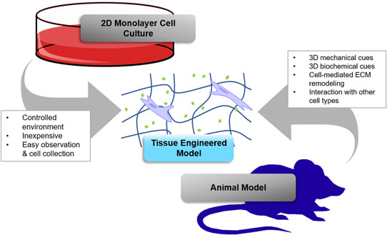
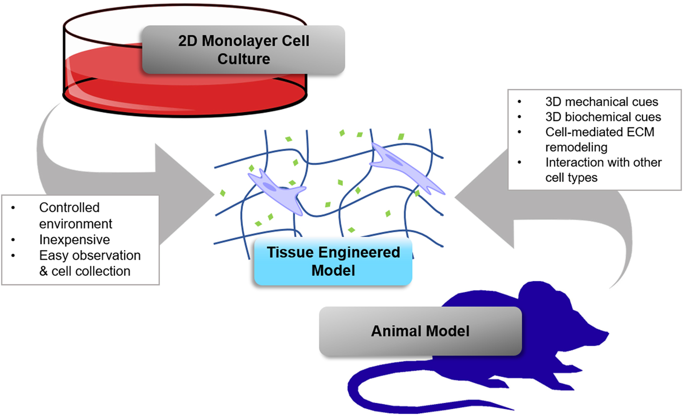
Tissue-engineered cancer models are a middle ground that emerged from research using 3D culture models in which cells were embedded in an extracellular matrix (ECM) material. Importantly, tissue-engineered 3D cultures recapitulate aspects of the in vivo environment better than conventional 2D culture methods. Moreover, they are simpler, cheaper, and provide more control over experimental conditions than animal models, which can enable greater insight into the disease mechanisms (Fig. 1). One of the most popular 3D culture materials used by biologists is Matrigel, which is a basement membrane gel made from the secreted ECM of Engelbreth–Holm–Swarm sarcoma cells. Notably, Matrigel has been used as a 2D and 3D substrate for the culture of various cancer cells. Seminal works by the Bissell laboratory and the Brugge laboratory have shown that non-cancerous breast epithelial cells form hollow spheres that mimic the physiology of breast acini in 3D culture in Matrigel,[Reference Petersen, Rønnov-Jessen, Howlett and Bissell9, Reference Debnath, Muthuswamy and Brugge10] whereas breast epithelial cancer cell lines and primary tumor cells form disorganized structures.[Reference Petersen, Rønnov-Jessen, Howlett and Bissell9] Matrigel is also commonly used in invasion assays to assess the invasiveness of tumor cells.[Reference Albini, Iwamoto, Kleinman, Martin, Aaronson, Kozlowski and McEwan11] Despite the benefits and progress that has been achieved with Matrigel, this material is ill defined and is well known to display batch-to-batch variability.[Reference Hughes, Postovit and Lajoie12] Moreover, the composition of Matrigel is not necessarily reflective of the ECM composition of all tissues. Collectively, these limitations have led to interest in using tissue-mimetic hydrogels that provide greater control and reproducibility over the tumor microenvironment. In the following sections, we will review tissue-mimetic hydrogel platforms and highlight examples of their use in cancer research.
The advantages of a tissue-engineered model of cancer. The tissue-engineered 3D culture model shares advantages with 2D conventional monolayer cell culture models and animal models. While this is not a comprehensive list of advantages of tissue-engineered models, it highlights some of the common considerations and highlights what each model has in common with tissue-engineered models.
Overview of tissue-mimetic hydrogels
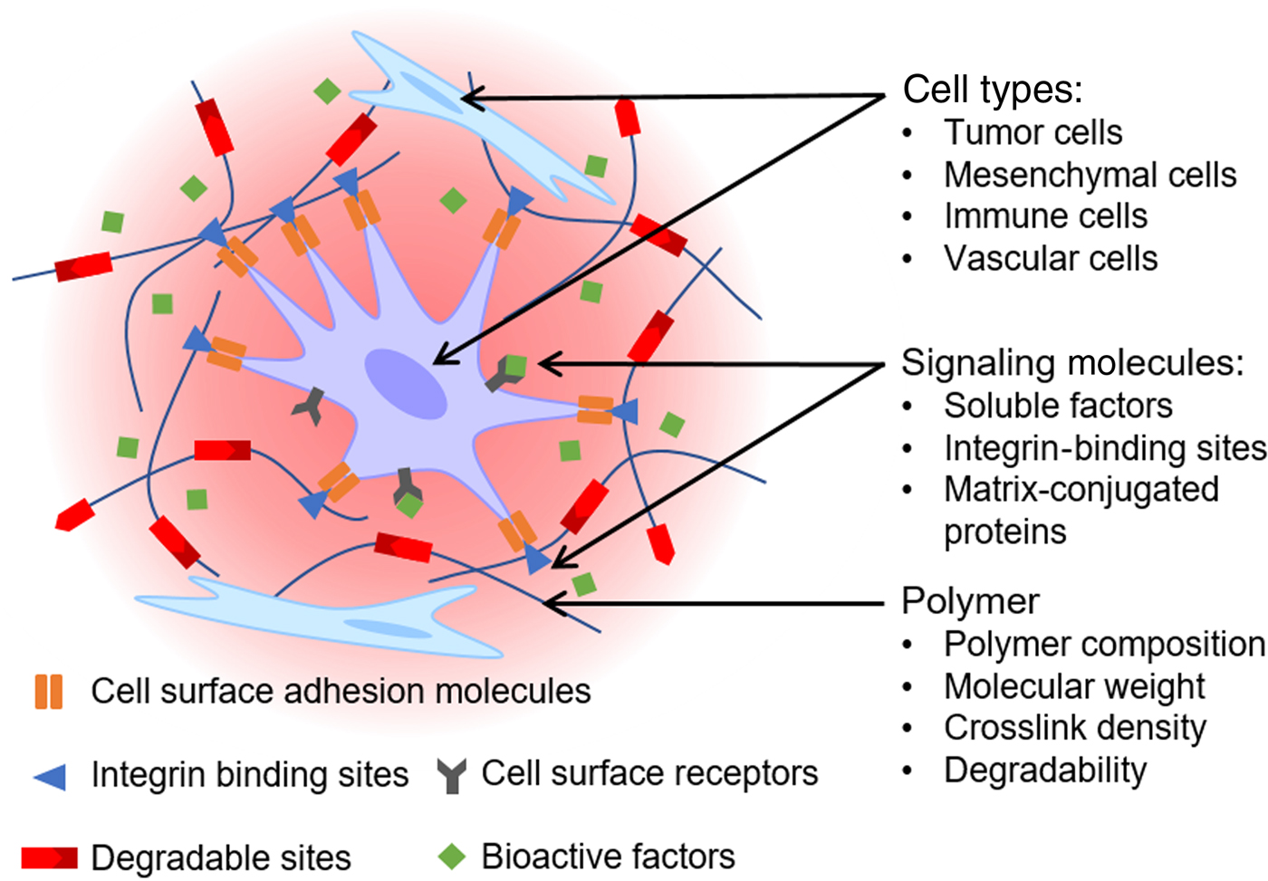
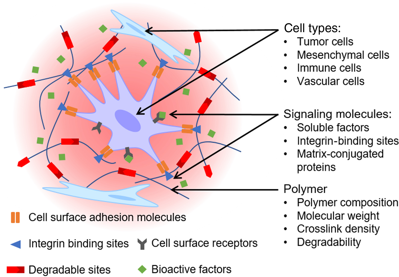
A comprehensive review of the polymers and crosslinking chemistries that can be used to create tissue-mimetic hydrogels is beyond the scope of this review. Instead, we will focus on the most widely used materials and those which are discussed in our selected examples of tissue-engineered 3D cancer models (Fig. 2).
Design features of a tissue-engineered 3D culture tumor model. Tuning hydrogel crosslink density can change the mechanical stiffness and the ease with which cells can migrate. Biochemical cell signaling molecules, including different cell adhesion moieties, growth factors, and cytokines, can also be incorporated into the model at controlled concentrations to simulate microenvironmental signaling. A variety of cell types can be used to create co-culture models to study the interaction between different cell types in the tumor microenvironment.
Hydrogels consist of hydrophilic polymers that are crosslinked, either chemically or physically, to form a 3D network that imbibes large amounts of water.[Reference Hoffman13] They can generally be classified as natural or synthetic, depending on the polymer used to make them, and both types have been used as tissue-mimetic platforms for cancer research. Natural hydrogels can be further sub-divided into ECM-derived and non-ECM derived. Moreover, a diverse toolkit for chemical crosslinking of both natural and synthetic hydrogels exists.[Reference Magin, Alge and Anseth14–Reference Lee and Mooney17] The most commonly used crosslinking reactions, and consequently the ones used most frequently to engineer 3D cancer models, are: (1) (meth)acrylate polymerization, (2) Michael-type thiol-ene click reactions, and (3) thiol-ene photoclick reactions. (Meth)acrylate polymerization occurs via a free radical-mediated chain-growth mechanism and is usually photopolymerized with cytocompatible initiators such as Irgacure 2959 or lithium acylphosphinate.[Reference Fairbanks, Schwartz, Bowman and Anseth18] Michael-type thiol-ene click reactions are useful because they enable facile incorporation of cysteine-containing peptides to provide cell adhesion motifs (e.g., RGD, YIGSR, IKVAV) and enzyme-sensitive crosslinks for cell-mediated remodeling.[Reference Azagarsamy and Anseth15, Reference Nimmo and Shoichet19] They proceed under mildly basic conditions and require an electron deficient alkene such as a (meth)acrylate, vinyl sulfone, or maleimide. Thiol-ene photoclick reactions offer the same advantages as the Michael-type thiol-ene reactions for peptide incorporation, but with the added advantage of spatiotemporal control via photopolymerization. The electron rich, ring-strained alkene norbornene is most commonly used as it undergoes step-growth rather than mixed-mode polymerization with thiols. Importantly, multiple studies have shown that thiol-ene photoclick crosslinking is more cytocompatible than (meth)acrylate photopolymerization.[Reference Lin, Raza and Shih20, Reference Shubin, Felong, Graunke, Ovitt and Benoit21]
Type I collagen (typically bovine or rat) is a popular commercially available ECM-derived natural hydrogel material that provides encapsulated cells with a nanoscale fibrillar network that they can attach to via integrin-binding motifs and remodel enzymatically via matrix metalloproteinases (MMPs). The mechanical properties of collagen hydrogels, which self-assemble in approximately 30 min at physiologic conditions, can be manipulated by the collagen concentration or through a variety of chemical and physical crosslinking strategies (e.g., with formaldehyde, genipin), as reviewed by Parenteau-Bareil et al.[Reference Parenteau-Bareil, Gauvin and Berthod22] However, these materials allow limited decoupling of biochemical and mechanical signals, and batch-to-batch variability is a concern. Consequently, many tissue engineers utilize gelatin in lieu of collagen. Gelatin, which is denatured collagen, also provides cell adhesion cites and can be remodeled by encapsulated cells. The most common implementation is gelatin-methacrylamide, or GelMA, which is prepared by functionalizing lysine amino groups in gelatin (e.g., with methacrylic anhydride).[Reference Loessner, Meinert, Kaemmerer, Martine, Yue, Levett, Klein, Melchels, Khademhosseini and Hutmacher23] GelMA hydrogels are generally photopolymerized, and their mechanical properties can be tuned by manipulating both the percent functionalization and the polymer concentration.
Other natural polymers that have been used to synthesize tissue-mimetic hydrogels for cancer models include hyaluronic acid and alginate. Hyaluronic acid is a polysaccharide found in the ECM of many tissues. Similar to gelatin, hyaluronic acid can be functionalized with polymerizable groups to varying degrees to control the crosslink density and properties.[Reference Xu, Jha, Harrington, Farach-Carson and Jia24] For example, (meth)acrylates have been added to enable both free radical chain-growth polymerization and Michael-type thiol-ene crosslinking, the latter of which has been leveraged to incorporate integrin-binding peptides and MMP-cleavable crosslinkers. Alternatively, some platforms have been based on thiolated hyaluronic acid, which is then crosslinked by thiol-ene Michael-type addition reaction (e.g., with a diacrylate polymer). Alginate is a polysaccharide derived from seaweed that can be used to synthesize physically and chemically crosslinked hydrogels.[Reference Augst, Kong and Mooney25] Because it is not an ECM-derived material, it lacks cell adhesive moieties, which provides an opportunity to introduce specific integrin-binding peptides. Alginate is most often physically crosslinked with divalent cations, namely calcium, in which case the calcium concentration controls crosslink density and mechanical properties. However, similar to hyaluronic acid, alginate can also be modified with polymerizable chemical groups such as (meth)acrylates.[Reference Jeon, Bouhadir, Mansour and Alsberg26]
The most widely used synthetic polymer for engineering tissue-mimetic hydrogels is poly(ethylene glycol) (PEG). PEG is considered to be a “blank slate” biomaterial due to its resistance to protein adsorption.[Reference Magin, Alge and Anseth14, Reference Zhu27] Consequently, cells are unable to bind to and interact with PEG hydrogels unless they are modified with bioactive components, which makes them a powerful platform for investigating how specific cues in the cancer microenvironment influence signaling pathways and susceptibility to treatment. Myriad strategies for synthesizing PEG hydrogels via chemical crosslinking have been reported in the literature. The most common strategy has been to end-functionalize linear PEG with (meth)acrylates and then perform free radical chain-growth polymerization. However, the Michael-type thiol-ene and thiol-ene photoclick reactions have become extremely popular in recent years because bioactive peptide functionalization is simpler with these chemistries. These step-growth polymerizations, which utilize multi-arm PEG star polymers synthesized by living polymerization methods, are also believed to produce more uniform polymer networks. Typically, Michael-type thiol-ene crosslinking is performed using PEG macromers functionalized with vinyl sulfone[Reference Lutolf, Lauer-Fields, Schmoekel, Metters, Weber, Fields and Hubbell28] or maleimide[Reference Phelps, Enemchukwu, Fiore, Sy, Murthy, Sulchek, Barker and Garcia29] groups, since the acrylate–thiol reaction produces esters with neighboring sulfides that are readily hydrolyzed under physiologic conditions.[Reference Rydholm, Anseth and Bowman30] The thiol-ene photoclick reaction is performed using norbornene-functionalized PEG.[Reference Fairbanks, Schwartz, Halevi, Nuttelman, Bowman and Anseth31] Regardless of the crosslinking chemistry, PEG hydrogel crosslink density and physical properties can be tuned by changing the PEG molecular weight or concentration.
Examples of tissue-engineered cancer models
The following sections will review selected examples of models of melanoma, glioblastoma, and breast cancer from the recent literature. While tissue-engineered models of other cancers have been developed (e.g., non-small cell lung cancer,[Reference Gill, Gibbons, Roudsari, Saik, Rizvi, Roybal, Kurie and West32, Reference Roudsari, Jeffs, Witt, Gill and West33] PDAC (pancreatic ductal adenocarcinoma),[Reference Ki, Lin, Korc and Lin34] hepatocellular carcinoma,[Reference Liang, Jeong, DeVolder, Cha, Wang, Tong and Kong35–Reference Lin, Ki and Lin37] oral squamous cell carcinoma,[Reference Fischbach, Kong, Hsiong, Evangelista, Yuen and Mooney38] prostate cancer,[Reference Sieh, Taubenberger, Rizzi, Sadowski, Lehman, Rockstroh, An, Clements, Nelson and Hutmacher39, Reference Xu, Gurski, Zhang, Harrington, Farach-Carson and Jia40] ovarian cancer[Reference Loessner, Stok, Lutolf, Hutmacher, Clements and Rizzi41–Reference Kaemmerer, Melchels, Holzapfel, Meckel, Hutmacher and Loessner43]), these examples were selected to highlight the role of materials innovation in developing hydrogel-based 3D tissue-engineered models.
Melanoma
Melanoma is the deadliest form of skin cancer, and the 19th most common form of cancer worldwide.[Reference Holmes44] It is a tumor of melanocytes, the pigment-producing cells of the skin. Incidence is highly linked to exposure to UVA and UVB radiation from the sun or tanning beds. Overall, the clinical burden of melanoma is increasing. In 2016, the lifetime risk of an American developing melanoma was 1 in 54,[Reference Glazer, Winkelmann, Farberg and Rigel45] and incidence rates of melanoma are increasing faster than any other solid tumor type.[Reference Holmes44] However, it should be noted that the incidence of pre-invasive melanoma is rising at a higher rate than invasive melanoma, which may indicate more effective methods of detection and may decrease mortality rates long term.[Reference Glazer, Winkelmann, Farberg and Rigel45] Additionally, melanoma is unusually common compared with other forms of cancer in young people. Incidence rates have been increasing in particular in young Caucasian women, with one study showing an eightfold increase from the 1970s to the 2000s.[Reference Reed, Brewer, Lohse, Bringe, Pruitt and Gibson46] It is thought that this trend is attributable to behavioral risk factors, in particular the use of UV tanning beds.
Several studies on tissue-engineered models of melanoma have been reported by the Anseth research group.[Reference Tokuda, Leight and Anseth47–Reference Tokuda, Jones and Anseth49] These have been based on the PEG thiol-ene photoclick hydrogel platform that is enzymatically degradable and incorporates a cell-adhesive RGD-containing peptide. As noted earlier, this platform allows for control of matrix stiffness by modulating the PEG molecular weight as well as the concentrations of PEG and crosslinker. In a fundamental study of cell–material interactions, Singh et al. leveraged this control and observed an effect of matrix stiffness in 2D culture on the proliferation rate of WM239A cells, a metastatic melanoma cell line.[Reference Singh, Schwartz, Tokuda, Luo, Rogers, Fujita, Ahn and Anseth48] They also made a 3D culture model by encapsulating WM239A cells in the PEG hydrogel and then surrounding them with human dermal fibroblasts seeded in a collagen gel for indirect co-culture. The WM239A cells proliferated and formed clusters in 3D culture, though the cluster size was significantly reduced in the fibroblast co-culture. This may suggest a realistic tumor–stroma interaction, as healthy fibroblasts are thought to inhibit tumor formation.[Reference Kalluri and Zeisberg50] However, after 4–5 days in co-culture, the WM239A cells appeared to protrude and invade into the surrounding hydrogel. Transformation of fibroblasts to a tumor-promoting phenotype is well reported in tumor biology research,[Reference Kalluri and Zeisberg50] which may explain this switch in this model. Altogether, these findings suggest that matrix stiffness influences tumor cell proliferation and 3D culture can, to an extent, recapitulate a physiologically relevant phenotype in tumor cells that includes interactions with stromal fibroblasts.
With that precedent, Tokuda et al. used a similar tissue-engineered melanoma model based on PEG thiol-ene click hydrogels to study the effects of BRAF-kinase inhibition.[Reference Tokuda, Leight and Anseth47] They found that a different metastatic melanoma cell line, A375, exhibited less sensitivity to the small-molecule BRAF inhibitor PLX4032 when cultured on soft but not stiff gels (Young's modulus ~0.6 kPa versus 1.6 and 13.1 kPa), while the early-stage radial growth melanoma line WM35 showed higher sensitivity to the same drug. Similar trends emerged when they advanced to 3D culture.[Reference Tokuda, Jones and Anseth49] Briefly, PLX4032 treatment induced higher levels of caspase 3 activity, a marker of apoptosis and an indicator of drug efficacy, in WM35 cells grown in soft gels (Young's modulus ~0.4 kPa) compared with conventional 2D culture on tissue culture-treated polystyrene. In contrast, A375 cells were even less sensitive to the drug in 3D culture conditions than in 2D hydrogel culture. Moreover, 3D spheroids of WM35 cells encapsulated in the same PEG hydrogels appeared less sensitive to PLX4032, as the spheroids showed significantly higher relative DNA content after treatment compared with single-cell 3D encapsulation conditions, suggesting higher proliferation. These findings suggest that early stage radial growth melanoma cells and late stage metastatic melanoma cells respond to PLX4032 differently, and that the biophysical cues of the model microenvironment influence this response.
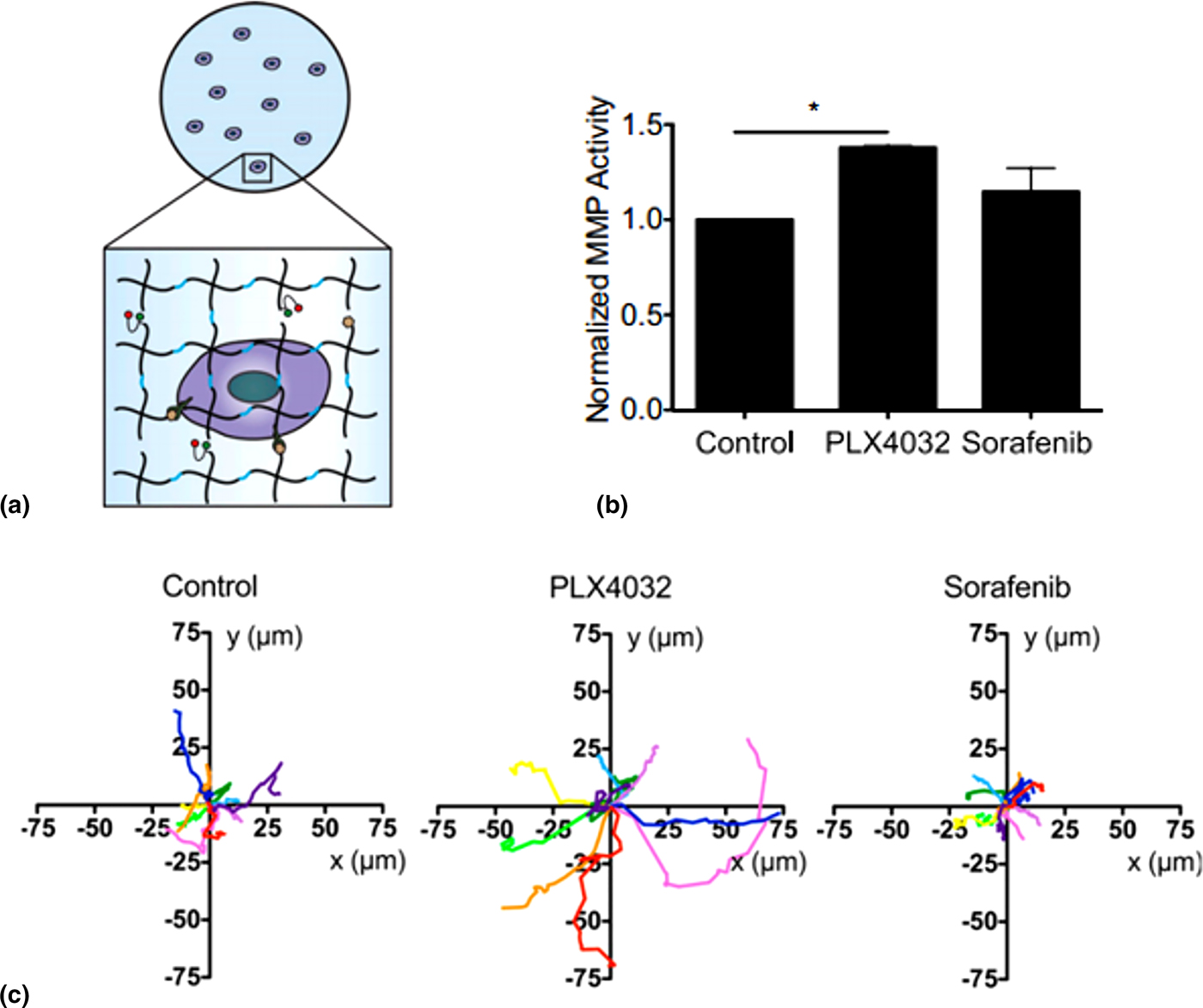
Leight et al. went a step further by incorporating a fluorogenic sensor for MMP activity into the PEG thiol-ene photoclick hydrogels and testing the effects of multiple BRAF inhibitors on several melanoma cell lines.[Reference Leight, Tokuda, Jones, Lin and Anseth51] The fluorogenic sensor enabled quantitative analysis of MMP expression. Their sensor, which was covalently attached to the hydrogel network, consisted of an MMP-cleavable peptide (the same GPQGIWGQ sequence) flanked with a fluorophore (fluorescein) and a quencher (dabcyl). When the peptide was cleaved by cell-secreted MMPs, separation of the fluorophore and quencher resulted in a fluorescent signal that was used to quantify the activity of cell-secreted MMPs in the gel. Four melanoma cell lines were tested, representing different points in tumor progression: WM35 (a radial growth line mentioned earlier), WM239A and A375 (two metastatic lines mentioned earlier), and WM115 (a vertical growth line). They also tested four small molecule BRAF-inhibitors: PLX4032 (tested by Tokuda et al.), sorafenib, AZD6244, and CI-1040. Importantly, they found that in 3D culture the metastatic line WM239A was highly drug resistant, with no significant change in metabolic activity, and exhibited higher levels of MMP activity compared with the control for all four drugs tested.[Reference Leight, Tokuda, Jones, Lin and Anseth51] In contrast, the A375 metastatic cell line exhibited lower metabolic activity after treatment with all four drugs but differences in levels of MMP activity. While A375 cells treated with sorafenib showed no significant increase in MMP activity compared with the control, PLX4032 cells showed significantly higher MMP activity (Fig. 3).[Reference Leight, Tokuda, Jones, Lin and Anseth51] Morphologically, PLX4032-treated A375 cells were elongated, with F-actin-rich protrusions into the surrounding matrix.[Reference Leight, Tokuda, Jones, Lin and Anseth51] In addition, PLX4032 treatment increased both the number and the speed of migrating A375 cells in the matrix (Fig. 3).[Reference Leight, Tokuda, Jones, Lin and Anseth51] These results suggest that while PLX4032 may be effective in reducing tumor cell metabolism, it may do so at the cost of increasing the invasive and migratory characteristics of the cells, especially in metastatic melanoma. This result would be nearly impossible to observe in vivo, which shows the promise of using tissue-engineered models for drug screening. It may also explain why melanoma patients taking PLX4032 were observed to relapse with metastatic lesions in clinical trials.[Reference Sosman, Kim, Schuchter, Gonzalez, Pavlick, Weber, McArthur, Hutson, Moschos, Flaherty, Hersey, Kefford, Lawrence, Puzanov, Lewis, Amaravadi, Chmielowski, Lawrence, Shyr, Ye, Li, Nolop, Lee, Joe and Ribas52] However, it should be noted that this model omitted the tumor stroma, which is a limitation of the study since tumor-associated endothelial cells, fibroblasts, and macrophages can be important sources of MMPs and contributors to tumor dissemination. Thus, to get an accurate picture of how drug treatment influences matrix remodeling in the tumor microenvironment, future work should also incorporate stromal cells in the model.
Engineered hydrogel scaffold enables real-time visualization and quantitation of MMP activity in tumor models. (a) Schematic representation of a cancer cell encapsulated in an MMP-degradable PEG-peptide hydrogel scaffold modified with a fluorogenic sensor of MMP activity. Melanoma cells treated with small-molecule BRAF inhibitor PLX4032 showed (b) increased MMP activity and (c) increased migration over melanoma cells treated with a different small-molecule BRAF inhibitor, sorafenib. Adapted from Leight et al.[Reference Leight, Tokuda, Jones, Lin and Anseth51] with permission from the National Academy of Sciences, USA.

Glioblastoma
Glioblastoma is the most aggressive form of brain cancer. Glioblastomas arise from the astrocytes of the brain and are highly metastatic, though they rarely metastasize outside the central nervous system. While it is one of the rarer forms of cancer, it is nonetheless deadly. The 5-year survival rate in 2010 was <5%.[Reference Thakkar, Dolecek, Horbinski, Ostrom, Lightner, Barnholtz-Sloan and Villano53] Recurrence after the first round of treatment is common, with virtually all patients experiencing disease progression within 7–10 months post-treatment.[Reference Omuro, Faivre and Raymond54] The median survival post-diagnosis is 15 months.[Reference Thakkar, Dolecek, Horbinski, Ostrom, Lightner, Barnholtz-Sloan and Villano53] Some genetic and environmental risk factors for glioblastoma have been studied, but none have been identified that account for a large portion of cases.[Reference Thakkar, Dolecek, Horbinski, Ostrom, Lightner, Barnholtz-Sloan and Villano53] Its aggressive and rapid metastasis and low sensitivity to available therapies make glioblastoma extremely difficult to treat and provide a strong impetus for tissue-engineering glioblastoma models.
A significant focus of glioblastoma research using tissue-mimetic hydrogels has been elucidating the connection between matrix stiffness and glioblastoma cell migratory phenotype. Ulrich et al. sought to accomplish this using polyacrylamide gels surface functionalized with plasma fibronectin for 2D culture substrate.[Reference Ulrich, de Juan Pardo and Kumar55] Using hydrogels with Young's moduli between 0.08 and 119 kPa, they found that cells from multiple glioma lines (U373-MG, U87-MG, U251-MG, SNB19, C6) isolated from both rat and human tumors exhibited greater spreading, faster migration, and faster proliferation on stiffer gels. Moreover, they showed that these effects could be attenuated by inhibiting molecular signaling pathways involved in mechanotransduction and cytoskeletal remodeling, including non-muscle myosin II and ROCK.[Reference Ulrich, de Juan Pardo and Kumar55] Pathak and Kumar also used a similar polyacrylamide gel platform to study glioblastoma cell migration in microchannels made using photolithography and found that the previously observed effects of stiff substrates on migration speed were enhanced in the microchannels, with cells in thinner microchannels migrating faster.[Reference Pathak and Kumar56] The result corroborating that glioblastoma migration speed is increased on stiffer substrates is particularly interesting, since the opposite trends have been observed in 3D models.
A number of studies have used natural polymer-based hydrogels to create tissue-engineered 3D glioblastoma models to investigate tumor angiogenesis. Verbridge et al. used RGD-functionalized alginate hydrogels for 2D culture and 3D encapsulation under hypoxic conditions to study angiogenic signaling in U87 MG (also known as U87) glioblastoma cells as well as OSCC-3 oral squamous cell carcinoma cells.[Reference Verbridge, Choi, Zheng, Brooks, Stroock and Fischbach57] They found that U87 cells exhibited elevated pro-angiogenic signaling via cytokine IL-8 in gels in both ambient and hypoxic conditions. Nguyen et al. investigated the use of a GelMA hydrogel “microwell” as a potential platform for high-throughput testing of tumor cultures and examined endothelial cell migration toward U87 glioblastoma cell spheroids.[Reference Nguyen, Fan, Akay and Akay58] Human umbilical vascular endothelial cells (HUVECs) were found to migrate through the gelatin matrix that held a U87 cell spheroid grown in a well at the center of the gel, suggesting angiogenesis.[Reference Nguyen, Fan, Akay and Akay58] Subsequent testing of the anti-angiogenic drug TNP-470 in this same platform showed fewer HUVECs in the vicinity of the glioblastoma spheroids, suggesting inhibition of angiogenic-like endothelial cell migration.[Reference Nguyen, Fan, Akay and Akay59] While additional molecular and cellular biological analyses are needed to verify that these models recapitulate the angiogenic signaling from tumor cells observed in vivo, microwell platforms like this one could accelerate the identification of novel angiogenesis inhibitors for clinical testing.
Tissue-engineered models are also being used to investigate the role of hyaluronic acid in the glioblastoma ECM, since this biomolecule is present at higher concentrations in the brain than most other tissues.[Reference Pathak and Kumar56] Ananthanarayanan et al. used hydrogels comprising hyaluronic acid methacrylate functionalized with cysteine-containing RGD peptides and crosslinked with dithiothreitol in a Michael-type addition reaction to investigate the effects of matrix stiffness on glioblastoma phenotype.[Reference Ananthanarayanan, Kim and Kumar60] They found that spheroids of glioblastoma cells displayed a more invasive phenotype marked by more infiltration into the surrounding matrix from the spheroid boundary in 3D culture in soft (150 Pa) gels compared with stiff gels. Importantly, this result was in contrast to 2D culture experiments on the hydrogels, in which the glioblastoma cells displayed more spread morphology, faster migration, and more proliferation on stiff gels (1 and 5 kPa), similar to what was observed by Ulrich et al.[Reference Ulrich, de Juan Pardo and Kumar55] These conflicting responses to matrix stiffness in 2D and 3D models may indicate that the dimensionality of the culture has a significant impact on the cellular response. However, it could also be an indication of a limitation of the 3D platform. The higher crosslinking and tighter mesh of the stiffer gels may be more difficult for the cells to migrate through, which may also explain why 3D migration was faster in a softer gel while the reverse was shown in 2D culture.
Hydrogels containing mixtures of hyaluronic acid and other polymers have also been investigated. Rao et al. used composite hydrogels comprising mixtures of type I collagen and thiolated hyaluronic acid to examine the effects of ECM composition on primary human glioblastoma-derived cell motility.[Reference Rao, DeJesus, Short, Otero, Sarkar and Winter61] They found that higher concentrations of hyaluronic acid correlated with a slower migration speed and more rounded cell morphology, as well as a higher elastic modulus of the gels.[Reference Rao, DeJesus, Short, Otero, Sarkar and Winter61] The Harley group has utilized hydrogels comprising a combination GelMA, acrylated PEG, and methacrylated hyaluronic acid crosslinked via radical-initiated chain polymerization.[Reference Pedron, Becka and Harley62–Reference Pedron and Harley64] In one study, they found that increasing the relative amount of hyaluronic acid in the gel induced U87 glioblastoma cells to increase expression of genes associated with malignancy, including the angiogenic factor VEGF and the hypoxia marker HIF-1.[Reference Pedron, Becka and Harley62] Interestingly, wild-type U87 cells showed decreasing levels of MMP-2 expression, a marker for invasiveness, in gels with higher concentrations of hyaluronic acid, while U87 cells modified to overexpress the epidermal growth factor receptor (EGFR) displayed the opposite trend. Pedron et al. suggested that this effect may be due to the “more malignant” phenotype of the EGFR+ cells, as EGFR overexpression is commonly seen in more metastatic forms of glioblastoma.[Reference Pedron, Becka and Harley62] In a separate study, they demonstrated that a less degradable matrix, with a higher ratio of PEG tetra-acrylate to GelMA, also increases U87 expression of VEGF, HIF-1, and MMPs in 3D culture,[Reference Pedron and Harley64] further demonstrating that microenvironmental composition is important in tissue-engineered glioblastoma models. Future studies may include comparisons of drug response in matrices with different concentrations of hyaluronic acid. Additionally, correlating features of the tumor cell phenotype and the composition of the surrounding matrix could be valuable in determining prognosis and treatment in a clinical setting.
PEG-based thiol-ene photoclick hydrogels have been used by the Yang research group to create 3D glioblastoma models.[Reference Pedron and Harley64, Reference Wang, Tong and Yang65] Their hydrogels were functionalized with RGD adhesive peptide, crosslinked with a mixture of PEG-dithiol and MMP-degradable crosslinker (GPQGIWGQCK), and also contained sodium hyaluronate. In their initial work, the sodium hyaluronate was physically trapped in the gel after crosslinking, and they studied the effects of matrix stiffness by creating soft (1 kPa) and stiff (26 kPa) hydrogels. They found that U87 cells encapsulated in soft gels showed greater proliferation, whereas cells in stiff gels exhibited denser aggregation as well as cellular protrusions into the surrounding gel, suggesting a propensity to invade.[Reference Wang, Tong and Yang65] These results are similar to previously referenced results in 3D culture platforms, but conflict with the rates of proliferation seen in 2D culture studies. However, the changes in cellular morphology may indicate a more invasive phenotype, which was further investigated through gene expression data. Cells in stiff gels showed increased expression of HA synthase 1 and MMP-1 genes and downregulation of hyaluronic acid synthase 2 and MMP-9 genes, all associated with matrix remodeling, compared with cells in soft gels,[Reference Wang, Tong and Yang65] and increased expression of Hras, RhoA, and ROCK1, which are associated with mechanotransduction.[Reference Wang, Tong and Yang65] Overall, these data suggest that the substrate stiffness influences the mechanosensing and ECM remodeling response of the glioblastoma cells. In a separate study using the same material platform, but with thiolated hyaluronic acid that can be chemically conjugated to the matrix, the gel stiffness was maintained between 1.2 and 2.0 kPa but the degradability of the matrix was adjusted by varying the ratio of MMP-degradable crosslinker peptide to PEG-di-thiol.[Reference Wang, Tong, Jiang and Yang66] They observed that U87 cells in degradable matrices (50% or 100% degradable crosslinker) exhibited a spread morphology and actin-dense structures extending into the surrounding gel, while cells in the non-degradable gels maintained a round morphology.[Reference Wang, Tong, Jiang and Yang66] Interestingly, cells in all gels exhibited increased expression of MMP-1 and MMP-9 as well as significantly increased expression of hyaluronic acid synthase 2 compared with pre-encapsulation levels, but the differences in MMP and hyaluronic acid synthase expression between gels of different levels of degradability were not significant. In addition, gene expression levels in these gels, normalized to pre-encapsulated gene expression, were similar to the soft gels of the previous study. Taken together, these results suggest that matrix stiffness may influence the nature of ECM remodeling in glioblastoma more strongly than hyaluronic acid presentation (free versus matrix conjugated) or matrix degradability.
The aforementioned studies utilized homogenous hydrogels of varying composition, but there has also been interest in incorporating spatial gradients of microenvironmental cues in 3D glioblastoma models to recapitulate tumor heterogeneity. For example, Pedron et al. used a microfluidic gradient mixing device to create gradients in chain-growth photopolymerized GelMA hydrogels used for 3D cell culture.[Reference Pedron, Becka and Harley63] The degree of methacrylation of the gelatin macromer, concentration of methacrylated hyaluronic acid added, and cell number were varied throughout bulk gels. They observed that cell proliferation of U87 cells increased in regions containing gelatin with a higher percent methacrylation, and consequently a higher stiffness. MMP expression levels as well as gene expression levels for fibronectin, HIF-1, and VEGF were also observed to vary across the gradient.[Reference Pedron, Becka and Harley63] In gels with varying concentration of hyaluronic acid, U87 cells displayed a tendency to aggregate more and showed decreased cell proliferation in regions with high hyaluronic acid concentrations. Varying the concentration of cells in the gel also created a gradient of pro-invasive and pro-angiogenic gene expression, as expression of genes for hypoxia marker HIF-1, pro-angiogenic growth factor VEGF, and ECM protein fibronectin all increased with increasing cell concentration. Since ECM density, ECM composition, and cellular composition of tumors can vary spatially, it may be necessary to use spatiotemporal patterning approaches like this to properly account for tumor heterogeneity.
Ultimately, tissue-engineered glioblastoma models may provide a better testing platform for cancer therapeutics and lead to better treatment strategies for patients. Work by Jiglaire et al. supports this promise.[Reference Jiguet Jiglaire, Baeza-Kallee, Denicolaï, Barets, Metellus, Padovani, Chinot, Figarella-Branger and Fernandez67] They used a commercially available hydrogel platform comprising a mixture of thiolated hyaluronic acid and thiolated gelatin crosslinked with PEG-diacrylate via a Michael-type addition for 2D and 3D culture of U87 glioblastoma cells, primary murine glioblastoma cells, and primary human patient-derived glioblastoma cells. They investigated the cell responses to radiation therapy as well as three common cancer chemotherapeutics: cisplatin, carmustin, and temozolomide. Notably, U87 cells showed more resistance to cisplatin, carmustin, and radiation in 3D culture than in 2D culture, but they were more sensitive to temozolomide in 3D culture than in 2D culture. While primary patient-derived cells showed variable responses to all treatments, cells from the same tumor often differed in response to treatment between 2D and 3D culture and generally displayed more sensitivity to therapy in 2D culture.[Reference Jiguet Jiglaire, Baeza-Kallee, Denicolaï, Barets, Metellus, Padovani, Chinot, Figarella-Branger and Fernandez67] While further studies are needed before this effect of 3D culture can be generalized to all patient-derived glioblastomas, this study suggests that 3D tumor models may prove useful for drug screening by simulating resistance to therapy. It would be particularly interesting to see similar studies performed using the tissue-engineered glioblastoma models described earlier, as these could provide greater insight on how the tumor ECM and biophysical cues affect drug efficacy, similar to what has been done with the melanoma models.
Breast cancer
Breast cancer is the most common form of cancer in American women, and an estimated 249,260 new cases were diagnosed in 2016 in women and men in the USA.[Reference Siegel, Miller and Jemal68] Breast cancer is a clinical and research priority worldwide; 1.7 million women across the globe were diagnosed with breast cancer in 2012.[Reference Woolston69] Overall, 5-year survival rates are good for breast cancer, with an average of 89.7% from 2006 to 2012, according to the National Cancer Institute's SEER data. Early detection methods have been critical in raising this survival rate from 75.2% in 1975. However, breast cancers detected post-metastasis remain deadly, with a 5-year survival rate of 26.3%. Early detection with mammography and screening does carry the risk of overdiagnosis, treatment when the disease would not have caused symptoms within the patient's lifetime. It is estimated that overdiagnosis has affected 1.3 million women in the USA in the past 30 years, since the implementation of mammography.[Reference Bleyer and Welch70] The advent of mammography has allowed for the detection of pre-cancerous lesions before the tumor breaches the basement membrane of the tissue, known as ductal carcinoma in situ (DCIS). DCIS presents a clinical challenge because there are no known markers of future metastasis.
The seminal work in 3D cancer models demonstrated that breast epithelial and epithelial carcinoma cells can form acini-like structures as they proliferate in Matrigel,[Reference Petersen, Rønnov-Jessen, Howlett and Bissell9, Reference Debnath, Muthuswamy and Brugge10] with highly ordered structures forming from non-cancer cells and markedly more disordered structures forming from malignant cells.[Reference Petersen, Rønnov-Jessen, Howlett and Bissell9] Understanding how microenvironmental cues lead to this behavior has been a major focus in tissue-engineered breast cancer models. As one example, Pradhan et al. utilized fibrinogen-conjugated PEG diacrylate crosslinked via a radical-initiated chain polymerization to encapsulate breast cancer cells.[Reference Pradhan, Hassani, Seeto and Lipke71] They adjusted the stiffness of the gels from 3.2 ± 0.5 kPa to 5.4 ± 0.5 kPa to 9.0 ± 1.4 kPa (Young's modulus) by introducing increasing concentrations of PEG diacrylate that was not fibrinogen-conjugated to the gel and observed that two breast cancer cell lines, MCF-7 and SK-BR-3, formed cell clusters after 15 days of culture and had comparable morphologies across gels of differing stiffness. These cells formed orderly spherical cell clusters, similar to the acini-like structures observed in Matrigel.[Reference Petersen, Rønnov-Jessen, Howlett and Bissell9, Reference Kenny, Lee, Myers, Neve, Semeiks, Spellman, Lorenz, Lee, Barcellos-Hoff, Petersen, Gray and Bissell72] However, MDA-MB-231 breast cancer cells, which are noted for their aggressive, invasive phenotype, did not cluster in the gels but rather showed a more disordered, spread morphology, with protrusions into the surrounding matrix.[Reference Pradhan, Hassani, Seeto and Lipke71] Collectively, these results suggest that the engineered PEG-fibrinogen platform is a suitable surrogate for Matrigel in breast cancer studies. Building on this work, a subsequent study used the PEG-fibrinogen platform to produce MCF-7 breast cancer cell spheroids via microsphere encapsulation in the hydrogels.[Reference Pradhan, Clary, Seliktar and Lipke73] They found that this approach resulted in greater uniformity and induced a more cancer-like cell morphology (i.e., less apico-basal polarity, more cellular and nuclear atypia, elevated nuclear to cytoplasmic ratio and nuclear density, and reduced cell–cell junction length) compared with self-aggregated spheroids produced by the hanging-droplet technique.[Reference Pradhan, Clary, Seliktar and Lipke73] Thus, hydrogel microsphere encapsulation could be a superior method for drug screening studies with breast cancer cell spheroids.
Several studies have used “blank slate” PEG-based hydrogels to investigate the role of different integrin-binding peptide motifs in breast cancer. Weiss et al. used plasmin-degradable Michael-type addition crosslinked PEG hydrogels to compare the effects of RGD and the laminin-derived YIGSR adhesion motif.[Reference Weiss, Bernabé, Shikanov, Bluver, Mui, Shin, Broadbelt and Shea74] They tested three cell types: MDA-MB-231 cells, a triple-negative breast cancer cell line, ErbB2.MCF10A cells, non-cancerous breast epithelial cells modified to overexpress the HER2 receptor also known as ErbB2, which is a common molecular feature of HER2+ breast cancer subtypes, and MCF10A cells, which were used as a control breast epithelial cell. The control MCF10A cells formed ordered spherical structures in the 3D PEG hydrogels, with some exhibiting a lumen reminiscent of acini. As with the PEG–fibrinogen hydrogel studies, these results were consistent with results previously observed in Matrigel experiments. Thus, engineered PEG-peptide hydrogels are also a good surrogate for Matrigel. Interestingly, the acini-like structures were formed more readily in RGD-functionalized gels than in YIGSR-functionalized gels or blank control gels, demonstrating the impact that differing integrin engagement has on cellular organization. However, the MDA-MB-231 cells and ErbB2.MCF10A cells exhibited less organization in 3D culture, and tended to form smaller and less developed structures in the YIGSR-functionalized gels. Overall, MDA-MB-231 cells were also more likely to form loose, non-spherical cellular aggregates than ErbB2.MCF10A cells in all matrices. The three cell types also showed distinctly different patterns of transcriptional factor activation in the different adhesive peptide-functionalized PEG gels. These results support previous results of abnormal breast cancer cell morphology observed in Matrigel culture, but they also indicate that matrix composition and breast cancer subtype significantly influence these morphological differences.
In another study, Taubenberger et al. used PEG hydrogels to investigate the effects of different cell-adhesive peptide motifs and co-culture with vascular cells on tumor cell phenotype of breast cancer cells.[Reference Taubenberger, Bray, Haller, Shaposhnykov, Binner, Freudenberg, Guck and Werner75] They specifically used a Michael-type addition crosslinked hydrogel comprising a PEG-based macromer functionalized with cysteine-terminated peptides with either a MMP-degradable or a cell adhesive motif and maleimide-functionalized heparin.[Reference Taubenberger, Bray, Haller, Shaposhnykov, Binner, Freudenberg, Guck and Werner75] They tested three cell adhesive peptides—RGD, the laminin-derived IKVAV motif, and the collagen type I-derived GFOGER motif—on two breast cancer cell lines, MCF-7 (ER+/PR+/HER2- subtype) and MDA-MB-231 (triple negative subtype). Interestingly, they observed that neither of the breast cancer cell lines exhibited significant differences in proliferation or invasiveness into the surrounding matrix with different adhesive peptides. However, the MDA-MB-231 breast cancer cells, unlike the MCF-7 breast cancer cell line, did not form spherical cell aggregates in the 3D culture. This result is similar to the results seen by Weiss et al. and may be a feature of the more invasive triple negative phenotype. Unfortunately, the MDA-MB-231 were excluded from subsequent experiments because of their non-spherical phenotype. However, tumor angiogenesis was studied for MCF-7 breast cancer cells, and they were compared with two prostate cancer cell lines (LnCaP and PC-3). Interestingly, regardless of the matrix composition, few endothelial-tumor cell interactions were observed when MCF-7 cells were in co-culture with HUVECs and mesenchymal stem cells (MSCs), which formed vessel-like structures in the hydrogel. This observation was in contrast to what was seen with PC-3 cells, which influenced angiogenesis significantly in hydrogels functionalized with IKVAV and GFOGER but not RGD. While additional studies are needed to elucidate the biologic mechanisms responsible for these differences, this study nonetheless demonstrates how the modularity of PEG-peptide hydrogels can be leveraged study the effects of microenvironmental cues on tumor cell phenotype.
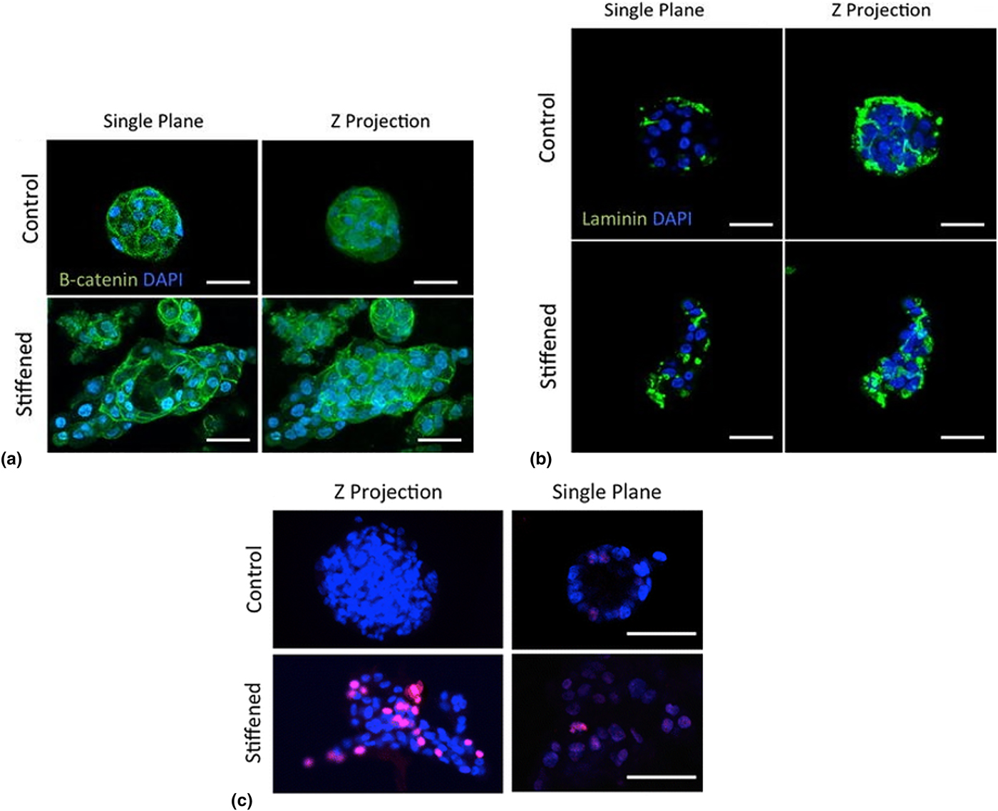
One exciting area of investigation that has not been highlighted in the other example sections is the development of mechanically dynamic microenvironments. Outside of cancer research, softening via photodegradable crosslinkers has been used extensively to create mechanically dynamic hydrogels and probe the effects of matrix stiffness in cell biology.[Reference Kloxin, Kasko, Salinas and Anseth76–Reference Yang, Tibbitt, Basta and Anseth78] However, matrix stiffening is more desirable for mimicking cancer progression, and platforms to achieve stiffening are not as well developed. Stowers et al. achieved this goal using an alginate-based platform.[Reference Stowers, Allen, Sanchez, Davis, Ebelt, van Den Berg and Suggs79] The key to their approach was the incorporation of 1,2-dipalmitoyl-sn-glycero-3-phosphocholine based liposomes loaded with CaCl2 and gold nanorods.[Reference Stowers, Allen, Sanchez, Davis, Ebelt, van Den Berg and Suggs79] Matrigel was also included to facilitate cell adhesion. Irradiation with an 808 nm laser induced surface plasmon resonance in the gold nanorods, resulting in localized heating that triggered the release of calcium ions to increase alginate crosslinking and stiffness. Their platform exhibited a high degree of stiffening over a physiologically relevant range, from an elastic modulus of 151 Pa pre-irradiation to 1074 Pa post-irradiation. Notably, non-malignant MCF10A breast epithelial cells encapsulated in gel initially formed acini-like structures, but after stiffening formed multicellular protrusions into the surrounding matrix with beta-catenin-staining cell-cell contacts (Fig. 4). Acini-like structures in stiffened gels also had larger cross-sectional area and higher levels of nuclear staining for the proliferation marker Ki67, and laminin staining indicated that the layer of basement membrane surrounding the outside of the structure became disorganized post-stiffening (Fig. 4). Collectively, these results indicate that stiffening induced a more tumor-like phenotype. However, the effects were dampened by treatment with small molecule inhibitors of Rac1 and ROCK, suggesting that dynamic material platforms like this one could provide new insight on the mechanisms of mechanotransduction that influence the progression of breast cancer.
Progressive matrix stiffening induces tumor-like morphology in non-cancer human mammary epithelial cells. (a) MCF10A non-cancer epithelial cells form less spherical, acini-like structure after matrix stiffening and (b) exhibit less acini-like organization of laminin-rich basement membrane and around cell clusters. (c) MCF10A cells exhibit greater nuclear staining of Ki67 (pink), a proliferative marker, after gel stiffening. Adapted from Stowers et al.[Reference Stowers, Allen, Sanchez, Davis, Ebelt, van Den Berg and Suggs79] with permission of Springer.

Challenges of studying cancer biology in 3D cultures
While 3D tissue-engineered models can provide a more physiologically relevant environment for in vitro culture, it is generally recognized that molecular and cell biological analyses become more difficult in these systems. Immunostaining and 3D fluorescence imaging techniques generally work well and can provide valuable insight on structure, organization, and cell–matrix interactions. However, implementing quantitative methods in 3D tissue-engineered culture systems to study intercellular and intracellular signaling pathways is not trivial since these methods were developed for simpler 2D culture systems. While methods for protein and nucleic acid detection and quantitation have been adapted to whole tissue samples, whole tissue samples have more cells and more ECM proteins than 3D tissue-engineered systems. This yields a stronger signal in protein and gene expression that is less vulnerable to significant degradation from common digestion methods used for quantitative protein or gene expression analysis, such as enzymatic digestion or phenol–chloroform extraction, compared with tissue-engineered models. Consequently, it is possible that signal loss from processing for molecular and cell biologic analyses could be limiting our understanding of cancer biology in 3D environments.
Perhaps the best evidence that the challenges of extending molecular biology tools to tissue-engineered systems could be limiting our understanding is a recent paper from Valdez et al.[Reference Valdez, Cook, Chopko, Wang, Brown, Kumar, Stockdale, Rothenberg, Renggli, Gordon, Lauffenburger, White and Griffith80] They developed a novel PEG hydrogel crosslinked with a peptide susceptible to enzymatic cleavage by both cell-secreted MMPs and Sortase A.[Reference Valdez, Cook, Chopko, Wang, Brown, Kumar, Stockdale, Rothenberg, Renggli, Gordon, Lauffenburger, White and Griffith80] Because Sortase A is a bacterial enzyme and is not produced by mammalian cells, this approach enabled on-demand exogenously controlled degradation of cell-laden hydrogels for protein expression analysis. Using a Sortase A variant engineered to have faster kinetics, they achieved complete degradation in approximately 5 min. In contrast to degradation with trypsin digestion and a commercially available collagenase digestion, which are common enzymatic digestion methods, they demonstrated that Sortase-mediated digestion was mild and preserved the integrity of cell-secreted cytokines in the matrix. Moreover, they detected significant differences between cytokines present in the matrix compared with the media. This finding suggests that only analyzing the media (an approach used from 2D studies) provides an incomplete picture of cellular signaling in 3D models, and studies that use this method may be missing important proteomic information. This work is a case study on how materials innovation can provide new insight on cancer biology.
Gene expression analysis is another staple in molecular biology analysis. Myriad kits for mRNA isolation from 2D cultures and bulk tissue samples are commercially available and are frequently used in conjunction with quantitative real-time reverse transcription polymerase chain reaction (qRT–PCR) to measure gene expression. However, isolating sufficient amounts of high-quality mRNA from 3D hydrogel cultures can be challenging. Work from the Gasparian et al. suggests that, at least in PEG-based hydrogels, this may be due to RNase-mediated degradation.[Reference Gasparian, Daneshian, Ji, Jabbari and Shtutman81] They showed that while low quantities and poor quality mRNA were obtained using standard procedures, the yield was drastically improved by flash freezing in liquid nitrogen and then adding sacrificial tRNA to protect the desired mRNA. While their study is useful for method development, it also raises the question of whether mRNA signals have been missing in previous work and to what degree RNA isolation methods impact signal strength and quality, especially with the advent of more sensitive RNA detection methods such as RNA sequencing (RNA-Seq).
Cell migration, which is of course fundamental to metastasis but is also important for evaluating cellular interactions (e.g., between fibroblasts and cancer cells), also has challenges in 3D. Live-cell microscopy techniques are frequently employed and used to perform real-time tracking of cell migration in 3D matrices, as in the study by Leight et al.[Reference Leight, Tokuda, Jones, Lin and Anseth51] Thus, methods and tools are not a limitation. However, a critical effector of cell migration in hydrogel cultures is mesh size, which is determined by the crosslink density and is inversely related to modulus. Typical pore sizes in tissue mimetic hydrogels being used for 3D culture are on the order of tens of nanometers. Thus, the polymer network constitutes a restrictive mesh to cells, which are microscale, and it must be degraded to permit cell spreading and migration. While this problem is generally circumvented in migration studies by choosing low-modulus matrices, it is potentially confounding since tumors are known to be stiffer than healthy tissues and it may be desirable to increase the matrix modulus to provide a more physiologically relevant microenvironment. Douglas et al. recently reported an intriguing solution to this problem when they demonstrated that ultrasoft microgels could be incorporated into stiff fibrin gels to form a connected network of tracks for cellular migration.[Reference Douglas, Fragkopoulos, Gaines, Lyon, Fernandez-Nieves and Barker82] Because this approach decouples cell migration from matrix degradation, it could allow migration to be studied in stiffer matrices that better replicate certain tumor microenvironments.
Another challenge in developing in vitro tumor models is accounting for the complex cellular milieu of tumor tissue. Increasing evidence supports that the tumor stroma, which includes all the cells of the tumor that are not the neoplastic tumor cells (i.e., fibroblasts, endothelial cells, adipocytes, MSCs, macrophages, and other vascular, mesenchymal, and immune cells), is crucial for the formation and progression of a tumor. Tumor vascularization and angiogenesis in particular require tumor–stroma interaction. Thus, tumor cell expression of pro-angiogenic signaling molecules is often investigated in 3D hydrogel cultures.[Reference Fischbach, Kong, Hsiong, Evangelista, Yuen and Mooney38] However, more compelling and physiologically accurate models include vascular cells such as HUVECs and quantify the production of pro-angiogenic signaling molecules as well as endothelial cell migration.[Reference Verbridge, Choi, Zheng, Brooks, Stroock and Fischbach57] More sophisticated models have been used to visualize the assembly of vessel-like structures in co-culture with tumor cells. For example, such co-culture models have included breast or prostate tumor cells, HUVECs, and MSCs[Reference Bray, Binner, Holzheu, Friedrichs, Freudenberg, Hutmacher and Werner83] or lung tumor cells, HUVECs, and HVP (human vascular pericytes).[Reference Roudsari, Jeffs, Witt, Gill and West33] However, only a few of the known intercellular interactions crucial to tumor angiogenesis have been targeted to date, and it may be important for future investigations to also consider how other stromal cells contribute to tumor vascularization.
Additionally, while it is widely recognized in tumor biology that metastasis requires the tumor stroma, investigation of the intercellular interactions of metastasis is lacking in tissue-engineered models. Many studies of 3D tissue-engineered tumor models report cancer cell MMP production as a marker of metastatic potential, but MMP expression by stromal cells has not been considered. Huang et al. demonstrated in a mouse model that stromal expression of MMP-9 is more critical to metastatic progression than cancer cell expression of MMPs.[Reference Huang, Van Arsdall, Tedjarati, McCarty, Wu, Langley and Fidler84] Within the stroma, tumor-associated macrophages in particular may be important to include in tissue-engineered models. In vivo studies have shown that macrophages are necessary for the formation of metastases. For example, Lin et al. prevented the recruitment of macrophages by tumors by knocking down the gene for macrophage colony-stimulating factor (CSF-1) in a MMTV-PyMT mouse model of breast cancer, which is known to exhibit lung metastases, and found that CSF-1 knockdown mice resulted in delayed histological progression and almost no metastases.[Reference Lin, Nguyen, Russell and Pollard85] According to Pollard and Condeelis, tumor-associated macrophages are not only important in metastasis but necessary for metastasis to occur.[Reference Condeelis and Pollard86] Incorporating stem cells like adipose-derived stem cells is also of interest for tissue-engineered models, as recently reviewed by Wittmann and Fischbach.[Reference Wittmann and Fischbach87]
Another challenge is that gradients of oxygen concentration and regions of hypoxia are common in tumors, especially larger tumors. Regions of hypoxia are thought to induce the development of cancer stem cells, cells with stem-like characteristics that are capable of regenerating full tumors in cases of recurrence and metastasis, and the emergence of these cells has been linked to the expression of hypoxia-induced factors, as reviewed in references.[Reference Heddleston, Li, Lathia, Bao, Hjelmeland and Rich88, Reference Keith and Simon89] Hypoxic gradients have been shown to induce cancer stem-cell-like phenotypes in glioblastoma cells[Reference Heddleston, Li, McLendon, Hjelmeland and Rich90] as well as in breast cancer cells in vitro and in mouse xenografts.[Reference Conley, Gheordunescu, Kakarala, Newman, Korkaya, Heath, Clouthier and Wicha91] The most common way of modeling hypoxia in vitro is by use of hypoxia chambers, which have a limited volume and are expensive. Recently, creative methods of inducing hypoxia in vitro in tissue-engineered tumor models have been explored. For example, Rodenhizer et al. developed a TRACER (tumor roll for analysis of cellular environment and response), made from a cellulose scaffold in a strip, filled with collagen I and seeded with cells, then wound around a spool.[Reference Rodenhizer, Gaude, Cojocari, Mahadevan, Frezza, Wouters and McGuigan92] Importantly, the physical arrangement of the cells and scaffold around the spool creates a gradient in oxygen concentration, with hypoxia in the inner layers, and the scaffold can be unrolled for analysis. They observed several metabolic changes with increasing hypoxia, including increased levels of lactate, a marker of aerobic glycolysis. They also found that the innermost layers of the scaffold contained less doxorubicin after the drug was added to the media and were more resistant to radiation treatment. Incorporation of oxygen-consuming enzymes can also be used to induce hypoxia in tissue-engineered hydrogel models. For example, Park et al. developed a hypoxia-inducing 3D culture system using ferulic acid-functionalized gelatin.[Reference Park and Gerecht93] Enzyme-mediated crosslinking of the ferulic acid molecules depleted the oxygen in the gel to create hypoxic conditions, which the authors reported lasted up to 50 h in gels with encapsulated cells due to cellular oxygen consumption. Lewis et al. subsequently used this platform to model hypoxia in soft tissue sarcoma.[Reference Lewis, Park, Tang, Xu, Pak, Eisinger-Mathason, Simon and Gerecht94] Primary samples of tumors showed enhanced invasion into hypoxic gels over non-hypoxic controls, with greater speed and range of migration. In addition, individual sarcoma cells encapsulated in the gel were able to survive and maintain low oxygen levels for 7 days in culture. The encapsulated individual cells also showed enhanced migration along hypoxic gradients in the gels, as well as more evidence of matrix remodeling than cells in non-hypoxic gels. This evidence of more invasive behavior of tumor cells, expected under hypoxic conditions, suggests that this material may offer a useful platform for future investigation on the effects of tumor hypoxia and drug screening.
ECM nanoarchitecture and dynamics are also important challenges for future research. Most of the hydrogels discussed in this review are chemically crosslinked gels that do not recapitulate the nanofibrillar structure of the ECM. Platforms such as peptide-amphiphile or PEG-conjugated peptide self-assembling nanofiber hydrogel matricies[Reference Hartgerink, Beniash and Stupp95, Reference Collier and Messersmith96] or electrospun methacrylated dextran[Reference Baker, Trappmann, Wang, Sakar, Kim, Shenoy, Burdick and Chen97] might be used in the future in tumor models to address this challenge. In addition, dynamic hydrogels that allow for spatiotemporal control over physicochemical properties could be used to mimic dynamic changes in the tumors. As discussed previously, Stowers et al. designed a controllably stiffened alginate platform to investigate the effects of dynamic matrix stiffness on breast cancer cells. However, other dynamically stiffening hydrogel platforms have been reported in the literature and could be useful for cancer research.[Reference Guvendiren and Burdick98, Reference Young and Engler99] In addition, while photodegradation is perhaps less relevant to tumor progression, photocleavage reactions could be leveraged to dynamically release molecules of interest[Reference Kloxin, Kasko, Salinas and Anseth76, Reference Rosales, Mabry, Nehls and Anseth100] in 4D experiments. This approach could provide a greater understanding of how various molecules of interest (e.g., hyaluronic acid in glioblastoma) influence tumor growth.
Conclusions and outlook
Tissue engineering holds great promise in creating cancer models. The diverse toolkit of tissue-mimetic hydrogels available now can be used to build models with more physiological relevance than current standard 2D culture models and greater control and reproducibility than current animal models. The potential of 3D tissue-engineered culture systems to recreate the physical, mechanical, biochemical, and cellular features of the tumor microenvironment ex vivo offers an exciting opportunity to fill the current gap in knowledge of how tumors create, maintain, and are sustained by their microenvironment. Several polymer strategies have been used to recreate the mechanically stiff matrix of the tumor, and material strategies for changing the stiffness spatially and temporally offer new insights into how mechanical features influence cancer promotion and progression. Additionally, modular scaffold designs have allowed for selective incorporation of cell-signaling molecules, cell-adhesive peptides, and degradable sites. This has enabled the investigation of microenvironmental biochemical features such as matrix degradability, specific cellular adhesion signaling motifs, and even the influence of differing concentrations of whole ECM proteins on tumor cells. Future models could potentially explore incorporating real-time sensing capabilities in the hydrogel platform, as has been explored with MMP activity.
In the future, it will likely be important to build additional microenvironmental features into cancer models to better account for tumor heterogeneity and complexity. To this end, emerging technologies in the field of tissue engineering will be enabling. Bioprinting, for example, has the potential to recreate the complex and heterogeneous spatial organization of cells and biochemical cues of the tumor microenvironment. Also, as more cell types are incorporated and sustained in 3D tissue-engineered culture models, better approximations of complex bi-directional cell-signaling networks will be able to be studied in vitro. However, moving forward, the balance between necessary and sufficient model complexity will have to be kept in mind. The value of increasing or reducing model complexity will have to be considered in the context of in vivo relevance, which will require continuing collaboration with cancer biologists. Ultimately, tissue-engineered tumor models will occupy a critical niche in studying how cancer works and be enabling tools for clinicians and scientists fighting the battle against cancer.
Acknowledgements
Support for this work was provided by a Strategic Areas Interdisciplinary Research Seed Grant to DLA, ESW, and RJO from Texas A&M Engineering Experiment Station, the Dwight Look College of Engineering, and the Texas A&M University Division of Research.