Introduction
Cognitive deficits are frequent and clinically highly relevant symptoms in patients with major depressive disorder (MDD). Impairments in attention, memory, and executive functioning, which at least partly persist during the process of remission of affective symptoms, have been robustly documented (Rock et al., Reference Rock, Roiser, Riedel and Blackwell2014). Cognitive dysfunction is not only associated with clinical variables such as onset and duration of the disorder (Listunova et al., Reference Listunova, Roth, Bartolovic, Kienzle, Bach, Weisbrod and Roesch-Ely2018), but also modulates processing of negative affective information that is subject to cognitive bias (Gotlib and Joormann, Reference Gotlib and Joormann2010). Currently, treatment of cognitive dysfunction in MDD patients is a major clinical challenge, since a substantial proportion of patients will not sufficiently respond to drug treatment or psychotherapy (Rock et al., Reference Rock, Roiser, Riedel and Blackwell2014; Baune and Renger, Reference Baune and Renger2014). Putative beneficial effects of cognitive remediation therapy (CRT) are therefore highly relevant and could lead to a general improvement of therapy in depression. CRT is already widely used in the treatment of schizophrenia, but also for other disorders in the community or hospital settings (Kim et al., Reference Kim, Bahk, Oh, Lee, Lee and Choi2018). In a meta-analysis, Motter et al. (Reference Motter, Pimontel, Rindskopf, Devanand, Doraiswamy and Sneed2016) found effects of computerized cognitive training, specifically for attention, working memory, and global functioning. In addition, further evidence suggests beneficial effects of CRT on functional outcome (Listunova et al., Reference Listunova, Roth, Bartolovic, Kienzle, Bach, Weisbrod and Roesch-Ely2018). Though accumulating evidence suggests that CRT can be a treatment option in MDD presenting with prominent cognitive impairment, the number of studies so far is small, and definitive statements considering the improvement of daily functioning or depressive symptom reduction cannot be made at present (Kim et al., Reference Kim, Bahk, Oh, Lee, Lee and Choi2018).
Meta-analytic evidence suggests that lateral and medial prefrontal, cingulate and temporoparietal regions together with striatal areas are particularly associated with cognition in MDD (Diener et al., Reference Diener, Kuehner, Brusniak, Ubl, Wessa and Flor2012). Yet, although cognitive impairment as well as putative neural correlates of cognitive dysfunction in MDD have been extensively described in the past decade (Rock et al., Reference Rock, Roiser, Riedel and Blackwell2014; Diener et al., Reference Diener, Kuehner, Brusniak, Ubl, Wessa and Flor2012), it is unclear how neural function in cognitively impaired patients with MDD may change as a function of targeted treatment (Meusel et al., Reference Meusel, Hall, Fougere, McKinnon and MacQueen2013). Given the urgent need for objective and reliable biological markers that have the potential to individually predict treatment response, expanding the knowledge on neural mechanisms underlying future clinically beneficial effects of CRT in MDD is essential.
Resting state fMRI (rs-fMRI) is a suitable method to examine intrinsic neural activity, i.e. neural activation in the absence of explicit (extrinsic) cognitive demand. Aberrant intrinsic neural activity patterns have been proposed as potential biomarkers for MDD (Sundermann et al., Reference Sundermann, Olde Lütke Beverborg and Pfleiderer2014). The fractional amplitude of low-frequency fluctuation (fALFF) analysis has been frequently used to investigate intrinsic neural activity in healthy persons and in individuals with mental disorders (Zou et al., Reference Zou, Zhu, Yang, Zuo, Long, Cao, Wang and Zang2008). In patients with MDD, previous studies have shown abnormal fALFF for MDD in frontoparietal regions (Wang et al., Reference Wang, Dai, Su, Wang, Tan, Jin, Zeng, Yu, Chen, Wang and Si2012; Lai and Wu, Reference Lai and Wu2015; Liu et al., Reference Liu, Guo, Liu, Long, Ma, Xue, Wang, Li, Hu, Zhang, Du, Zeng, Liu, Wooderson, Tan, Zhao and Chen2013; Jing et al., Reference Jing, Liu, Ma, Yan, Zhuo, Zhang, Wang, Li and Wang2013). Several cross-sectional studies indicate an association between intrinsic brain activity and cognition, especially attention, executive function, and memory (Huang et al., Reference Huang, Lu, Yu, Li, Zhang, Hu, Zhou, Hu, Wei, Huang, Weng and Xu2017; Tadayonnejad et al., Reference Tadayonnejad, Yang, Kumar and Ajilore2015). Nevertheless, despite the clinical significance of cognition in MDD, very few studies so far have attempted to specifically link cognitive dysfunction to intrinsic neural activity in MDD patients. Even more important, there is a considerable dearth of longitudinal studies investigating cognition in MDD after cognitive remediation. Recently, Han et al. found a reduction of depressive symptoms after cognitive training over 8 weeks in patients with depressive symptoms and chronic traumatic brain injury. Clinical improvement in patients was found to be associated with a reduction of intrinsic neural connectivity in prefrontal regions over time (Han et al., Reference Han, Martinez, Chapman and Krawczyk2018). The neural correlates associated with such improvements in MDD are unclear at present.
In this study, we investigated the longitudinal effects of CRT on brain activity at rest, as well as relationships between intrinsic neural activity and cognitive function in MDD. Changes in intrinsic neural activity in cognitively impaired patients with MDD were assessed before and after CRT which was conducted over 5 weeks. Participants received either individualized or generic CRT and groups were combined in the analysis. We investigated regionally specific effects as well as changes on the neural network level using univariate statistical data analysis methods in conjunction with multivariate techniques. We expected that: (i) patients with MDD and cognitive deficits improve cognition after CRT. (ii) CRT is accompanied by changes of intrinsic neural activity, predominantly in regions associated with cognition in MDD (Diener et al., Reference Diener, Kuehner, Brusniak, Ubl, Wessa and Flor2012), and in particular in lateral prefrontal and parietal areas. (iii) After CRT, intrinsic neural activity in patients will show a trend toward ‘normalization’, i.e. toward activity levels exhibited by healthy controls (HC). (iv) Activity changes following CRT are associated with improved cognitive performance.
Method
Participants
The conducted study is part of a project evaluating the effects of CRT in a sample of cognitive impaired patients with MDD. Results and details on the randomization process from this RCT will be published elsewhere (Listunova et al., in preparation). All patients were randomly (stratified permuted-block randomization) and observer-blind assigned to a passive control group receiving treatment as usual (TAU; n = 19) or a cognitive training group receiving either individualized (IT; n = 22) or generic training (GT; n = 21). Drop outs in the IT and GT group were n = 2 and n = 3, respectively. For individualized training, the patient's individual cognitive profile was identified and then the three most severely impaired cognitive functions were specifically trained. For generic training, a fixed set of six cognitive domains was trained independently from the patient's individual deficits. Rater blindness was verified by guessing treatment allocation. Patients and trainers were not blinded with respect to treatment allocation.
For a sub-sample of 42 patients, MRI data were obtained. The final sample with complete data at baseline and follow-up included 37 MDD patients (TAU: n = 12; cognitive training group: n = 25). Drop-outs were due to >80% missed training sessions (n = 2), discontinuation of the training (n = 2), or missed follow-up (n = 1). Full structural and rs-fMRI data-sets were available for 20 patients receiving training (IT: n = 10; GT: n = 10) due to technical problems including fMRI data artifacts, and 10 patients receiving TAU due to missed rs-scanning at baseline or technical problems at follow-up. All patients were recruited from different sources: announcements, general practitioners, psychiatric practices. Diagnosis of MDD was confirmed by a blinded trained psychologist (LL) according to ICD-10 criteria (World Healthy Association, Dilling et al., Reference Dilling, Mombour, Schmidt and Schulte-Markwort2011). A total of 10 patients were on antidepressants (venlafaxine: n = 3; duloxetine: n = 1; escitalopram: n = 2; citalopram: n = 1; sertraline: n = 1; vortioxetine: n = 1; trimipramine: n = 1). Due to the small sample size (n = 10), TAU data were not considered for further analyses. Since we were primarily interested in treatment outcome and related neural activity changes regardless of CRT modality, IT and GT groups were combined in subsequent analyses.
Inclusion criteria for all participants were: aged between 18 and 60 years, IQ>80 according to the ‘Multiple Choice Vocabulary’ test (MWT-B; Lehrl, Reference Lehrl2005), no comorbid mental disorder or a history of psychosis, no documented or suspected major brain damage or other neurological diseases, and no contraindications for MRI. The severity of cognitive dysfunction in patients was defined as performance below a percentile rank of 16 for at least two cognitive functions, as indicated below (section ‘Neuropsychological assessment’). Patients scoring ⩾ 20 points on the Hamilton Rating Scale for Depression 24 (HAMD; Guy and Bonato, Reference Guy and Bonato1970) were excluded to avoid confounds arising from severe depressive symptoms. No restrictions for regular treatment services were given. All patients received a sociodemographic interview and the Mini International Neuropsychiatric Interview (MINI; Sheehan et al., Reference Sheehan, Lecrubier, Sheehan, Amorim, Janavs, Weiller, Hergueta, Baker and Dunbar1998). For past MDD diagnoses the Structured Clinical Interview for DSM-IV Disorders (SKID-I; Wittchen et al., Reference Wittchen, Wunderlich, Gruschitz and Zaudig1997) was used. Depressive symptoms were assessed via the German version of the Beck Depression Inventory (BDI; Hautzinger et al., Reference Hautzinger, Keller and Kühner2006) and HAMD.
Twenty-six right-handed HC participated in the study. Only participants with a negative family history of heritable neurological or mental diseases were included. Baseline- and follow-up MRI data were collected for 19 HC. Full structural and rs-fMRI data-sets were available for 18 HC, since one control participant missed his follow-up appointment.
The authors asserted that all procedures contributing to this work complied with the ethical standards of the relevant national and institutional committees on human experimentation and with the Helsinki Declaration of 1975, as revised 2008. All procedures involving human subjects were approved by the Ethics Committee of the Medical Faculty at Heidelberg University, Germany. All participants gave their written informed consent prior to inclusion in the study and received monetary compensation for their participation.
Neuropsychological assessment
Neuropsychological baseline assessment included the Trail Making Test A (task performance measure (TPM): reaction times, RT) and Symbol Coding (TPM: accuracy) for estimating information processing speed, alertness (WAFA; TPM: RT), divided attention (WAFG; TPM: RT, missed stimuli), and selective attention (WAFS; TPM: RT, false alarms) for estimating attention, verbal memory (California Verbal Learning Test; Niemann et al., Reference Niemann, Sturm, Thoene-Otto and Willmes2008; TPM: learning sum score, immediate recall and delayed recall accuracy), and figural memory (FGT; TPM: learning sum score, immediate recall and delayed recall accuracy) for estimating learning and memory as well as working memory (verbal n-back task; TPM: incorrect responses), inhibition (Go-Nogo; TPM: RT, false alarms), cognitive flexibility (Trail Making Test B, Langensteinbach version; TPM: RT), and planning (Tower of London, ToL, Freiburg version; TPM: number of correct responses) for estimating executive function. If not other specified, assessments were included in the ‘Vienna Test System’ (VTS, Schuhfried, 2012). For all tests, raw scores were transformed into z-scores and polarized into one direction with higher z-scores indicating better cognitive performance. Domain scores were averaged to form neurocognitive composite scores. A trainable tests composite score (TTCS; mean of WAFA, WAFS, WAFG, Nback, ToL, Go-Nogo) and a general composite score (GCS; mean of attention, learning and memory, executive functioning, and information processing speed) were calculated. Neuropsychological assessments were chosen according to the National Institute of Mental Health's measurement and Treatment Research to Improve Cognition in Schizophrenia (MATRICS) recommendation (Nuechterlein et al., Reference Nuechterlein, Green, Kern, Baade, Barch, Cohen, Essock, Fenton, Frese, Gold, Goldberg, Heaton, Keefe, Kraemer, Mesholan-Gately, Seidman, Stover, Weinberger, Young, Zalcman and Marder2008).
Cognitive training
Training sessions were offered three times per week and last for 60 min, as well as one 30 min compensatory training and transfer session per week over 5 weeks in total. Patients had to attend at least 12 training sessions out of 15 possible sessions. CogniPlus® (Schuhfried, 2008) was used as a scientifically based training system for training cognitive functions. The system can identify the participant's cognitive ability and adapts the difficulty level of the training automatically. For generalized training, divided attention (ability to perform different tasks simultaneously; module: DIVID), selective attention (ability to respond quickly to relevant stimuli and to suppress inappropriate responses; module: SELECT), alertness (ability to temporarily increase and sustain intensity of attention; module: ALERT), working memory (ability to retain information and continuously update it for a short period of time; module: NBACK), response inhibition (ability to inhibit inappropriate behavior; module: HIBIT), planning, and actions skills (ability to plan actions and execute; module: PLAND) were trained (for a detailed description see https://www.schuhfried.com/cogniplus/training/). For IT, the three domains with the lowest percentile rank score at baseline were trained. In each training session, three different randomly stratified domains were trained for 20 min each. Transfer sessions included psychoeducation on cognition, strategy coaching, recap of the past week, and transfer of the training to everyday life. Additionally, participants received a semi-structured ‘cognitive diary’ for daily monitoring of cognition, sleep, and mood, as well as information and working sheets in each transfer session.
Patients were trained in small groups of three to a maximum of five persons with a trained psychologist continuously being present. The psychologist provided general instructions at the beginning of a training session and was available for questions regarding the instructions throughout the session. Training and transfer sessions were held by trained clinical psychologists in the Department of General Psychiatry, Heidelberg University. HC did not receive cognitive training.
MRI data acquisition
MRI scans were conducted before and after the intervention (i.e. after 5 weeks of cognitive training) using a 3 T Siemens Magnetom TIM Trio MR Scanner located at the Department of Neuroradiology at Heidelberg University. Rs-fMRI scanning was carried out in darkness with the instruction for participants to relax without falling asleep, to move as little as possible, to keep eyes closed, and not to think about something special. Adherence was verified by verbal contact immediately after the rs-fMRI scan and as part of a post-scanning exit interview. A total of 200 T2*-weighted echo-planar-imaging whole-brain-volumes in an axial orientation were acquired with following parameters: TR = 2000 ms, TE = 30 ms, flip angle = 90°, slice thickness = 4 mm, distance factor between slices = 1 mm, field of view 200 = mm.
MRI data preprocessing
Data preprocessing was conducted using the Data Processing Assistant for Resting-State fMRI (DPARSF, Yan and Zang, Reference Yan and Zang2010, http://rfmri.org/DPARSF) which is based on Statistical Parametric Mapping (SPM 12, http://www.fil.ion.ucl.ac.uk/spm) and the toolbox for Data Processing & Analysis of Brain Imaging (DPABI, Yan et al., Reference Yan, Wang, Zuo and Zang2016, http://rfmri.org/DPABI) implemented in MATLAB (R2017a, the Math Works, Natick, MA, USA). The origin of the original images was manually set on the anterior commissure. Further preprocessing included slice timing and head motion correction, spatial normalization, and smoothing. Spatial normalization (3 × 3 × 3 mm) was performed using the standard SPM 12 EPI template. A 4 mm full-width half-maximum Gaussian filter was applied for spatial smoothing. In order to regress out nuisance covariates, head motion parameters, white matter signal, and cerebrospinal fluid signal were considered. FALFF images were calculated using temporal bandpass (0.01–0.1 Hz) filtering in order to remove low-frequency drifts and physiological high-frequency noise.
Data analysis
Independent t tests for continuous variables and χ2 test for categorical variables were used to analyze group differences in demographics, BDI, and HAMD scores, as well as paired t tests with Bonferroni correction for multiple testing for analyzing effects of CRT using SPSS 19.0 software (SPSS Inc, Chicago, Illinois, USA).
For rs-fMRI second- level analyses, univariate analyses were used to investigate the regionally-specific effects of CRT on intrinsic neural activity. Using the SPM software package, difference images were calculated as post-treatment v. baseline fALFF maps. In order to explore fALFF differences between the two groups (patients and HC) at baseline and for longitudinal changes, second-level random-effects two-sample t tests were calculated. Age, gender, and medication status were used as nuisance covariates. To further account for potential confounds of head motion, mean framewise displacement (FD) values across the scan for each subject were computed, as suggested by Power and colleagues (2012). Derivatives of the six rigid-body realignment parameters estimated during standard volume realignment were used for FD calculation. A radius of 50 mm was employed to convert angle rotations to displacements.
One-sample t tests using age, gender, medication status, and FD as nuisance variables were used to examine longitudinal changes within the groups (patients and HC). Following strong a-priori hypotheses, for all second-level analyses, a significance threshold of p < 0.005 (uncorrected for multiple comparisons) was used at the whole brain level. A threshold of p < 0.05 at cluster level for small volume correction (SVC; FWE-corrected) was set. Regions of interest for SVC were set based on the meta-analysis of Diener et al. (Reference Diener, Kuehner, Brusniak, Ubl, Wessa and Flor2012) and included brain regions associated with cognition in MDD (bilateral inferior frontal gyrus (IFG), right superior frontal gyrus, bilateral middle frontal gyrus (MFG), medial frontal cortex, right temporal gyrus, left inferior parietal lobule (IPL), right precentral gyrus (PCG), bilateral insula, right caudate, left putamen, and left anterior cingulate). The AAL atlas included in the WFU PickAtlas version 3.0 (ANSIR Laboratory, Wake Forest University School of Medicine; Maldjian et al., Reference Maldjian, Laurienti, Burdette and Kraft2003; Maldjian et al., Reference Maldjian, Laurienti and Burdette2004; Tzourio-Mazoyer et al., Reference Tzourio-Mazoyer, Landeau, Papathanassiou, Crivello, Etard, Delcroix, Mazoyer and Jolijot2002) was used for anatomically localizing coordinates of significant clusters. Effect sizes were calculated using Cohen's d for all significant results.
MarsBaR Toolbox (0.44; Brett et al., Reference Brett, Anton, Valabregue and Poline2002) was used to extract rs-fMRI values for correlational analyses. Pearson/Spearman correlations (two-tailed) were performed to test significant associations between abnormal fALFF values of detected brain regions in the longitudinal (n = 4) analysis and cognitive performance (n = 6) in MDD. For completeness, correlations between baseline measures of brain activity (n = 7) in MDD and cognitive performance (n = 6) were also computed.
Besides regional effects, as investigated using the analysis strategy described above, we were interested in the effects of CRT on distinct neural systems. For this purpose, we explored functional network strength (FNS) using a multivariate data analysis strategy, i.e. ‘source-based morphometry’ (SBM; Xu et al., Reference Xu, Groth, Pearlson, Schretlen and Calhoun2009). SBM toolbox essentially takes advantage of Independent Component Analysis (ICA) to extract spatially independent patterns of data across the whole sample. SBM decomposition of subjects-by-voxel data matrix results in spatial maps, which are the sources of covariance, and a mixing matrix that indicates the contribution for each subject for each source. We have previously applied SBM to structural data to investigate neural effects associated with electroconvulsive therapy in MDD (Wolf et al., Reference Wolf, Nolte, Hirjak, Hofer, Seidl, Depping, Stieltjes, Maier-Hain, Sambataro and Thomann2016), as well as effects of psychotherapy in patients with substance-use disorders (Fahmy et al., Reference Fahmy, Wasfi, Mamdouh, Moussa, Wahba, Schmitgen, Kubera, Wolf, Sambataro and Wolf2019), where SBM has been shown to be sensitive to treatment related changes of neural network strength. This technique has also been used previously on functional imaging data, including SPECT (Premi et al., Reference Premi, Calhoun, Garibotto, Turrone, Alberici, Cottini, Pilotto, Gazzina, Magoni, Paghera, Borroni and Padovani2017).
Using the individual fALFF maps, a spatial ICA was computed. We employed the SBM algorithm (see Xu et al., Reference Xu, Groth, Pearlson, Schretlen and Calhoun2009 for methodological details) as implemented in the ‘Group ICA for fMRI Toolbox’ (GIFT; http://mialab.mrn.org/software/gift). The Infomax algorithm was used to estimate a set of independent components (IC). The ICASSO algorithm (http://research.ics.aalto.fi/ica/icasso/; Himberg et al., Reference Himberg, Hyvarinen and Esposito2004) was used to increase component reliability and consistency; here, we repeated the ICA estimation 50 times with bootstrapping and permutation. To avoid component underestimation, we employed a higher model order where the robustness of ICA estimation was quantified using a quality index (I q) ranging from 0 to 1 (Himberg et al., Reference Himberg, Hyvarinen and Esposito2004). A model order of eleven stable components was identified where all components were associated with an I q>0.9 indicating stable decomposition (Allen et al., Reference Allen, Erhardt, Damaraju, Gruner, Segall, Silva, Havlicek, Rachakonda, Fries, Kalyanam, Michael, Caprihan, Turner, Eichele, Adelsheim, Bryan, Bustillo, Clark, Feldstein Ewing, Filbey, Ford, Hutchison, Jung, Kiehl, Kodituwakku, Komesu, Mayer, Pearlson, Phillips, Sadek, Stevens, Teuscher, Thoma and Calhoun2011). Each fALFF image was then converted into a one-dimensional vector arrayed into a subject-by-voxel matrix. The matrix was decomposed into one mixing and one source matrix. The mixing matrix represents the relationship between participants (controls and MDD) and components. Between-group comparisons were performed using ICA loading parameters (indices from the mixing matrix) representing the contribution of every IC per participant and source. For component visualization the source matrix was reshaped back to a 3D-image, scaled to unit standard deviations (Z) and thresholded at Z > 2.0. Maps from components exhibiting significant differences between the controls and patients at baseline and those who showed a significant response in terms of FNS after CRT (see below) were overlaid onto a Montreal Neurological Institute (MNI) normalized anatomical template. Anatomical denominations and stereotaxic coordinates were derived from clusters above a threshold of Z = 3.0 by linking the SBM output to the Talairach Daemon data base (http://www.talairach.org/daemon.html).
CRT-effects on FNS were analyzed offline using SPSS 19.0 software (SPSS Inc). Since our intent was the identification of CRT effects in patients, we limited our analyses to the networks that had an effect of diagnosis with an exploratory test. Then, we tested the independent effect of treatment using rigorous statistical testing. Analyses were performed as follows: First, we sought to specify those networks which would exhibit baseline differences (before CRT) between patients and HC. Two-sample t tests were calculated on every column of the mixing matrix. For these analyses a nominal significance threshold of p < 0.05 was chosen, uncorrected for multiple comparisons. Next, for those networks found to be significant, paired t tests were performed, where the significance threshold was set to p < 0.05, corrected for multiple comparisons using the Bonferroni method.
Finally, within the patient group, exploratory correlation analyses between FNS differences pre/post CRT and cognition were calculated. Spearman correlations (two-tailed) were performed to test significant associations between abnormal FNS and cognitive performance in MDD. As nominal level of significance, a threshold of p < 0.05 (uncorrected) was defined.
Results
Demographics and clinical scores
The groups did not differ with regard to gender, age, or education (Table 1).
Demographics and clinical scores for patients with MDD and healthy controls (HC)

M, mean; SD, standard deviation. t1, baseline; t2, follow-up. *Significant differences for comparisons between groups, uncorrected for multiple comparisons
Cognitive remediation therapy
Only participants with complete scans for both time points and complete training for MDD patients were included (n = 20). Over the 5 weeks, patients with MDD participated in a total of 14 sessions (mean: 14.4, SD: 8.1). In MDD patients, cognitive performance improved significantly for the majority of the tested domains after CRT (Table 2; original T score units are shown in online Supplementary Table S5).
Neuropsychological results for patients with MDD

Depicted are z-transformed neuropsychological raw scores with higher scores indicating better neurocognitive performance. M, mean; SD, standard deviation. t1, baseline; t2, follow-up. TTCS, Trainable Test Composite Score; GCS, General Composite Score. p Values Bonferroni-corrected for multiple testing. *Significant differences
Rs-fMRI
Baseline comparisons
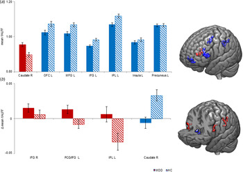
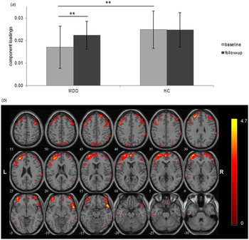
Patients with MDD exhibited increased fALFF in the right caudate, as well as lower fALFF in the left medial orbitofrontal cortex (OFC), left MFG, left IFG, left IPL, left insula, and left precuneus (Table 3, Fig. 1a). With respect to FNS, one (from a total of eleven) IC was identified that showed significantly (T 36 = −2.71, p = 0.010) reduced FNS in MDD compared to controls at baseline. This component included bilateral medial, inferior and middle frontal gyri, right superior temporal gyrus and IPL, left precuneus, and precentral cortex (see Table 4 and Fig. 2).
(a) Mean fALFF values for significant baseline comparison between patients with MDD and healthy controls (HC) for right caudate, left orbitofrontal gyrus (OFC), left middle frontal gyrus (MFG), left inferior frontal gyrus (IFG), left inferior parietal lobule (IPL), left insula, and left precuneus and for (b) significant difference scores (follow-up minus baseline) of mean fALFF values for right inferior frontal gyrus (IFG), left precentral gyrus(PCG)/IFG, left inferior parietal lobule (IPL), and right caudate in patients with MDD and HC. Red: MDD>HC; blue: MDD<HC.

Structural network showing a response to CRT in patients with MDD. (a) Mean values of the bilateral prefrontal network in patients with MDD and healthy controls at baseline and follow-up. (b) Shown is the spatial pattern of a predominantly bilateral prefrontal network and the magnitude of network strength (ICA loadings) (left) in healthy controls and patients with MDD. This network significantly differed between controls and patients at baseline and showed a significant change following CRT (see main text for further details). For illustration purposes, the SBM output image was thresholded at Z > 3.0 and rendered onto the anatomical template implemented in GIFT. The color bar indicates Z-values. R = right. The numbers correspond to the respective slice number.

Brain regions, which survived FWE correction at p < 0.05 after small volume correction for the respective ROI

MNI coordinates (x, y, z) for primary peak locations. L, left hemisphere; R, right hemisphere.
A bilateral prefrontal system showed reduced network strength between HC and MDD at baseline, as well as a significant increase of network strength in MDD patients after CRT

For the network shown in Fig. 2, voxels >Z = 3.5 were converted from MNI to Talairach coordinates and coupled with the Talairach Daemon database to provide anatomical labels. Maximum Z-values and stereotaxic coordinates (x, y, z) are provided for each hemisphere (left = L, right = R). The volume of voxels in each area is provided in cubic centimeters (cc)
Longitudinal changes in MDD
Compared to baseline, patients with MDD showed increased fALFF of in the right IFG after cognitive training (Table 3, Fig. 1b). With respect to FNS, the prefrontal network, as described above, showed increased FNS after CRT in MDD (T 19 = −4.38, p < 0.001).
Longitudinal changes in HC
HC showed an increase of fALFF values in the right caudate after 5 weeks (Table 3). For the prefrontal network, significant FNS increases over time were not found in HC (T 17 = 0.14, p = 0.890).
Brain–behavior relationships
For baseline measures, significant positive correlations within the MDD group were found between fALFF values of the right caudate and learning and memory (r = 0.47, p = 0.036) as well as information processing (r = 0.46, p = 0.041). There was a positive relationship between left medial OFC activity and learning and memory (r = 0.48, p = 0.031), as well as a positive correlation between left MFG activity and learning and memory (r = 0.51, p = 0.019).
With respect to longitudinal changes in MDD, a significant correlation was found between difference scores of fALFF values of right caudate and the difference score of learning and memory (r = −0.499, p = 0.025). Baseline fALFF values of left IPL (r = 0.47, p = 0.038), left insula (r = 0.47, p = 0.04), and left precuneus (r = 0.52, p = 0.019) were also positively correlated with difference scores of attention. FNS differences pre/post CRT and cognition did not reveal any significant correlations.
Discussion
This study examined longitudinal changes in intrinsic neural activity in patients with MDD who received CRT over a 5-week period. Regarding our primary hypotheses, results were mainly confirmative: First, patients with MDD and cognitive deficits improved cognition after CRT. Second, CRT was accompanied by changes of intrinsic neural activity, predominantly in the right inferior frontal cortex, the IPL, and caudate. CRT also led to increased FNS in a predominantly bilateral prefrontal network. Third, intrinsic neural activity in patients showed a trend toward ‘normalization’ after CRT. Fourth, longitudinal changes of intrinsic activity (left IPL, left insula, left precuneus, and right caudate) in patients were partly associated with improved cognitive performance.
At baseline, patients with MDD showed larger fALFF values at the right caudate and lower fALFF values at the left medial OFC, left MFG, left IFG, left IPL, left insula, and left precuneus compared to HC. The medial OFC is associated with reward and learning processes (Kringelbach and Rolls, Reference Kringelbach and Rolls2004), and decreased fALFF values in medial OFC in MDD were also suggested by Wang et al. (Reference Wang, Dai, Su, Wang, Tan, Jin, Zeng, Yu, Chen, Wang and Si2012). Our correlations further suggest an association of left medial OFC and cognition, especially learning and memory, in MDD. Decreased fALFF values in MDD were previously also shown for the MFG (Huang et al., Reference Huang, Lu, Yu, Li, Zhang, Hu, Zhou, Hu, Wei, Huang, Weng and Xu2017). Here, we found an association of intrinsic activity of the MFG and learning and memory, suggesting a relationship of disturbed MFG baseline activity and cognitive function in MDD. Increased activity in this region has been reported in executive, inhibition, feedback, and emotion processing tasks in MDD compared to HC (Aron et al., Reference Aron, Robbins and Poldrack2014; Diener et al., Reference Diener, Kuehner, Brusniak, Ubl, Wessa and Flor2012). Huang et al. found decreased fALFF values in MDD compared to HC in left IFG (2017), which is in line with our results. The IPL has an integrative function in memory processing and executive functioning (Vilberg and Rugg, Reference Vilberg and Rugg2008) and is part of the so-called default mode network (DMN, Grodd and Beckmann, Reference Grodd and Beckmann2014). Wang et al. found decreased ALFF values in bilateral IPL and decreased fALFF values in the right IPL (2012). IPL hypoactivation at baseline might be associated with DMN disturbance leading to memory impairment (Wang et al., Reference Wang, Dai, Su, Wang, Tan, Jin, Zeng, Yu, Chen, Wang and Si2012). The insula is associated with multiple cognitive functions and language (Gasquoine, Reference Gasquoine2014). Contrary to our results, the extant studies that report abnormal insula activity mostly describe alterations of the right insula with increased fALFF values in anxious depression (Liu et al., Reference Liu, Ma, Song, Fan, Wang, Lv, Zhang, Li, Wang and Wang2015), but also decreased intrinsic activity in MDD (Liu et al., Reference Liu, Ma, Yuan, Song, Jing, Lu, Tang, Fan, Walter, Liu, Wang and Wang2017). Similar to our findings, the precuneus, a further key node of the DMN, has been found to be associated with decreased activity in MDD during active cognitive processing (Diener et al., Reference Diener, Kuehner, Brusniak, Ubl, Wessa and Flor2012). Decreased fALFF values were found in the right precuneus in MDD compared to HC by Jing et al. (Reference Jing, Liu, Ma, Yan, Zhuo, Zhang, Wang, Li and Wang2013) which could indicate an involvement of the precuneus in the pathophysiology of depression. The caudate nucleus as part of the dorsal striatum plays an important role in executive function, reward, and emotion processing (Arsalidou et al., Reference Arsalidou, Duerden and Taylor2013). Previous studies support low frequency differences in the striatum in MDD (Huang et al., Reference Huang, Lu, Yu, Li, Zhang, Hu, Zhou, Hu, Wei, Huang, Weng and Xu2017; Tadayonnejad et al., Reference Tadayonnejad, Yang, Kumar and Ajilore2015). In contrast, hypoactivity in the right caudate has been associated with both cognitive and affective processing (Diener et al., Reference Diener, Kuehner, Brusniak, Ubl, Wessa and Flor2012).
Examining the effects of CRT in MDD, longitudinal comparisons proved the effectiveness of cognitive training. Regarding rs-activity, an increase in the right IFG after cognitive training in patients with MDD was observed. As described above, the IFG is a key region subserving predominantly executive control. Cognitive training could lead to an increase in intrinsic activity in this specific region. Comparing both groups, fALFF values in the left pars opercularis of the IFG were found to increase over time in MDD, which could be related to training of verbal and working memory. In MDD, this region has been meta-analytically related to cognitive processing (Diener et al., Reference Diener, Kuehner, Brusniak, Ubl, Wessa and Flor2012). Interestingly, Vianin et al. previously showed increased IFG activity after cognitive remediation in patients with schizophrenia (Vianin et al., Reference Vianin, Urben, Magistretti, Marquet, Forari and Jaugey2014). In this regard, transdiagnostic effects of CRT are conceivable, though this notion should be considered as speculative at this stage of research.
CRT did also modulate left IPL and caudate nucleus activity. As suggested by baseline measures, the IPL and caudate clearly play a role in disturbed cognitive processes in MDD. Elevated activity in the caudate nucleus, as found in our data, together with its association with cognition may suggest neural compensatory mechanisms that have been frequently implied in MDD (Lin et al., Reference Lin, Lee, Huang, Chen, Ho, Liu, Chen, Lee and Wu2019; Garrett et al., Reference Garrett, Kelly, Gomez, Keller, Schatzberg and Reiss2011). Yet, in the absence of an active task, this notion is preliminary and awaits extension by task-based fMRI data. It is important to note, though, that at follow-up, increased neural activity of the left IPL and right caudate was found in HC after 5 weeks without intervention. Dynamic changes in intrinsic activity are conceivable, as the brain is constantly adapting, although resting state methods have shown good test-retest stability (Li et al., Reference Li, Kadivar, Pluta, Dunlop and Wang2012). For instance, over a period of 1 month, Chen and colleagues (Reference Chen, Xu, Zhou, Wang, Yang, Wang, Dong, Yang, Zang, Zuo and Weng2015) found a tendency of fALFF for moderate intra-individual variability. Multivariate network extraction may yield better re-test reliability (Zuo and Xing, Reference Zuo and Xing2014), and in this regard, it is noteworthy that FNS did not differ in controls over time, in contrast to MDD.
Apart from regional changes of intrinsic activity, in MDD patients we also observed changes in neural network function after CRT. In particular, a predominantly bilateral prefrontal network exhibited lower FNS at baseline and ‘normalized’ (i.e. approaching FNS levels of HC) network activity after CRT, which is in line with our regional findings. The observation of reduced prefrontal network function in MDD is in line with early studies (Rogers et al., Reference Rogers, Bradshaw, Pantelis and Phillips1998; Dolan et al., Reference Dolan, Bench, Brown, Scott and Frackowiak1994), as much as it is in line with recent studies on intrinsic neural activity in MDD (Brakowski et al., Reference Brakowski, Spinelli, Dörig, Bosch, Manoliu, Holtforth and Seifritz2017; Dutta et al., Reference Dutta, McKie and Deakin2014; Zhong et al., Reference Zhong, Pu and Yao2016). It is noteworthy that several treatment modalities, including electroconvulsive therapy, repetitive transcranial magnetic stimulation, and psychotherapeutic interventions, demonstrated converging effects on intrinsic activity of the prefrontal cortex (Brakowski et al., Reference Brakowski, Spinelli, Dörig, Bosch, Manoliu, Holtforth and Seifritz2017). Altered functional connectivity at rest in the prefrontal cortex has been also associated with treatment response (Seeberg et al., Reference Seeberg, Kjaerstad and Miskowiak2018). In our study, correlations between FNS and cognition did not reveal significant associations in MDD. As highlighted by others (e.g. Listunova et al., Reference Listunova, Roth, Bartolovic, Kienzle, Bach, Weisbrod and Roesch-Ely2018), effects of CRT may well extend beyond the improvement of cognition. Thus, other parameters, such as different levels of functional outcome may be considered as well. Yet, given only very limited data considering overall functional outcome, robust conclusions cannot be derived from the present findings. We assume that CRT might not only modulate cognition, but also other components like depressiveness or functional outcome.
Several potential limitations of this study need to be considered. These include the relatively modest sample size, methodological constraints, practice effects, psychotropic medication, and the lack of information regarding MDD patients with pronounced cognitive deficits who did not receive training, but may have changes of intrinsic activity over time. The lack of correction for multiple comparisons in the correlational analyses is a further limitation of this study. Nevertheless, we believe that the neurobiological plausibility of our findings and the regional overlap between our study and other MRI studies on cognitive dysfunction in MDD do indicate the importance of the identified neural changes following CRT. Effects of medication cannot be fully excluded, as 50% of the patients reported intake of psychotropic drugs, even if we covaried for medication status. However, as drug regimens were heterogeneous, class-specific effects are unlikely, and since all patients were in stable partial remission and medication regimes were kept constant across the entire training period, we consider medication effects as unlikely.
In conclusion, the current study revealed positive effects of CRT on intrinsic neural activity in MDD. We identified several regions which seem to be involved in cognitive dysfunction in MDD, and that were associated with post-treatment effects. Our findings need replication and extension, as much as complementary data regarding the effects of cognitive training on brain structure or extrinsic activity, i.e. activation elicited by cognitive activation tasks.
Acknowledgements
The authors cordially thank all participants for their time and interest in this study. We thank Schuhfried GmbH for providing us neuropsychological diagnostic and training tools.
Funding
This work was supported by the German Research Foundation (DFG, grant number WO 1883/4-1 to R.C.W. and RO 3418/6-1 to D.R.E.)
Conflict of interest
D.R.E. has contracts for the development of neuropsychological diagnostic and training tools with Schuhfried GmbH.
Ethical standards
The authors assert that all procedures contributing to this work comply with the ethical standards of the relevant national and institutional committees on human experimentation and with the Helsinki Declaration of 1975, as revised in 2008.