I. INTRODUCTION
Rare-earth oxysulfate (Ln2O2SO4) was widely explored as oxygen storage materials, Reference Machida, Kawamura, Ito and Ikeue1,Reference Machida, Kawano, Eto, Zhang and Ikeue2 catalysts for water–gas shift (WGS) and reverse WGS reactions, Reference Lessard, Valsamakis and Flytzani-Stephanopoulos3,Reference Valsamakis and Flytzani-Stephanopoulos4 polycrystalline solid electrolytes, Reference Yamamoto, Tamura and Imanaka5 efficient host lattice for down-conversion, Reference Lian, Sun, Liu, Yu and Li6,Reference Lian, Liang, Wang and Liu7 and up-conversion photoluminescence (PL). Reference Chen, Chen, Liu, Ma, Luo, Li, Ma and Qiu8 As host lattice the PL behaviors of rare-earth activators in Y2O2SO4 Reference Song, Shao, Du, Cao, Hui, Xing and Xiong9,Reference Kijima, Shinbori, Sekita, Uota and Sakai10 and Gd2O2SO4 Reference Lian, Sun, Liu, Yu and Li6,Reference Lian, Liang, Wang and Liu7 have been intensely studied across the whole oxysulfate family; however, such study in the La2O2SO4 host is yet rather insufficient. La3+ has the smallest electronegativity (∼1.11) Reference Li, Li, Sun and Ishigaki11 among all the rare-earth elements, which will probably make the charge transfer (CT) from the 2p orbital of O2− to the 4f orbital of Eu3+ much easier, and thus enhance the intensities of the CT/PL bands. Moreover, comparatively, La2O3 is cheaper in price and more abundant. Therefore, it would be of scientific interest to prepare the Eu3+ doped La2O2SO4 phosphor and investigate the PL properties of the phosphors.
Until now, some techniques have been established to synthesize oxysulfates, including the conventional solid-state reaction, Reference Srivastava, Setlur, Comanzo, Gao, Hannah, Hughes and Happek12 direct heating of commercial Ln2(SO4)3·nH2O salts, Reference Machida, Kawamura, Ito and Ikeue1 and oxidation of Ln2S3 under controlled conditions. Reference Yamamoto, Tamura and Imanaka5 These methods, however, yet have difficulty in controlling the morphology of the final products, and frequently, micron-sized particles are obtained. Though the biomolecule-assisted hydrothermal synthesis Reference Chen, Chen, Liu, Ma, Luo, Li, Ma and Qiu8 and electrospinning Reference Song, Shao, Du, Cao, Hui, Xing and Xiong9 are recently developed intending to achieve morphology control, either template, or environmentally unfriendly sulfur dioxide need to be used.
In this regard, a facile and clean route was used in this work to synthesize Ln2O2SO4 with the sulfate-type layered rare-earth hydroxide (LRH) of Ln2(OH)4SO4·2H2O as precursor, taking advantage of the fact that the LRH compound has already had the exact Ln:S molar ratio of Ln2O2SO4 and thus Ln2O2SO4 can be directly yielded via proper annealing without using any hazardous sulfurization reagents. It was found in practice, however, that the LRH tends to crystallize as micron-sized thick plates and hence rounded oxysulfate phosphor particles, usually better luminescent and desired for most practical applications, are hardly obtainable. Reference Wang, Li, Zhu, Li, Sun and Sakka13 The crystallization behavior of LRH is heavily dependent on the ionic size of Ln3+. For La3+, as shown in this work, the LRH can even be crystallized at the low temperature of ∼100 °C to yield fine platelets of up to ∼500 nm in lateral and 30 nm in thickness, which are readily disintegrated into better dispersed and finer particles upon calcination. We systematically investigated herein the structural features of the (La1−x Eu x )2(OH)4SO4·2H2O (x = 0–0.09) LRH phase and morphology evolution of the LRH upon calcination; the proper heating temperature range for pure phase of La2O2SO4, and also the photoluminescent properties of the derived (La1−x Eu x )2O2SO4 phosphors, including the excitation, emission, quantum yield of luminescence, and fluorescent decay kinetics.
II. EXPERIMENTAL
The starting reagents including the La(NO3)3·6H2O (>99.99% purity), Eu(NO3)3·6H2O (>99.99% purity), (NH4)2SO4 (>99.5% purity), and NH3·H2O (ultrahigh purity) were purchased from Kanto Chemical Co., Inc., Tokyo, Japan. In a typical synthesis, ∼0.8 g of (NH4)2SO4 was dissolved in a 0.1 M nitrate solution containing 6 mmol of (La1−x Eu x )3+ (x = 0–0.09) to make a clear solution, followed by dropwise addition of NH3·H2O to adjust the pH value to be 9. Reference Wang, Li, Zhu, Li, Sun and Sakka13 The resultant suspension, after being homogenized by constant stirring for 15 min, was transferred into a glass bottle of 100 mL capacity. The glass bottle was tightly sealed and then put into an electric oven, which was preheated to 100 °C, for 24 h of hydrothermal reaction. After cooling down to room temperature, the product was collected via centrifugation, washed with Milli-Q filtered water for three times to remove by-products, rinsed with absolute ethanol, followed by drying in the air at 70 °C for 24 h to yield a white LRH powder. Calcination of LRH was performed in the air with a heating rate of 5 °C/min at the ramp stage and a holding time of 1 h at the predetermined temperature.
Phase identification was performed via x-ray diffractometry (XRD; Model RINT2200, Rigaku, Tokyo, Japan) operated at 40 kV, 40 mA using nickel-filtered Cu-Kα radiation. Morphologies of the products were observed by field emission scanning electron microscopy (FE-SEM; Model S-5000, Hitachi, Tokyo) operated at 10 kV. Fourier transform infrared spectroscopy (FTIR; Model 4200, JASCO, Tokyo) of the LRH and its calcination products were performed by the standard KBr method. PL spectra and fluorescence decay kinetics of the phosphors were recorded at room temperature using an FP-6500 fluorospectrophotometer (JASCO, Tokyo, Japan) equipped with a 60-mm-diameter integrating sphere (Model ISF-513, JASCO, Tokyo, Japan) and a 150 W Xe lamp as the excitation source. The quantum efficiency of luminescence was analyzed by comparing response of the phosphor to excitation with a diffusive white standard. The external quantum efficiency and the internal quantum efficiency of the phosphors were deduced from the following equations Reference Ohkubo and Shigeta14,Reference Wang, Wang, Kohsei, Yoshimura, Izumi, Hirosaki and Xie15 using a built-in analysis software,
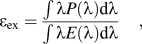 $${\varepsilon _{{\rm{ex}}}} = {{\int {\lambda P\left( \lambda \right){\rm{d}}\lambda } } \over {\int {\lambda E\left( \lambda \right){\rm{d}}\lambda } }}\quad ,$$
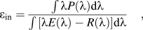 $${\varepsilon _{{\rm{in}}}} = {{\int {\lambda P\left( \lambda \right){\rm{d}}\lambda } } \over {\int {\left[ {\lambda E\left( \lambda \right) - R\left( \lambda \right)} \right]{\rm{d}}\lambda } }}\quad ,$$
where E(λ)/hν, R(λ)/hν, and P(λ)/hν are the number of photons in the excitation, reflectance, and emission spectra of the samples, respectively.
III. RESULTS AND DISCUSSION
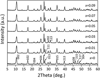
Figure 1 shows XRD patterns of the products after the hydrothermal reaction, where the diffraction peaks can all be indexed to the sulfate-type (LRH) of Ln2(OH)4SO4·2H2O in each case. Reference Geng, Ma, Matsushita, Liang, Michiue and Sasaki16 The compound crystallizes in a face-centered monoclinic system (A2/m, no. 12) with α = γ = 90° and the β angle approximately equaling to 90°. The crystal structure can be viewed as an alternative stacking of the corrugated two-dimensional host layers formed by LnO9 polyhedra and SO4 2− layers along the [001] direction (c-axis). Reference Geng, Ma, Matsushita, Liang, Michiue and Sasaki16 The appearances of the (002) and (004) diffractions in the low-angle region are thus indicative of a layered structure while the sharp and strong non-(00l) diffractions [e.g., (111)] confirm that the Ln3+-containing host layers are well organized and crystalline. With increasing Eu3+ doping both the (00l) and the non-(00l) diffractions shift to larger angles. The calculated lattice spacing of d 111 [Fig. S1(a)] accordingly successively decreases due to the incorporation of smaller Eu3+ ions into the host layers to form solid solution (for 9-fold coordination, La3+ and Eu3+ have ionic radii of 0.1216 and 0.1120 nm, respectively). Reference Shannon17 Since the (002) diffraction is closely related to the gallery height (1/2c sin β) of the layered structure, the decreasing d 002 value [Fig. S1(b)] along with increasing Eu3+ doping thus suggests gradually contracted interlayer distance. This is in accordance with previous studies, Reference Geng, Ma, Matsushita, Liang, Michiue and Sasaki16 which showed that the gallery height tends to shrink with decreasing ionic radius of Ln3+, following lanthanide contraction, and is reasonable from the fact that the interlayer SO4 2− ions are directly coordinated to the Ln3+ centers in the main layers to form Ln–O–S bonds. Reference Geng, Ma, Matsushita, Liang, Michiue and Sasaki16
XRD patterns of the (La1−x Eu x )2(OH)4SO4·2H2O LRHs synthesized at 100 °C and pH of 9.

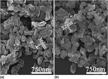
Typical morphologies of the obtained LRHs are shown in Fig. 2. It is found that the compounds crystallize as nanoplates having lateral sizes of up to 500 nm and thicknesses of 20–30 nm. In addition, Eu3+ doping does not obviously change the overall morphology of the LRHs.
Typical FE-SEM images of the (La1−x Eu x )2(OH)4SO4·2H2O (x = 0–0.09) powders synthesized at 100 °C and pH of 9, with (a) x = 0, (b) x = 0.09.

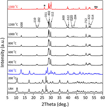
(La0.95Eu0.05)2(OH)4SO4·2H2O was chosen as an example to investigate the proper temperature range in which the phase pure (La0.95Eu0.05)2O2SO4 can be obtained (Fig. 3). The product calcined at 200 °C exhibits a similar XRD pattern with that of the LRH precursor, suggesting no essential change in chemical composition or crystal structure. While, when treated at 300 °C, the product began to show some diffraction peaks different from the LRH precursor and oxysulfate can be assigned from the results of FTIR as the dehydration product of (La0.95Eu0.05)2(OH)4SO4, and crystal structure analysis of which is underway. The targeted oxysulfate, (La0.95Eu0.05)2O2SO4, has been to be obtained at the temperature as low as 400 °C, via dehydroxylation of the anhydrous intermediate. With increasing calcinations temperature up to 1200 °C, the individual diffraction peaks become better resolved and the peak intensity gets enhanced, indicating lattice perfection and crystallite growth. Calcination at higher temperatures such as 1300 °C yielded a phase mixture of (La0.95Eu0.05)2O2SO4 and (La0.95Eu0.05)2O3 (labeled with asterisks) due to partial decomposition of the sulfate ions. Therefore, proper heating temperature to produce pure phase of (La,Eu)2O2SO4 should be in the range of 400–1200 °C.
XRD patterns of the products calcined from the (La0.95Eu0.05)2(OH)4SO4·2H2O LRH at different temperatures for 1 h in air.

One interesting finding which should be noted is that, though the oxysulfates of Y and Lu are either only stable in a limited temperature range or unstable, Reference Shoji and Sakurai18 the La-based oxysulfate exists in the very wide temperature range of 400–1200 °C. This makes La2O2SO4 a suitable host for luminescence, since high-temperature calcination effectively helps to improve PL intensities. Moreover, crystallite sizes of the phosphors assayed from the Scherrer equation are shown in Table I. Coarsening of the crystallites from ∼15 to ∼45 nm was observed for the products calcined from 400 to 1200 °C.
Optical properties and crystallite sizes of the (La0.95Eu0.05)2O2SO4 red phosphors calcined at different temperatures.

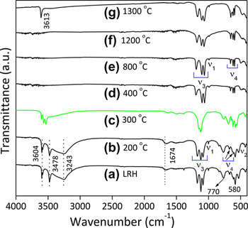
Besides the XRD patterns, the calcination transformation process of (La0.95Eu0.05)2(OH)4SO4·2H2O into (La0.95Eu0.05)2O2SO4 can be further confirmed by the FTIR characterization, which is shown in Fig. 4. In spectrum (a), the vibration at ∼3243 cm−1 and the shallow, that shoulder near 1674 cm−1, are assignable to the O–H stretching vibrations (ν1 and ν3) and the H–O–H bending mode (ν2) of hydration water in the structure, respectively, Reference Gadsden19 while the two separated sharp peaks at 3604 and 3478 cm−1 are indicative of hydroxyl (OH−) groups. The vibrations of SO4 2− (ν1, ν2, ν3, and ν4) Reference Gadsden19 are all well resolved in spectra (a) and (b) as indicated in the figure. In spectrum (c), the vibration of hydration water at ∼3243 cm−1 is hardly found, suggesting the occurrence of dehydration. The bands of hydroxyls and sulfate are still clearly observable but with vibration behaviors different from the original LRH and the 200 °C product. These phenomena suggest that dehydration has changed coordination spheres of the rare-earth ions. The vibrations of hydroxyls are no longer observable from spectra (d)–(f), indicating complete dehydroxylation to form (La0.95Eu0.05)2O2SO4 in this temperature range. The above observations are in accordance with the results of XRD (Fig. 3). Similar vibrations of sulfate ions are observed for the LRH and the oxysulfates, indicating similar configurations of sulfate ions. Expansion of the ν3 band from ∼1205–1075 to ∼1230–1030 cm−1 and obviously deeper and sharper splitting of the ν4 band are nonetheless observed for the oxysulfates indicting enhancement of SO4 2− coordination to the rare-earth centers after dehydration and dehydroxylation. The 1300 °C product contains La2O3 impurity (Fig. 3), which is chemically unstable in air and readily reacts with water vapor to form hydroxide via surface reaction, Reference Imanaka, Masui and Kato20 and hence hydroxyl vibration at 3613 cm−1 appears in spectrum (g).
FTIR spectra of the (La0.95Eu0.05)2(OH)4SO4·2H2O LRH and its calcination products.

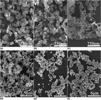
Morphology evolution of the LRH upon calcination is shown in Fig. 5, where it can be seen that the products calcined at 400 and 600 °C tend to preserve the two-dimensional nanoplate morphologies. Calcination at 800 °C leads to partial collapse of the nanoplates into rounded nanoparticles, and at the higher temperature of 1000 °C, basically all the nanoplates have been disintegrated into rounded nanoparticles of ∼100–200 nm. At the even higher temperature of 1200 °C, the nanoparticles apparently underwent coarsening to generate bigger particles of ∼350–650 nm. Rapid particle growth took place at 1300 °C, where micron-sized (2–5 µm) particles/crystallites with sharper edges were resulted.
FE-SEM micrographs of the products derived from (La0.95Eu0.05)2(OH)4SO4·2H2O by calcination at (a) 400, (b) 600, (c) 800, (d) 1000, (e) 1200, and (f) 1300 °C.

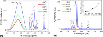
Figure 6 shows photoluminescence excitation (PLE) and PL spectra of the (La0.95Eu0.05)2O2SO4 red phosphors calcined at different temperatures. The PLE spectra consist of the broad and strong band (centered at 284 nm) which arises from electronic transition from the completely filled 2p orbital of O2− to the partially filled 4f orbital of Eu3+ (CT), and the sharper peaks are attributed to the intra-4f 6 electronic transitions of Eu3+ at longer wavelengths (350–500 nm). (Gd,Eu)2O2SO4 and (Y,Eu)2O2SO4 red phosphors were reported to have CT bands centered at ∼270 nm, Reference Lian, Sun, Liu, Yu and Li6,Reference Song, Shao, Du, Cao, Hui, Xing and Xiong9 which is shorter than the ∼284 nm observed for (La0.95Eu0.05)2O2SO4 in this work. CT transitions occur when a valence electron is transferred from the ligand to an empty or incompletely filled orbital of the lanthanide ions. Reference Li and Zhang21 Higher covalency of the Ln–O bond means that the electrons spread out over wider orbitals, which reduces the interactions among the electrons and diminishes the electronegativity difference between the constituting ions. Reference Đorđević, Antićn, Nikolić and Dramićanin22 This in turn lowers the energies needed for electronic transition between energy levels. Red-shift of the CT transition in La2O2SO4 is thus understandable from the lower electronegativity of La3+. Under CT band excitation, the characteristic 5D0 → 7F J (J = 0–4) transitions of Eu3+ were observed with the strongest one located at 617 nm as labeled in the figure. Owing to the shielding by 5s 25p 6 electrons, the 4f 6 energy levels of Eu3+ are hardly affected by crystal field Reference Li and Sakka23 and thus the position of Eu3+ emission would theoretically not be affected by host lattice. The oxysulfate phosphor obtained in this work, nonetheless, shows a 7 nm red-shift of the main 5D0 → 7F2 emission of Eu3+ when compared with the classic red phosphor of Y2O3:Eu3+ (emission at 610 nm). Reference Li, Li, Sun and Ishigaki11 The Wyckoff position of Ln3+ was reported to be solely of 8f in monoclinic La2O2SO4 while 8b and 24d in cubic Y2O3. Reference Machida, Kawano, Eto, Zhang and Ikeue2,Reference Dhananjaya, Nagabhushana, Nagabhushana, Rudraswamy, Shivakumara and Chakradhar24 Both the lattices have relatively low site symmetries for Ln3+, and thus the so called J-mixing effect is particularly applicable. Reference Souza and Couto Dos Santos25 The higher symmetry of O–Ln–O in monoclinic La2O2SO4 than in cubic Y2O3 produces less J-mixing effect in the former and thus red-shifts the 5D0 → 7F2 emission if one assumes that Eu3+ exclusively replaces Ln sites. Reference Song, Shao, Du, Cao, Hui, Xing and Xiong9 Increasing temperature of calcination did not appreciably change peak positions of the PL bands but led to successively stronger PL intensities. A 160% increase in the intensity of the 617 nm emission was observed from 400 to 1200 °C. The external and internal quantum efficiencies and the percentage of excitation absorption (η, η = (εex/εin) × 100%) of the phosphors under O–Eu CT excitation are tabulated in Table I, where it is seen that raising the synthesis temperature from 400 to 1200 °C effectively improved the external and internal quantum efficiencies from ∼16 to ∼40% and from ∼33 to ∼54%, respectively. Notably, an over 16% increment in the external quantum efficiency was observed from 1000 to 1200 °C, which is understandable from the combined contribution of two aspects, that is, improved internal quantum efficiency (10%, Table I) by lattice perfection and the significantly increased excitation absorption (∼21%, Table I) by reduced surface scattering along with obvious crystallite/particle growth (Fig. 5). Commission International de L'Eclairage (CIE) color coordinates of the phosphors (Table I) calcined at different temperatures were found to be stable at around (0.65, 0.35), corresponding to a vivid red color. Since the 5D0 → 7F2 electric dipole transition is hypersensitive to the Eu3+ local environment while the parity-allowed 5D0 → 7F1 magnetic dipole transition is not, the intensity ratio 5D0 → 7F2/5D0 → 7F1 has been widely adopted to probe the local symmetry of Eu3+. Reference Ji, Huang, Xia, Molokeev, Jiang, Lin and Atuchin26 We analyzed the I 617/I 595 values for the (La0.95Eu0.05)2O2SO4 phosphors calcined at different temperatures and the results are shown in Table I. The ratio was found to slowly decrease from ∼9.7 to ∼7.7 with increasing calcination from 400 to 1200 °C, which is mainly due to the decreased number of lower-symmetric surface Eu3+ ions along with particle/crystallite growth.
PLE (a) and PL (b) spectra of the (La0.95Eu0.05)2O2SO4 red phosphors calcined from (La0.95Eu0.05)2(OH)4SO4·2H2O at different temperatures. The PLE spectra are obtained by monitoring the 617 nm emission, and the PL spectra are measured under UV excitation at 284 nm.

Luminescence decay kinetics of a phosphor can be described by the Inokuti–Hirayama formula, Reference Inokuti and Hirayama27 as follows:
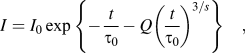 $$I = {I_0}\,{\rm{exp}}\,\left\{ { - {t \over {{\tau _0}}} - Q{{\left( {{t \over {{\tau _0}}}} \right)}^{{3 \mathord{\left/ {\vphantom {3 s}} \right. \kern-\nulldelimiterspace} s}}}} \right\}\quad ,$$
where τ0 is the intrinsic decay time, t the decay time, I the relative intensity. The s values of 3, 6, 8, and 10 correspond to exchange, dipole–dipole, dipole–quadrupole, and quadrupole–quadrupole interactions, respectively. The energy transfer parameter Q is defined as:
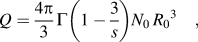 $$Q = {{4\pi } \over 3}\Gamma \left( {1 - {3 \over s}} \right){N_0}\mathop {{R_0}}\nolimits^3 \quad ,$$
where Γ(x) is the gamma function, N 0 the concentration of acceptor, R 0 the critical distance defined as donor–acceptor separation. In this work, the s value was analyzed to be 3, as shown later in Fig. 8(b), and thus Eq. (3) can be reduced to a single exponential. Fitting the fluorescence decay data of the strongest red emission with the single exponential equation indeed yielded good linear relationship for the semilog (log I–t) plots [Figs. S2(a) and S2(b)], and the derived lifetime values are summarized in Table I. It can be seen that the lifetime stays stable at 2.0 ms for the 400–800 °C products and decreases to 1.88 and 1.17 ms for the 1000 and 1200 °C products, respectively. Shortened lifetime at a higher temperature of synthesis was also previously observed by other researchers. Reference Lu, Li and Sakka28
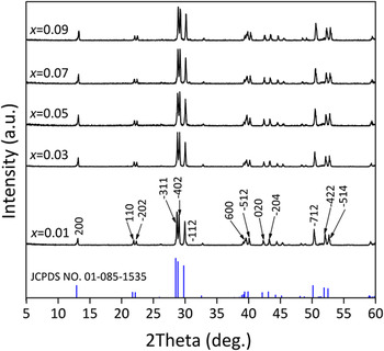
Figure 7 illustrates XRD patterns of the oxysulfate phosphors calcined at the same temperature of 1200 °C with a holding time of 1 h but with different Eu3+ concentrations. All the products can be indexed to the monoclinic La2O2SO4 phase. The diffraction peaks continuously shift to larger angles with increasing Eu3+ content, indicating contracted cell dimensions. The cell parameters derived from the software Cellcalc are shown in Table SI, where it is seen that the constants a, b, and c all decrease with increasing Eu3+ doping and follow Vegard's law (Fig. S3), implying a successful incorporation of Eu3+ into the host lattice to form solid solution. The crystallite sizes calculated from the Scherrer equation are similarly 50 nm for the x = 0.01–0.09 powders.
XRD patterns of the (La1−x Eu x )2O2SO4 red phosphors calcined at 1200 °C for 1 h.

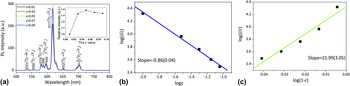
Figure 8 displays the effects of Eu3+ content on PL of the oxysulfate phosphors. The PL [Fig. 8(a)] and PLE (Fig. S4) spectra are essentially identical to those shown in Fig. 6, that is, the PLE spectra are dominated by the CT band centered at ∼284 nm and the PL spectra dominated by the red emission of Eu3+ at ∼617 nm. The PL intensity increases with increasing Eu3+ content until 5 at.% and then decreases, revealing an optimal concentration of ∼5 at.% (x = 0.05). The value agrees well with that reported before Reference Lian, Liang, Wang and Liu7 and also the theoretical value analyzed later [Fig. 8(c)]. The PLE spectra also imply that the phosphors can be effectively excited by the longer UV light of ∼395 nm (the 7F0,1 → 5L6 intra-f transition of Eu3+) and the blue light of ∼465 nm (the 7F0,1 → 5D2 intra-f transition of Eu3+, also around the wave length of blue LED chips), though the emission efficiency would be lower than that under CT excitation. The CIE color coordinates calculated from the PL spectra (Table II) are all around (0.65, 0.35). The fluorescence lifetime was also obtained by applying single-exponential fitting [Fig. S5(a)], as also seen from the good linear relationships between log I and t on the semilog plots [Fig. S5(b)]. The resultant lifetime tends to decrease with increasing Eu3+ content (Table II). This is basically due to the gradually shortened separation distance among the Eu3+ activators, which may result in the formation of a resonant energy transfer network to advance energy transfer from the inside to the nonradiative centers on particle surfaces and thus leads to a shorter lifetime. The lifetime obtained in this work also agrees with the values of 1.2–1.6 ms reported before. Reference Wang, Li, Zhu, Li, Sun and Sakka13 The internal quantum efficiency (εin, Table II) reached its maximum of ∼53.9% at 5 at.% of Eu3+, in accordance with the PL intensity [Fig. 8(a)]. The percentage of excitation absorption (η, Table II) tends to steadily increase with increasing Eu3+ doping, as expected, and the external quantum efficiency (εex, Table II), interplayed from η and εin, is ∼40% for x = 0.05. The I 617/I 595 intensity ratio remains almost constant at 7.6 ± 0.2 for the phosphors of different Eu contents (Table II).
PL spectra of the (La1−x Eu x )2O2SO4 red phosphors calcined at 1200 °C (a, excitation at 284 nm), where the inset is the relative intensity of the 617 nm emission as a function of the Eu3+ content; (b) and (c) show the plots of log(I/c) against log(c) and log(1 − c), respectively, where the number in bracket represents standard deviation.

Optical properties of the (La1−x Eu x )2O2SO4 red-emitting phosphors.

The mutual interaction type of luminescence quenching in solid-state phosphors can be concluded by analyzing the s value in the equation, Reference Lian, Liang, Wang and Liu7,Reference Huang and Lou29,Reference Dai, Song, Wang, Bai, Dong, Qin, Qu and Zhang30
 $$\log \left( {{I \mathord{\left/ {\vphantom {I c}} \right. \kern-\nulldelimiterspace} c}} \right) = \left( { - {s \mathord{\left/ {\vphantom {s d}} \right. \kern-\nulldelimiterspace} d}} \right)\log \,c + \log \,f\quad ,$$
where I is the emission intensity, c is the Eu3+ content, d is the sample dimension (d = 3 for energy transfer among the activators inside particles), f is a constant independent of activator concentration. The s values of 3, 6, 8, and 10 are for the mechanisms of exchange interaction, dipole–dipole, dipole–quadrupole, and quadrupole–quadrupole electric interactions, respectively. The log(I/c)–log(c) plot is given in Fig. 8(b) for the phosphors, from which a slope (−s/3) of −0.86 is derived via linear fitting, leading to an s value of 2.58 (close to 3). This indicates that the observed luminescence quenching is dominantly resulted from exchange interactions for the energy transfer among Eu3+ ions.
According to Ozawa, Reference Ozawa31 the starting concentration of luminescence quenching via exchange interactions can be approximated as 1/(1 + Z), where the Z value can be derived from the equation
 $$I = bc{\left( {1 - c} \right)^z}\quad ,$$
where I is the emission intensity, c the activator content, b an independent constant, and Z the nearest lattice sites for the activator. The equation proved to be widely applicable by previous studies. Reference Zhang, Lu, Yang, Han, Xu and Zhang32–Reference Fouassier, Saubat and Hagenmuller34 In this work, the Z can be obtained from the following transformed logarithm:
 $$\log \left( {{I \mathord{\left/ {\vphantom {I c}} \right. \kern-\nulldelimiterspace} c}} \right) = Z\,\log \left( {1 - c} \right) + \log \,b\quad .$$
Fitting the log(I/c)–log(1 − c) plot [Fig. 8(c)] according to Eq. (5) yields a slope of Z = 21.99 and thus an optimal Eu3+ concentration of ∼4.3 at.% [1/(1 + Z) = 0.043]. The value is nearly identical to that (5.0 at.%) experimentally determined in this study which further indicate that the exchange interaction is responsible for the quench observed [Fig. 8(a)].
IV. CONCLUSIONS
In summary, sulfate-type LRHs of (La1−x Eu x )2(OH)4SO4·2H2O (x = 0–0.09) with well-dispersed nanoplate morphology have been obtained at 100 °C and pH of 9 via 24 h of hydrothermal reaction. With the layered compound as a green precursor, (La1−x Eu x )2O2SO4 red phosphors yielded through simple dehydration and dehydroxylation in ambient air. The influences of calcination temperature and Eu3+ content on PL of the phosphors were studied in terms of excitation, emission, quantum efficiency, fluorescence decay kinetics, asymmetry factors (I 617/I 595), and color coordinates of emission. It was found that (La1−x Eu x )2O2SO4 phosphors exist in the wide temperature range of 400–1200 °C for calcination in air. O–Eu CT excitation bands were observed at ∼284 nm and under CT excitation the phosphors exhibit the strongest 5D0 → 7F2 red emission of Eu3+ at ∼617 nm. Raising the calcination temperature effectively improves the PL intensity and quantum efficiency of the phosphors. The optimal Eu3+ concentration was experimentally found to be ∼5 at.%, and concentration quenching of luminescence was suggested to be resulted from exchange interactions among the Eu3+ activators. The phosphors were shown to have relatively stable color coordinates of around (0.65, 0.35), independent of calcination and Eu3+ concentration. Fluorescence decay of the 617 nm emission follows a single-exponential behavior, and the lifetime was found to decrease with increasing Eu3+ content or calcination temperature.
Supplementary Material
To view supplementary material for this article, please visit http://dx.doi.org/10.1557/jmr.2016.185.
ACKNOWLEDGMENTS
This work is supported in part by the National Natural Science Foundation of China (Grants No. 51172038, 51302032, and U1302272), the Fundamental Research Fund for Central Universities (Grants No. N140204002 and L1502046), and Grants-in-Aid for Scientific Research (JSPS KAKENHI No. 26420686). X.J. Wang acknowledges the financial support from the China Scholarship Council for overseas Ph. D. study.