Introduction
The treatment goals for early-stage glottic squamous cell carcinoma are twofold: tumour eradication and preservation of a functional larynx. Surgery for early-stage lesions of the glottic larynx has improved over the last few decades with the development of minimal-invasive procedures, particularly endoscopic laser surgery.Reference Silver, Beitler, Shaha, Rinaldo and Ferlito1 However, for stage II neoplasms, open larynx preservation surgery (when feasible) has yielded better results than transoral laser surgery, with local control rates reaching 95 per cent. However, this procedure is associated with severe hoarseness and swallowing difficulties which severely affect the functional outcome.Reference Hartl2 The alternative treatment of radiotherapy alone has yielded failure rates of up to 50 per cent.Reference Frata, Cellai, Magrini, Bonetti, Vitali and Tonoli3 In fact, tumour–node–metastasis (TNM) stage T2 glottic carcinomas include glottic tumours with (1) supraglottic and/or subglottic spread and/or (2) impaired vocal fold mobility.Reference Edge, Byrd, Compton, Fritz, Greene and Trotti4 Combining these heterogeneous criteria into a single prognostic category has resulted in heterogeneous treatment outcomes for these ‘early-stage’ lesions.Reference McCoul and Har-El5
This study aimed to assess the value of local tumour extension and impaired vocal fold mobility as prognostic factors, based on results obtained for patients treated for T2N0 squamous cell carcinoma of the glottic larynx in a large monocentric cohort (at our institution). An original objective was to assess the influence of vocal fold mobility on survival.
Materials and methods
Patients
This study was approved by our institutional review board. A retrospective analysis of patients treated at our centre between 2001 and 2012 for T2N0 squamous cell carcinoma of the glottic larynx was performed. The main inclusion criteria were age over 18 years, no previous medical history of squamous cell carcinoma, no previous radiotherapy to the head and neck, no other cancer site, and a diagnosis of stage T2N0 squamous cell carcinoma of the glottic larynx made after an initial consultation by direct laryngoscopy under general anaesthesia with biopsy. Data were collected on vocal fold mobility at the initial consultation (normal, T2a; impaired, T2b) and local tumour extension on endoscopy (anterior commissure, posterior commissure, epiglottis, ventricle, ventricular fold, subglottis). Subglottic extension was defined as the tumour extending more than 1 cm under the lateral margin of the ventricle, in accordance with the American Joint Committee on Cancer definition. Pretreatment investigation also included computed tomography (CT) scanning of the head and neck and a chest CT scan. Each patient was examined at a multidisciplinary consultation before treatment. Follow up during and after treatment included neck examinations and indirect laryngoscopy at regular intervals.
Statistical design and analysis
The primary objective was to assess the difference in overall disease control between T2a and T2b cancer patients. Therefore, the primary end-point was disease-free survival. Secondary end-points were local control, overall survival, laryngeal preservation, laryngectomy-free survival and the results of each treatment strategy employed in T2a and T2b patient groups.
The results are expressed as percentages or medians (range), and groups were compared using non-parametric tests: chi-squared tests or Fisher's exact tests for qualitative data and the Wilcoxon test for quantitative data. Analyses of disease-free survival, local control, larynx preservation, laryngectomy-free survival and overall survival were based on the Kaplan–Meier method, and curves were compared using the log-rank test. Statistical analyses were performed with Instat version 3 and Prism version 6.04 for Windows (GraphPad Software, La Jolla, California USA).
Results
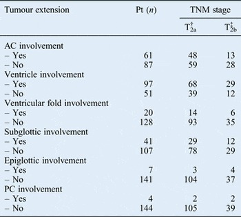
A total of 148 patients (130 men and 18 women) met the criteria. The median age at diagnosis was 60 years (range 28–82 years). Vocal fold mobility was normal in 107 patients (T2a) and impaired in 41 (T2b). Details of local tumour extension and treatment are summarised in Table I. There was no difference between T2a and T2b patient groups in the sex ratio, mean age, performance status or rate of local spread to each site.
Local extension of stage II glottic squamous cell carcinoma*

* Total n = 148; assessed by endoscopy at our institution. †Normal vocal fold mobility. ‡Impaired vocal fold mobility. Pt = patients; TNM = tumour–node–metastasis; AC = anterior commissure; PC = posterior commissure
Patients
Surgery was the first treatment for 72 patients (T2a, 52; T2b, 20): transoral laser surgery, 12; horizontal partial laryngectomy, 10; supracricoid partial laryngectomy, 48; and total laryngectomy, 3 (all performed because of a positive frozen section analysis after supracricoid laryngectomy). One patient had undergone total laryngectomy one month after supracricoid partial laryngectomy because the definitive analysis revealed positive inferior surgical margin findings. Twenty-four patients underwent bilateral neck dissection, 36 underwent unilateral neck dissection and 12 did not undergo neck dissection. Histopathological examination showed that the inner thyroid cartilage had been invaded in five patients (6.9 per cent), resulting in pathological stage T3: 1 out of 52 patients with clinically staged T2a tumours and 4 out of 20 patients with clinically staged T2b tumours. Four patients had perineural invasion (5.6 per cent). Only three patients had lymph node invasion (4.2 per cent) without extracapsular spread: one had a single positive homolateral lymph node in zone IIa, one had a single homolateral lymph node exhibiting a 0.3-mm micrometastasis in zone III, and one had a positive node in zone IIa and three positive nodes in zone VIa. Two patients had undergone further total laryngectomy for chronic refractory aspirations. Five patients (6.9 per cent) had local recurrence within an average of 10.2 months after partial laryngectomy; all had undergone salvage total laryngectomy. However, four had died as a result of a new local recurrence and/or distant metastasis. Single lymph node recurrence had occurred in a patient treated with transoral laser surgery without neck dissection whose tumour had invaded the ventricle without impaired vocal fold mobility. He had undergone salvage neck dissection with adjuvant radiotherapy.
Forty-two patients (34 with T2a tumours, 8 with T2b tumours) had been treated with radiotherapy alone: these were mainly patients with bulky, clinically more favourable lesions without impaired vocal fold mobility who refused surgery or patients with a medical or surgical contraindication such as subglottic extension. An alternative surgical option was total laryngectomy because of subglottic extension (eight T2a tumour patients). Another alternative surgical option was supracricoid partial laryngectomy (26 T2a tumour patients). Radiotherapy was not administered to the latter because of patient refusal (n = 11) or medical contraindications (n = 15). We had proposed radiotherapy alone to these patients, as recommended by the guidelines for stage II disease.Reference Pfister, Ang, Brizel, Burtness, Busse and Caudell6 Of the T2b tumour patients, two underwent total laryngectomy because of subglottic extension and six underwent supracricoid partial laryngectomy. Partial laryngectomy was not performed because of patient refusal (n = 3) or medical contraindications (n = 3). These patients were treated with radiotherapy without concurrent chemotherapy because of patient refusal (n = 2) or medical contraindications (n = 6). There was no difference in the distribution of patients with medical contraindications to concurrent chemotherapy between the T2a and T2b groups. These patients had received a median conventional dose of 69 Gy in 33 fractions (2 Gy per fraction, 1 fraction per day, 5 days per week). Nineteen patients had received prophylactic bilateral neck lymph node radiotherapy (zones II, III, IV and VI) at a median dose of 50 Gy. Eleven patients in all (10 with T2a tumours, 1 with a T2b tumour; 26.2 per cent) had developed a local recurrence within an average of 20.4 months after therapy. One patient with recurrence in the anterior commissure without cartilage involvement and with normal vocal fold mobility had undergone salvage partial supracricoid laryngectomy; the other 10 patients had undergone salvage total laryngectomy. All patients had pathologically staged N0 disease. No patient had undergone re-irradiation. Two patients who underwent salvage total laryngectomy developed a new local recurrence (at one and seven months, respectively) soon after salvage surgery, and died at seven and nine months, respectively, post-surgery. One patient had lymph node recurrence in zone III 16 months after the end of radiotherapy for a T2a glottic tumour that had invaded the infrahyoid epiglottis. He had been treated with induction chemotherapy, radical neck dissection without residual tumour and re-irradiation at a conventional dose of 64 Gy.
Twenty-four patients (18 with T2a tumours, 6 with T2b tumours) had been treated with concomitant first-line chemoradiotherapy with the intent of preserving the larynx as an alternative to total laryngectomy. The alternative surgical procedure was total laryngectomy for all patients. They received a median conventional dose of 70 Gy in 33 fractions (2 Gy per fraction, 1 fraction per day, 5 days per week). Chemotherapy comprised cisplatin for 16 patients, carboplatin for 3 patients and cetuximab for 5 patients. Twenty-one patients had received prophylactic bilateral neck lymph node radiotherapy (zones II, III, IV and VI); the median dose was 50 Gy. Nine patients (37.5 per cent) developed a local recurrence within an average of 23.4 months after therapy: one patient had undergone salvage partial supracricoid laryngectomy with bilateral neck dissection, and the remaining eight patients had undergone salvage total laryngectomy (five with bilateral neck dissection and three with unilateral neck dissection). Only one patient had lymph node invasion with extracapsular spread. There was no regional lymph node recurrence.
Ten patients (three with T2a tumours, seven with T2b tumours) had first received induction chemotherapy. The alternative surgical option for all of these patients was total laryngectomy. Five patients had received cisplatin–fluorouracil chemotherapy, two received carboplatin–fluorouracil and three received taxane–cisplatin-fluorouracil. Two patients had later received concurrent chemoradiotherapy of weekly cisplatin with a dose of 70 Gy to the larynx and prophylactic bilateral neck lymph node radiotherapy at a dose of 50 Gy; six patients had received a conventional 70 Gy dose of radiotherapy to the larynx and prophylactic bilateral neck lymph node radiotherapy at a median dose of 50 Gy. The remaining two patients had no local response after induction chemotherapy and the larynx had become progressively immobile. They had undergone total laryngectomy and bilateral neck dissection: one had lymph node tumour invasion with extracapsular spread. One patient who received radiotherapy and 1 who received chemoradiotherapy had developed local recurrence at 6 and 17 months, respectively, after the end of therapy. Both had undergone salvage total laryngectomy.
Prognostic factors and treatment outcomes for the whole cohort
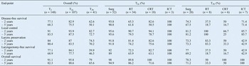
The two- and five-year estimates of end-points (disease-free survival, local control, larynx preservation, laryngectomy-free survival and overall survival) for stage II disease patients are shown in Table II. Regarding the tumour report before treatment, invasion of the infrahyoid epiglottis or the posterior commissure were the only types of tumour spread associated with reduced overall survival (p = 0.002 and p = 0.028, respectively) and reduced disease-free survival (p = 0.028 and p = 0.056 respectively), but in fewer patients (n = 7 and n = 4, respectively). In the whole cohort, there was no difference in disease-free survival between treatment options for patients with subglottic involvement and those with anterior commissure involvement (p = 0.637 and p = 0.227, respectively). Neck dissection during tumour surgery was not associated with a change in disease-free or overall survival.
Two- and five-year estimates of efficacy end-points by initial vocal fold mobility and treatment strategy*

* For stage II glottic squamous cell carcinoma patients treated at our institution. Surg = surgery; RT = radiotherapy; CRT = chemoradiotherapy; ICT = induction chemotherapy
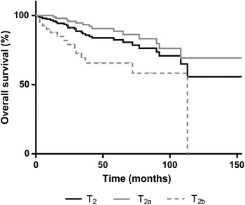
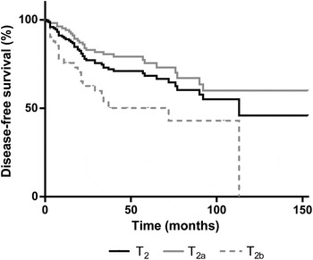
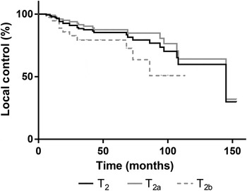
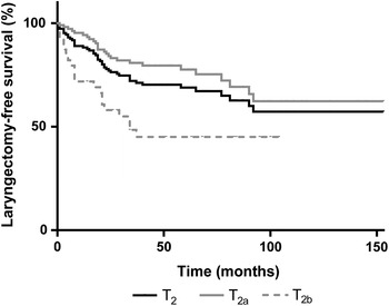
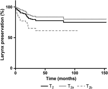
However, impaired vocal fold mobility was strongly associated with decreased disease-free survival (p = 0.0009; hazard ratio 0.3965, 95 per cent confidence interval (CI) 0.1655 to 0.6276), laryngeal preservation (p = 0.0037; hazard ratio 0.3682, 95 per cent CI 0.1253 to 0.6631), local control (p = 0.0362; hazard ratio 0.4490, 95 per cent CI 0.1445 to 0.9243), laryngectomy-free survival (p = 0.0003; hazard ratio 0.3588, 95 per cent CI 0.1339 to 0.5449) and overall survival (p = 0.0004; hazard ratio 0.2854, 95 per cent CI 0.08035 to 0.4777; see Figs 1–5).
Kaplan–Meier curves for overall survival of all stage II glottic squamous cell carcinoma (T2) patients after treatment, stratified according to an initial diagnosis of normal (T2a) or impaired vocal fold mobility (T2b).

Kaplan–Meier curves for disease-free survival of all stage II glottic squamous cell carcinoma (T2) patients after treatment, stratified according to an initial diagnosis of normal (T2a) or impaired vocal fold mobility (T2b).

Kaplan–Meier curves for local control of all stage II glottic squamous cell carcinomas (T2) after treatment, stratified according to an initial diagnosis of normal (T2a) or impaired vocal fold mobility (T2b).

Kaplan–Meier curves for laryngectomy-free survival of all stage II glottic squamous cell carcinoma (T2) patients after treatment, stratified according to an initial diagnosis of normal (T2a) or impaired vocal fold mobility (T2b).

Kaplan–Meier larynx curves for larynx preservation for all stage II glottic squamous cell carcinoma (T2) patients after treatment, stratified according to an initial diagnosis of normal (T2a) or impaired vocal fold mobility (T2b).

Treatment of T2a tumour patients
For patients with normal vocal fold mobility (T2a), disease-free survival and local control were better after surgery and induction chemotherapy than after radiotherapy or chemoradiotherapy (p = 0.0140). There was no difference between radiotherapy and chemoradiotherapy as a first treatment in disease-free survival (p = 0.7917), local control (p = 0.5087), laryngectomy-free survival (p = 0.6660), laryngeal preservation (p = 0.7028).
Laryngectomy-free survival was better after surgery and induction chemotherapy (p = 0.0305) than after radiotherapy or chemoradiotherapy but larynx preservation was not (p = 0.1261). There was no difference in overall survival between the four treatment options (p = 0.184).
Treatment of T2b patients
In patients with impaired vocal fold mobility (T2b), disease-free survival, local control, larynx preservation and laryngectomy-free survival were better in those treated with surgery or induction chemotherapy than in those treated with radiotherapy and chemoradiotherapy (p = 0.041, p < 0.0001 and p < 0.0001, respectively). There was no difference in disease-free survival (p = 0.5922), local control (p = 0.3045), laryngectomy-free survival (p = 0.5440), laryngeal preservation (p = 0.2657) and overall survival (p = 0.1992) between radiotherapy and chemoradiotherapy as a first treatment.
Overall survival was better after surgery or induction chemotherapy than after radiotherapy (p = 0.038), but there was no significant difference after chemoradiotherapy.
Discussion
The prognosis of T2 lesions is mainly local spread, and treatments must focus on local control. At our centre, 27.7 per cent of patients treated for a stage II carcinoma of the glottic larynx had impaired vocal fold mobility, which is consistent with the proportions in most previously published series.Reference McCoul and Har-El5, Reference Garden, Forster, Wong, Morrison, Schechter and Ang7 The five-year overall survival rate of 82.3 per cent and five-year disease-free survival rate of 68.3 per cent in our case series are also consistent with previous reports.Reference Garden, Forster, Wong, Morrison, Schechter and Ang7, Reference Khan, Koyfman, Hunter, Reddy and Saxton8 Therefore, these tumours have a good overall prognosis, and the treatment goal is preservation of a functional larynx whenever possible. In a recent meta-analysis pooling 7600 patients, Higgins et al. compared transoral laser surgery vs radiotherapy alone for early-stage glottic cancers, and found no difference in local control or laryngectomy-free survival.Reference Higgins, Shah, Ogaick and Enepekides9 However, overall survival was better following surgery, as observed in our case series. No studies have compared surgery with induction chemotherapy for treating stage II tumours. Moreover, the small number of patients treated with induction chemotherapy (n = 10) in our series does not allow conclusions to be drawn, despite a trend towards higher local control, larynx preservation, laryngectomy-free survival, overall survival and disease-free survival compared with radiotherapy. Concurrent chemoradiation has never demonstrated better local control or better overall survival compared with radiotherapy alone for stage II glottic T2 tumours.Reference Kosugi, Suzuki, Konishi, Nonaka, Mineta and Sakahara10, Reference Hirasawa, Itoh, Naganawa, Ishihara, Suzuki and Koyama11 This combined modality is therefore not recommended for stage II glottic cancers but is ‘to be used for selected patients with stage III, T2N+ [(node-positive)] cancers when total laryngectomy is the only surgical option, when the functional outcome after larynx preservation surgery is expected to be unsatisfactory or when surgical expertise in such a procedure is not available’.Reference Pfister, Laurie, Weinstein, Mendenhall, Aldestein and Ang12 However, there are no data specifically comparing these treatments for T2b tumours. Interestingly, only 3 of the 72 (4.2 per cent) patients who underwent surgery had lymph node invasion. Moreover, only 1 of the 72 patients who underwent surgery and 1 of the 42 treated with radiotherapy alone had regional lymph node recurrence. Thus, most stage II neoplasms of the glottic larynx seem to remain local.
However, local spread (including subglottic and/or supraglottic extension) had no effect on treatment outcomes for T2N0 glottic cancer in our case series. The correlation between tumour extension before treatment and local control after treatment has been investigated by numerous authors searching for predictive factors to influence the choice of primary treatment. Subglottic extension is a classic contraindication to larynx preservation surgery related to cricoid cartilage conservation.Reference Hartl2 Levy et al. reported that using radiotherapy alone for these lesions as an alternative to total laryngectomy is not associated with decreased survival, the local control rate or larynx preservation compared with other laryngeal sites.Reference Levy, Blanchard, Janot, Temam, Bourhis and Daly-Schveitzer13 Some authors have reported a decreased local control rate for stage I cancers after spreading to the anterior commissure without decreased larynx preservation rate or survival.Reference Hakeem, Tubachi and Pradhan14–Reference Mendenhall, Amdur, Morris and Hinerman17 Hartl et al. found that CT was not effective for predicting histologically confirmed thyroid cartilage invasion.Reference Hartl, Landry, Bidault, Hans, Julieron and Mamelle18 This technique overestimated cartilage invasion for lesions with local spread to the anterior commissure and underestimated invasion for lesions with decreased vocal fold mobility. However, according to Mendenhall et al., anterior commissure invasion does not significantly affect local control after radiotherapy.Reference Mendenhall, Amdur, Morris and Hinerman17 This is consistent with our findings, in that anterior commissure or subglottic region involvement was not associated with reduced survival or a difference in survival between treatments. Glottic cancer spread to the posterior commissure or epiglottis is rare; however, the small number of patients in our case series with local extension to the posterior commissure (n = 4) or epiglottis (n = 7) prohibits making conclusions based on our results.Reference Buckley and MacLennan19 Shvero et al. reported significantly decreased disease-free survival in patients with T1 glottic lesions with disease extension to the posterior commissure, but no data are currently available concerning the prognostic value of posterior commissure or epiglottic spread for T2 glottic cancers.Reference Shvero, Shvili, Mizrachi, Shpitzer, Nageris and Koren20 The current study found that no region of local spread had a clear impact on treatment outcomes for T2N0 glottic cancers.
On the other hand, vocal fold mobility was strongly associated with treatment outcomes. Actually, impaired vocal fold mobility was a strong prognostic factor for all efficacy end-points: disease-free survival, local control, larynx preservation, laryngectomy-free survival and overall survival. Previous studies have reported differing treatment outcomes for stage II cancers according to vocal fold mobility. Since Martensson and colleagues' study in 1967, only five have reported survival rates.Reference McCoul and Har-El5, Reference Martensson, Fluur and Jacobsson21 However, a recent meta-analysis by McCoul and Har-El failed to demonstrate a significant effect of vocal fold mobility on combined absolute and disease-specific survival.Reference McCoul and Har-El5 In contrast, our results demonstrated a difference in survival between T2a and T2b tumour patients, maybe because we studied a large monocentric cohort. Moreover, local control and disease-free survival were better for patients with lesions associated with normal vocal fold mobility (T2a) who were treated with surgery rather than with radiotherapy or chemoradiotherapy because of the higher rate of local recurrence after non-surgical treatments. There was no difference in overall survival, probably because of the lower success rate for salvage procedures after initial surgery than after radiotherapy. For patients with impaired vocal fold mobility (T2b), all efficacy end-points were better after surgery rather than radiotherapy, probably because of the lower success rate of salvage therapy for T2b than for T2a lesions, as previously reported.Reference McCoul and Har-El5, Reference Gorphe, Blanchard, Temam and Janot22 The lack of a difference in efficacy end-points between radiotherapy and chemoradiotherapy is consistent with the low success rate of salvage surgery after failure of these treatments in T2b tumour patients. Therefore, in our study, vocal fold mobility was the main prognostic factor associated with treatment outcomes and was associated with survival.
• T2 glottic carcinomas can have supraglottic and/or subglottic spread with/without impaired vocal fold mobility
• Impaired vocal fold mobility is reported to be associated with decreased local control
• In this study, no type of local spread affected treatment outcomes for T2N0 glottic cancer patients
• Impaired vocal fold mobility was a strong prognostic factor for all efficacy end-points
The objective of this study was not to compare treatment efficacy. The great variety of treatment approaches used makes conclusions about the efficacy of different treatment approaches for T2a and T2b tumours difficult. Of course, this is a retrospective study with numerous biases; only prospective, randomised studies can rigorously assess the outcomes of each treatment strategy and the influence of vocal fold mobility. However, no prospective, randomised studies of these lesions have been published; therefore, successive classification and treatment recommendations have been based only on retrospective case series of stage II glottic squamous cell carcinoma. However, our primary goal was to assess the influence of vocal fold mobility on treatment outcomes and survival in two distinct populations: this goal was achieved.
Conclusion
To our knowledge, this is the first study to assess the influence of vocal fold mobility on disease-free survival and the overall survival for T2N0 glottic carcinoma. The data of this study are consistent with previous reports that T2 glottic carcinoma with impaired vocal fold mobility (T2b) is an intermediate-stage lesion with a poorer survival rate compared with early-stage T2 lesions with normal vocal fold mobility (T2a). As the classification of T2 glottic larynx cancers has not changed since the first editions of the Union for International Cancer Control (1972) and American Joint Committee on Cancer (1977) guidelines, a formal subdivision of T2 into T2a and T2b tumours could be considered in future revisions.
Acknowledgements
The authors would like to thank Mrs L Saint Ange for English editing and Dr G Succo for editorial assistance.