Introduction
The grey wolf is widely distributed in North America and Eurasia, including Italy, where the wolf population declined until the 1970s, when it was estimated to comprise only 100 individuals. From the end of the 1970s to today, the population increased to approximately 1600–1900 individuals, but fluctuations occur every year due to natural and human factors (Mattioli et al., Reference Mattioli, Canu, Passilongo, Scandura and Apollonio2018). Wolf populations have expanded through a unidirectional migration from the Apennines to the Western Italian and French Alps (Fabbri et al., Reference Fabbri, Miquel and Lucchini2007). Canis lupus Linnaeus, 1758 is regarded as a protected species (Council Directive 92/43/EEC on the Conservation of Natural Habitats and of Wild Fauna and Flora). The Piedmont Region carries out wolf re-colonization throughout the Italian Alps, by the development of a monitoring and research program on the natural increase of wolves; the program aims to collect the technical data necessary to protect the species and confirm its impact on economic activities (Canavese & Marucco, Reference Canavese and Marucco2016). Within the regional Piedmont territory, wolves are concentrated mainly in the Turin and Cuneo provinces, which are alpine sectors, but certain packs maintain transboundary territories that cross over from the Italian to the French Alps (Marucco et al., Reference Marucco, Marboutin, Duchamp, Avanzinelli, Potocnik and Rauer2013).
Information on helminth infections in carnivores, which occupy the highest positions in food chains, is still incomplete. However, such information is essential because parasites have roles in the stabilization of food-web dynamics by regulating host (e.g. specific parasites) abundance or by connections among a variety of host species (e.g. heteroxenous parasites). Diseases are recognized as significant issues for wild carnivore conservation, particularly where there is potential for outbreaks of common pathogens transmitted between domestic and wild canids (Alexander et al., Reference Alexander, McNutt, Briggs, Standers, Funston, Hemson, Keet and van Vuuren2010).
Wolves share several of their parasite species with other canids, including helminths of medical and veterinary importance (Craig & Craig, Reference Craig and Craig2005). Nevertheless, there is little information regarding the health status or parasitic fauna of wolves in Italy, and specific data regarding the epidemiological situation of parasites in Italian wolves are scarce. Reports concerning Echinococcus granulosus (Batsch, 1786) Rudolphi, 1805 and Trichinella Railliet, 1895 were published in the 1980s and 2000s (Arru et al., Reference Arru, Garippa and Fico1986; Guberti et al., Reference Guberti, Stancampiano and Francisci1993, Reference Guberti, Bolognini, Lanfranchi and Battelli2004). More recently, new research concerning Thelazia callipaeda Railliet & Henry, 1910 (Otranto et al., Reference Otranto, Cantacessi, Mallia and Lia2007) and Angiostrongylus vasorum (Baillet, 1866) Kamensky, 1905 (Eleni et al., Reference Eleni, De Liberato, Azam, Morgan and Traversa2014) have been published. Guberti et al. (Reference Guberti, Stancampiano and Francisci1993) reported 12 species of intestinal helminths, the most complete study concerning wolves in Italy. Therefore, knowledge about wolf helminths can contribute to analysing specific risks and to planning appropriate methods for intervention, primarily in the Italian Alps where no details regarding helminth fauna have been published.
This study aims to report helminth species infecting the gastrointestinal tract of free-ranging wolves in the north-western Italian Alps, Piedmont Region.
Materials and methods
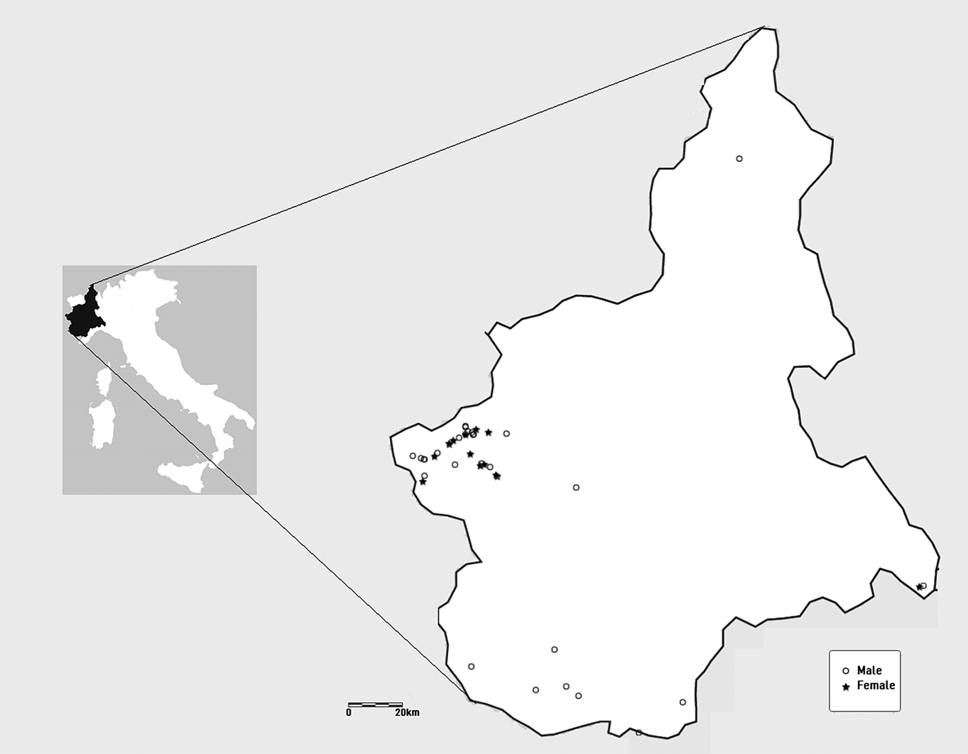
Forty-two (n = 42) road-killed grey wolves were collected between November 2003 and December 2014 in the Piedmont Region (fig. 1). The carcasses were maintained at 4°C until post-mortem examination and necropsy. The 28 males and 14 females were sorted into two age classes: juveniles (not exceeding their second spring) and adults. In the course of necropsy, the gastrointestinal tract was removed, labelled and frozen at −20°C. Before parasitological examination, the packages were defrosted overnight at room temperature.
Map showing locations of where the 42 wolves used in the current study were collected from in Piedmont, north-western Italy, between November 2003 and December 2014.

The helminths were collected as described by Amato & Amato (Reference Amato, Amato, Von Matter, Straube, Accordi, Piacentini and Cândido2010). The stomach and small and large intestines were individually opened and washed with tap water for helminth collection and identification. The mucosa was scraped exactingly to remove the contents entirely. Using a stereomicroscope, the contents of each organ were examined in aliquots of 50 ml in Petri dishes.
Nematodes were cleared in lactophenol and mounted between a slide and coverslip for identification (Amato & Amato, Reference Amato, Amato, Von Matter, Straube, Accordi, Piacentini and Cândido2010). Nematodes were identified using keys and descriptions (Anderson et al., Reference Anderson, Chabaud and Willmott2009; Gibbons, Reference Gibbons2010). Cestoda were compressed and fixed in alcohol-formalin-acetic acid (AFA) solution (alcohol 70°GL 92%, formalin 5%, acetic acid 3%), and stained with Langeron's Carmine. The specimens were mounted in Canada balsam and identified using standard taxonomic keys (Esch & Self, Reference Esch and Self1965; Khalil et al., Reference Khalil, Jones and Bray1994; Loos-Frank, Reference Loos-Frank2000; Padgett et al., Reference Padgett, Nadler, Munson, Sacks and Boyce2005; Hrckova et al., Reference Hrckova, Miterpakova, O'Connor, Snabel and Olson2011).
The parameters for prevalence, mean abundance and mean intensity were calculated according to Bush et al. (Reference Bush, Lafferty, Lotz and Shostak1997). Fisher's exact test was used to test prevalence data against age, sex and season (winter or summer). Calculations were performed using the R software package (www.r-project.org), and statistical significance was inferred if the specific P-value of the test was ≤0.05.
Results and discussion
The wolves examined included 28 males (17 adults, 11 juveniles) and 14 females (eight adults, six juveniles). The mean weight of the carcasses for the females was 24.9 kg (standard deviation (SD) 5.06; range 17.50–31.80 kg), for the males 27.41 kg (SD 8; range 15–37 kg), for the adults 32.54 kg (SD 3.85; range 25–37 kg) and for the juveniles 20.74 kg (SD 3.85; range 15–25.25 kg).
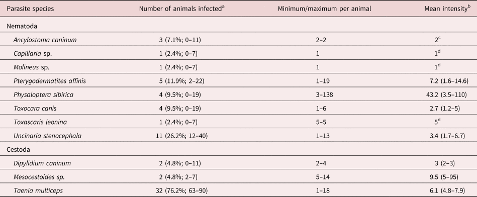
Thirty-six of the 42 wolves examined contained helminths. The total prevalence of infection was 85.7%. Of the recorded parasites, there were nine species of nematode and three cestode species (table 1). Co-infections involving two or more helminth species per animal were found in 50% of the cases that presented two or more nematodes and one cestode (e.g. Physaloptera sibirica Petrov & Gorbunov, 1931, Uncinaria stenocephala Railliet, 1884 and Taenia multiceps Leske, 1780); two nematodes (e.g. Toxocara canis (Werner, 1782) Stiles, 1905 and U. stenocephala), two cestodes (e.g. Mesocestoides sp. Vaillant, 1863 and T. multiceps), one nematode and two cestodes (e.g. U. stenocephala, Dipylidium caninum (Linnaeus, 1758) Leuckart, 1863 and T. multiceps), one nematode and one cestode (e.g. T. canis and T. multiceps); and three nematodes (e.g. Pterygodermatites affinis Jägerskiöld, 1904, P. sibirica and U. stenocephala).
Summary statistics of gastrointestinal helminths isolated from 42 wolves of Piedmont, by post-mortem examination.

a Prevalence and 95% confidence limits in parentheses.
b Confidence limits in parentheses.
c Intensity is constant, confidence limits were not calculated.
d There is only one infected host in the sample, confidence limits cannot be calculated.
In general, the most prevalent helminths were T. multiceps (76.2%), followed by nematodes U. stenocephala (26.2%) and P. affinis (11.9%); whereas, P. sibirica demonstrated the highest mean intensity (43.2%), followed by Mesocestoides sp. (9.5%) and P. affinis (7.2%). Taenia multiceps, P. sibirica and U. stenocephala were, respectively, the parasites with the highest mean abundance.
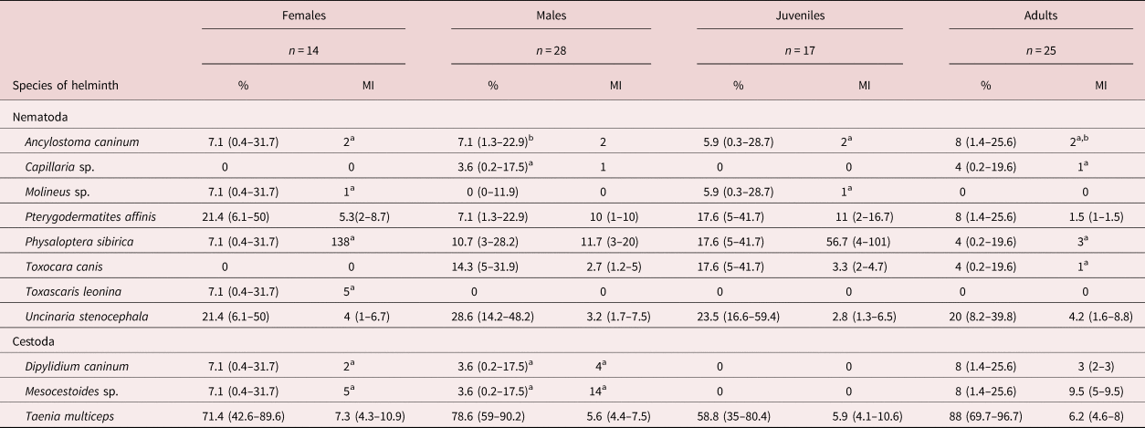
Our data presented higher prevalences of the same parasite species in adults compared to juveniles and in females compared to males (table 2), but the mean intensity was higher in females and juveniles, although not statistically significant (P > 0.05). On the other hand, due to the small proportion of positive animals for each helminth species, and the low number of parasites in the sample group, it was not possible to explore the effects of sex, age and season against mean intensity for each parasite species. Further, no significant differences in any helminth species were found relative to sex, age or season in terms of their prevalence (P > 0.05).
Prevalence and mean intensity (MI) of helminth infections in wolves, according to sex and age.

a There is only one infected host in the sample, confidence limits cannot be calculated.
b Intensity is constant, confidence limits were not calculated.
In accordance with other studies in Europe, U. stenocephala is among the most prevalent and frequently reported nematode species. Shimalov & Shimalov (Reference Shimalov and Shimalov2000) reported a prevalence of 15.4%. However, Popiolek et al. (Reference Popiolek, Szczesnaa, Nowaka and Myslajeka2007) and Bagrade et al. (Reference Bagrade, Kirjusina, Vismanis and Ozolins2009) reported prevalences of 37% and 41.2%, respectively. Our data are in agreement with previous investigations, yet U. stenocephala was more prevalent than Ancylostoma caninum (Ercolani, 1859) Hall, 1913. From a literature review based on coprological exams and necropsy findings, both nematode species are found in wolves in several areas of Europe and North America. Differences in prevalence between the two species (U. stenocephala, 26.2% vs. A. caninum, 7.1%) are comparable to those prevalences reported in similar studies.
Until the present study, there had been no reports of P. affinis parasitizing wolves in Europe or North America. In this case, it was found in 11.9% of samples. The prevalence of infection is within the range of values reported for red foxes in Italy, varying from 8% (Capelli et al., Reference Capelli, Stancampiano, Magi, Poglayen and Guberti2003) to 19.4% (Cerbo et al., Reference Cerbo, Manfredi, Trevisiol, Bregoli, Ferrari, Pirinesi and Bazzoli2008). We suggest that in the area studied, and more generally in the Alps (where the population of wolves is expanding), suitable ecological conditions exist for P. affinis to utilize the wolf as a new host successfully. Pterygodermatites affinis depends on the frequency of ingestion by carnivores of intermediate (insect) or paratenic (small mammals, reptiles and birds) hosts. The stomachs of some wolves contained parts of small mammals, reptiles and arthropods, but primarily comprised bones of cervids. Similarly, P. sibirica has been reported parasitizing carnivores and mustelid species such as Martes martes (Segovia et al., Reference Segovia, Torres, Miquel, Sospedra, Guerrero and Feliu2007) in the Iberian Peninsula, and Vulpes vulpes and Meles meles in the Western Italian Alps (Ferroglio et al., Reference Ferroglio, Ragagli and Trisciuoglio2009), but not as yet wolves. We did find P. sibirica in wolves from high-altitude areas with colder climates. This finding is notable because P. sibirica constitutes a new component for helminth fauna in Italian wolves.
Molineus Cameron, 1923 is a common roundworm in carnivores. For example, Molineus patens Dujardin, 1845 and Molineus legerae Durette-Desset & Pesson, 1987 were found in mustelids and canids (Manfredi et al., Reference Manfredi, Giacometti, Fraquelli and Piccolo2003; Sato & Suzuki, Reference Sato and Suzuki2006; Segovia et al., Reference Segovia, Torres, Miquel, Sospedra, Guerrero and Feliu2007; Popiolek et al., Reference Popiolek, Jarnecki and Luczynski2009), but in wolves, it is the first report of this genus. The new data contribute to the knowledge of the geographical distribution and variety of hosts for this parasite.
The ascarid worms Toxascaris leonine (Linstow, 1902) Leiper, 1907 and T. canis presented low prevalence in our samples. The prevalence of T. leonine (2.4%) is similar to the prevalence determined for Poland (Kloch et al., Reference Kloch, Bednarska and Bajer2005; Popiolek et al., Reference Popiolek, Szczesnaa, Nowaka and Myslajeka2007). Toxocara canis is one of the most common parasites of both wild and domestic canids, and it is a recognized worm in both Italian canids and wolves (Guberti et al., Reference Guberti, Stancampiano and Francisci1993). Thus, our data confirm the persistence of T. canis infecting wolves in Italy.
Three species of cestodes were found in this investigation: T. multiceps (P = 76.2%), D. caninum and Mesocestoides sp., all three with prevalence P = 4.8%. The prevalence reported for T. multiceps, which frequently infects a variety of canids worldwide, was higher than the prevalence determined in previous studies carried out in Europe (Segovia et al., Reference Segovia, Guerrero, Torres, Miquel and Feliu2003; Moks et al., Reference Moks, Jogisalu, Saarma, Talvik, Jarvis and Valdmann2006), particularly compared to those values reported in Italy by Guberti et al. (Reference Guberti, Stancampiano and Francisci1993). We attribute this occurrence to recent conservation efforts to promote both wolves and their prey. The preservation of prey and predator may have led to parasite life-cycle stabilization and subsequent host–parasite relationship consolidation.
The lower prevalence of Mesocestoides sp. and D. caninum is consistent with data on the species reported by Guberti et al. (Reference Guberti, Stancampiano and Francisci1993) – 8.7% in Italy; Shimalov & Shimalov (Reference Shimalov and Shimalov2000) – 7.7% in Belarus; and Bagrade et al. (Reference Bagrade, Kirjusina, Vismanis and Ozolins2009) – 5.9% in Latvia. The low prevalence of D. caninum reported in the present study is consistent with the findings of Guberti et al. (Reference Guberti, Stancampiano and Francisci1993) in Italy (2%), and of Shimalov & Shimalov (Reference Shimalov and Shimalov2000) in Belarus (15.4%). Concerning Mesocestoides sp. infection data observed during the investigation, we can correlate parasitism with the dietary preferences of wolves. The infective phase of Mesocestoides occurs in several small vertebrates (amphibians, reptiles and small mammals); however, the stomach contents of the examined wolves predominantly comprised deer bones. The Mesocestoides species could not be identified because not all of the specimens were complete.
Most of the helminth species occurring in wolves are known to be parasites of both domestic and wild animals, but it is difficult to draw conclusions about the wolves' epidemiological role because the populations are small, and they have an extensive home range. As the populations grow and disperse to new areas, there may be exchanges of parasites between wolves and other carnivore species. The parasite diversity recorded in wolves from the study area was smaller compared to results reported for other study locations, such as Belarus, where 24 species of helminths were recorded (Shimalov & Shimalov, Reference Shimalov and Shimalov2000), but the mean of species is similar to other European areas and Italy, where between four and 14 species were recorded (Guberti et al., Reference Guberti, Stancampiano and Francisci1993; Kloch et al., Reference Kloch, Bednarska and Bajer2005; Moks et al., Reference Moks, Jogisalu, Saarma, Talvik, Jarvis and Valdmann2006; Popiolek et al., Reference Popiolek, Szczesnaa, Nowaka and Myslajeka2007; Fiocchi et al., Reference Fiocchi, Gustinelli, Gelmini, Rugna, Renzi, Fontana and Poglayen2016; Bindke et al., Reference Bindke, Springer, Boer and Strube2017). Nevertheless, we have confirmed the stability of parasitic relationships between wolves and helminths over the last few decades. We have also increased knowledge concerning the geographic distribution of P. affinis, as well as reporting wolves as hosts to both P. sibirica and Molineus sp.
Helminths such as T. canis and D. caninum are commonly found in both domestic and wild carnivores, and preventive measures, treatment and epidemiology are well described in the literature. Physaloptera spp. is a well-known agent of gastritis, vomiting and weight loss in dogs, but little is known about its pathogenic role in wildlife. Thus, we emphasize that further studies are required to evaluate the pathogenicity of P. sibirica in wild animals and its effects on host life-history traits.
Financial support
This work was supported by the Coordination of Improvement of Higher Education Personnel (CAPES), Ministry of Education, Brazil (process number 99999.003555/2014-03).
Conflicts of interest
None.