INTRODUCTION
Injuries of peripheral nerves are a clinical problem both very serious and common, occurring 3% of all polytrauma cases (Noble et al., Reference Noble, Munro, Prasad and Midha1998). Especially defects extending over a distance which cannot be repaired by direct coaptation is posing a problem. Nerve grafting is currently state of the art, but donor site morbidity must be taken into consideration and results are inferior to coaptation. Recently, artificial nerve conduits consisting of various materials (Millesi, Reference Millesi1984; Rutkowski et al., Reference Rutkowski, Miller, Jeftinija and Mallapragada2004; Sinis et al., Reference Sinis, Schaller, Schulte-Eversum, Schlosshauer, Doser and Dietz2005; Nie et al., Reference Nie, Zhang, Tian, Jiang, Dong and Chen2007) to avoid the harming procedure of nerve graft harvesting have been under evaluation. These works have shown that cell coating of the bioartificial tube can enhance its regenerative potential, especially when the coating consists of Schwann cells.
Previous experiments have shown that conduits coated with Schwann cells genetically modified toward greater motility may improve regeneration (Gravvanis et al., Reference Gravvanis, Lavdas, Papalois, Tsoutsos and Matsas2007). However, it is still under debate whether genetic modification of Schwann cells should aim toward overexpression of neurotrophic factors or not. One the one hand, Li et al. (Reference Li, Ping, Jiang and Liu2006) report better nerve regeneration using a conduit filled with Schwann cells overexpressing GDNF. On the other hand, Streppel et al. (Reference Streppel, Azzolin, Dohm, Guntinas-Lichius, Haas and Grothe2002) could show that there is a significant increase in the expression of neurotophic factors at the site of injury that could be already sufficient. The finding of Haastert et al. (Reference Haastert, Grosheva, Angelova, Guntinas-Lichius, Skouras and Michael2009) that there is no benefit from Schwann cells overexpressing FGF-2 in facial nerve regeneration is in accordance with this observation. Nevertheless, even the strong upregulation of neurotrophic factors described by Streppel et al. still could be insufficient for optimal healing. In order to prove this, scientists keep on working with cells overexpressing neurotrophic factors.
For genetic modification of Schwann cells, non-viral and viral methods exist. Concerning non-viral vectors, chemical and physical methods are in use. As to chemical vectors, there are DNA-ensheathing microparticles such as cationic liposomes, polymers and DEAE dextran (Dean, Reference Dean2005; Bergen et al., Reference Bergen, Park, Horner and Pun2008). These agents are complexing DNA to neutralize its charge and to thereby facilitate uptake via the cell membrane, typically by endocytosis. Physical methods promote entry of uncomplexed DNA into the target cell. They include highly experimental approaches such as microinjection of individual cells and delivery of DNA by kinetic energy (‘particle gun’). In electroporation, cell membrane permeability for DNA is increased by applying an electric voltage to a single cell or united structure (Jordan et al., Reference Jordan, Collins, Terefe, Ugozzoli and Rubio2008). A similar effect can be achieved with ultrasound waves. The AMAXA© nucleofection method combines electroporation with a cell-specific transfection solution. Promising results with this method have been reported for various cell types (Lakshmipathy et al., Reference Lakshmipathy, Pelacho, Sudo, Linehan, Coucouvanis and Kaufman2004; Haastert et al., Reference Haastert, Mauritz, Chaturvedi and Grothe2007; Marchenko and Flanagan, Reference Marchenko and Flanagan2007; Nakayama et al., Reference Nakayama, Sato, Shinohara, Matsubara, Yokomine and Akasaka2007; Magg et al., Reference Magg, Hartrampf and Albert2009). Advantages of non-viral transfection methods are lack of pathogenity and high organ specificity compared to viral vectors. Drawbacks are low transfection rate, unstable transfection and cytotoxicity of the particles or the physical impact. Concerning viral vectors, lentiviral, adenoviral and adeno-associated viral (AAV) vectors are in use (Joung et al., Reference Joung, Kim, Hong, Kwon and Kwon2000; Abdellatif et al., Reference Abdellatif, Pelt, Benton, Howard, Tsoulfas and Ping2006). Viral vectors comprise high transfection rate and stable transfection. On the other hand, producing the DNA-virus compound is often difficult and organ specificity of the vector is low for systemic application. Furthermore, immune reactions to the vector may occur and application in higher species is ethically doubtful.
It is aim of this study to evaluate the feasibility of two non-viral methods, FuGENE HD© lipofection (Roche, Germany) and AMAXA© (Lonza Cologne AG, Germany) nucleofection, for Schwann cell transfection in an in vitro model of rat Schwann cells with green fluorescent protein (GFP) as reporter gene. Transfection efficiencies and cell tolerance are observed.
OBJECTIVE
The use of Schwann cell genetically modified to produce neurotrophic factors as a coating of scaffolds for bridging peripheral nerve defects is promising. It is therefore the aim of this study to determine the efficiency of non-viral genetic transfection of rat Schwann cell using the two methods of FuGENE HD© lipofection and AMAXA© nucleofection. Culture purity is determined by immunocytochemistry, cell viability is determined by light microscopy including trypan blue staining and transfection efficiency is determined by fluorescence microscopy.
METHODS
Nerve harvesting and in vitro predegeneration
Sciatic and median nerves were harvested from adult (6–8 weeks old) Lewis rats (Harlan, Blackthorne, UK). Good laboratory animal practice was observed according to the international standards for animal experimentation (EC Council Directive of November 1986, 86/609/EEC) and following approval by local government. The animals were euthanized by CO2. They were brought in face-down position on the operation table for nerve harvesting. An incision was made distal to the knee directed to the upper leg and the spine, another incision on the upper arm. At both locations muscle tissue was dissected and the two muscle bellies at the upper leg were bluntly dissected to expose the sciatic nerve. Further dissection was performed under the operation microscope (Stereomicroscope 2000, Zeiss, Oberkochen, Germany). By using microscissor and microtweezers (FST, Bad Oeynhausen, Germany) the sciatic nerves were harvested. The animals were then brought to supine position and both median nerves were exposed from the axilla to the cubital fossa where the nerve branches into the flexor muscle group of the forearm. The nerve was then harvested on a distance as long as possible. For further ex vivo preparation under the operation microscope, the dissected median and sciatic nerves were then given into a Petri dish (Greiner Bio-one, Frickenhausen, Germany) together with PBS solution (PAA Laboratories, Pasching, Austria) to remove connective tissue using 10× magnification. After thorough removal of the connective tissue, nerve material was given into a six-well plate (Becton Dickinson, Heidelberg, Germany) together with the predegeneration medium and cut into 1 mm pieces. The medium (D-10) consisted of Dulbecco's modified Eagle medium (DMEM), 10% fetal calf serum and 1% Penicilline/Streptomycine (Pen/Strep, all reagents from PAA, Pasching, Austria). Predegeneration was performed with a duration of 7 days (Mauritz et al., Reference Mauritz, Grothe and Haastert2004; Kraus et al., Reference Kraus, Taeger, Kohler, Manoli, Haerle and Werdin2010).
Dissociation and plating of nerve tissue
For dissociation of the nerve fragments, the predegeneration medium was removed and the nerves were kept for 20 h at 37°C in dissociation solution consisting of D-10 with 0.125% collagenase IX (Sigma-Aldrich, Taufkirchen, Germany) and 1.25 U/ml dispase (Roche, Grenzach-Wyhlen, Germany). The cells were separated by titration and plated on poly-l-ornithine and laminin (10 ng/ml, both from Sigma-Aldrich, Munich, Germany) coated six well plates in melanozyte growth medium (PromoCell, Heidelberg, Germany) supplemented with 2 µM Forskolin (Sigma-Aldrich, Taufkirchen, Germany), 5 µg/ml bovine pituitary extract (PromoCell, Heidelberg, Germany) and 10 ng/ml FGF (Sigma-Aldrich, Taufkirchen, Germany). The cells were transferred every third day on fresh coated six well plates.
Schwann cell characterization
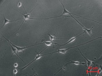
Schwann cell morphology was determined by means of light microscopy using 100× magnification. Optically Schwann cells were identified as elongated bipolar or tripolar cells with a bright oval soma whereas fibroblasts show a flattened non-luminescent appearance with a large nucleus (Fig. 1).
Culture of Schwann cells at 20× magnification. Schwann cells are characterized by their typical bipolar to tripolar shape and bright and lucid cell body. Fibroblasts, in contrast show flattened, non-luminescent cell bodies with large round nuclei.

After one subculture/trypsination cycle (72 h), Schwann cells were seeded on poly-l-ornithine and laminin coated 48 well plates (Corning, Kaiserslautern, Germany). Immunostaining was performed using a monoclonal rabbit antibody against S-100β (Swant, Bellinzona, Switzerland) a polyclonal antibody binding to p75 (Promega, Madison, WI, USA). P75 was chosen as a marker of neuronal cells whereas S100β was chosen as a marker of myelinating Schwann cells. Neither p75 nor S100β are expressed by fibroblasts.
After immobilization of the cultured cells with 2% paraformaldehyde, the cell membranes were permeabilized for 10 min with 0.1% Triton in PBS. Following a wash out step with PBS, the cells were treated with 1% BSA in PBS to avoid non-specific antibody binding. Rabbit anti-p75 and rabbit anti-S100β were diluted in PBS containing 0.1% BSA to their final concentration of 1:500 and 1:5000, respectively. Both antibodies were incubated together with the cells at 4°C over night. A second antibody goat-anti-rabbit, conjugated with the Alexa Fluor® 488 dye (Invitrogen, Karlsruhe, Germany), was used for detection under a fluorescence microscope (Axiovert 200, Zeiss, Jena, Germany). DAPI (Invitrogen, Karlsruhe, Germany) was added to the second antibody for staining cell nuclei. NCL-Ki67p (Promega, Mannheim, Germany) as a proliferation-specific antibody was used to prove cell division.
FuGENE HD© lipofection
Four microlitres of pMAX eGFP were diluted in 96 µl medium followed by addition of 5 µl of the transfection reagent. The solution was mixed with a vortexer and liposomes were allowed to constitute for 15 min at room temperature. 100 µl of the transfection solution were added dropwise to the Schwann cell culture in a six well plate. After 24 h cultivation, the medium was replaced.
AMAXA© nucleofection
Cells were transfected with ‘nucleofector kit V’. Plasmid pMAX eGFP (0.5 µg/μl) was mixed with transfection solution V and stored on ice. Schwann cells were trypsinated and centrifugated with 1000 U/min for 6 min, room temperature. The pellet was taken up in 1 ml medium and Schwann cell number was determined using trypan blue (Sigma-Aldrich, Munich, Germany) staining and counting in Neubauer's chamber. A total of 1 × 10⁶ cells were resuspended in 90 µl transfection solution T and mixed with 10 µl pMAX eGFP. The cell suspension was transferred into the electroporation chamber free from air bubbles (distance between the electrodes: 2 mm) and transfected using program T30 (recommended for Schwann cells by manufacturer). Cells were plated on freshly laminin and poly-l-ornithine coated six well plates and cultured for 24 h at 37°C, 5% CO2.
Cell counting
Cell counting was performed under light microscopy by means of a Neubauer chamber. The number of p75 and S100β positive cells based on the total cell number was determined by counting of about 40 cells per well after immunostaining.
Determination of transfection rate
Transfection rate was determined using a fluorescence microscope (Axiovert 200, Zeiss, Germany) at 200× magnification. Total cell number and number of fluorescent cells per field of view were counted. Transfected cell to total cell number ratio was calculated. Photos were taken using AxioCam MRm (Zeiss, Germany).
RESULTS
Immunocytochemical characterization
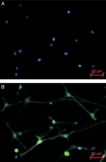
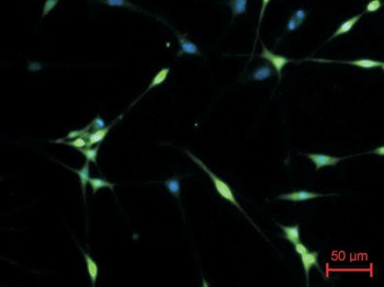
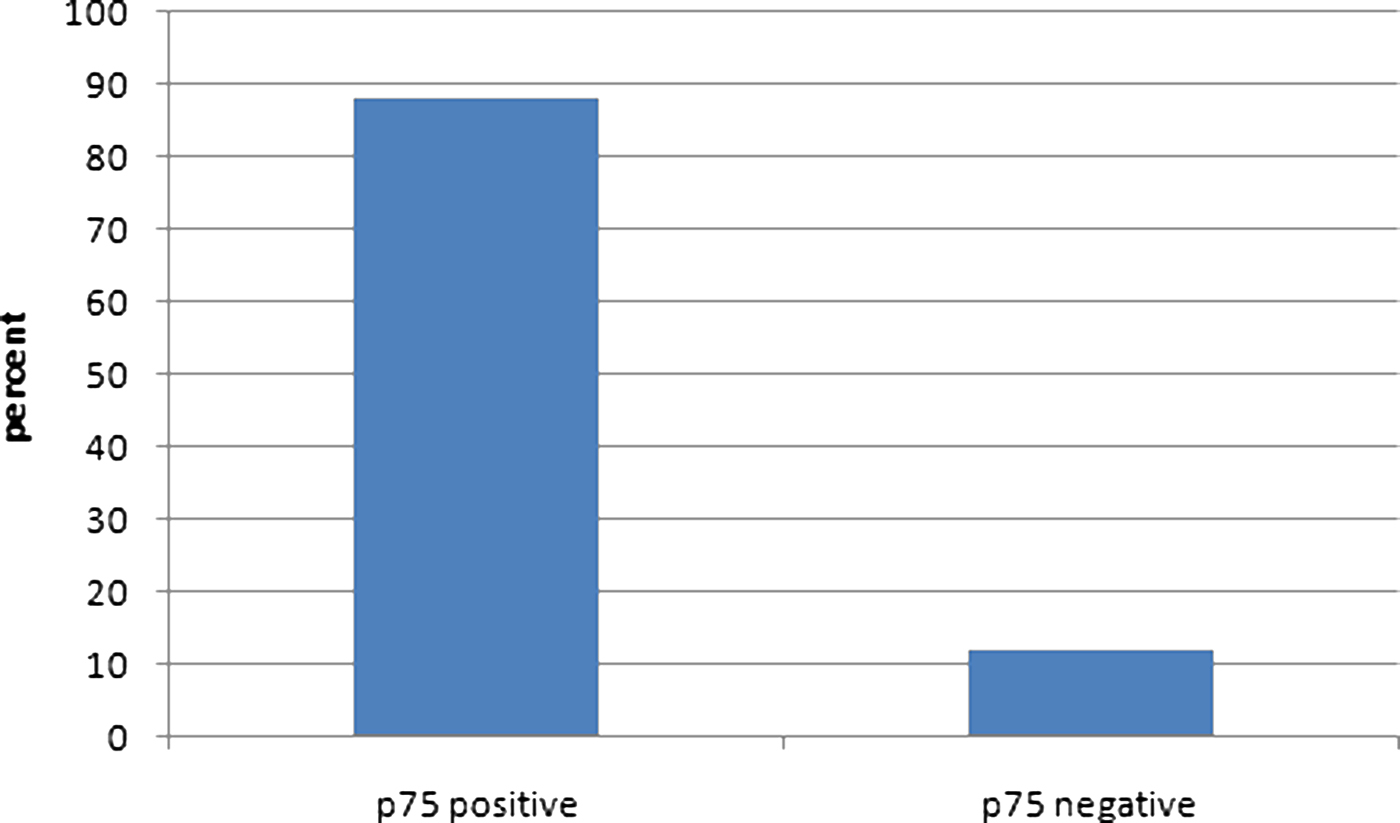
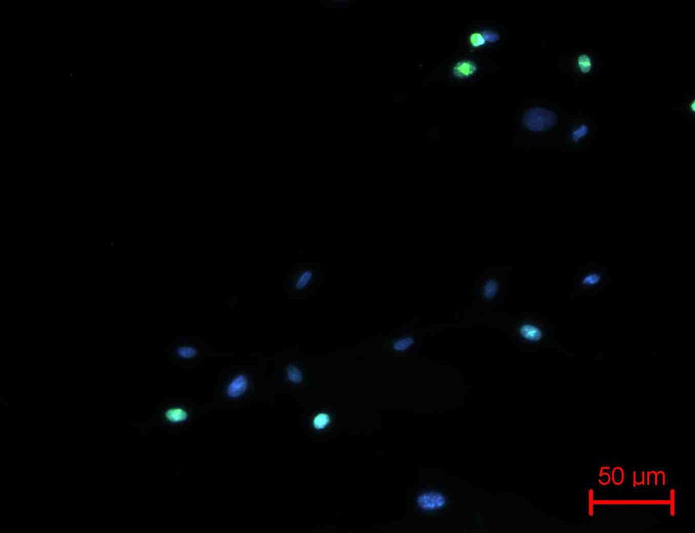
Purity of Schwann cell culture was 88% as determined by immunohistologic staining and optical counting in Neubauer's chamber. Schwann cells were characterized after one passage by the use of fluorescence microscopy using DAPI (for total cell count) and antibodies against p75 and S100β (Figs 2 and 3). Culture purity was determined by calculating the ratio of p75 positive to p75 negative cells in three independent fields of view (Fig. 4). Purity was approximately 88% with 12% marker-negative cells, most likely fibroblasts. Schwann cell proliferation was proven by staining with NCL-Ki67p antibody (Fig. 5).
(A) Immunofluorescence imaging of Schwann cells at 20× magnification using DAPI-staining for determining total cell count and (B) additional p75-staining specific for Schwann cells.

Immunofluorescence imaging of Schwann cells at 20× magnification using DAPI-staining and additional S100β-staining as another Schwann cell specific marker.

Purity of the Schwann cell culture measured by the ratio of p75 positive to p75 negative cells.

Immunofluorescence imaging of Schwann cells at 20× magnification using anti-NCL-Ki67p antibody for visualization of cell proliferation.

FuGENE HD© lipofection
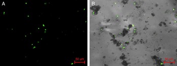
By comparison of transfected cell to total cell number per field of view each, transfection rate was determined at 2% (Fig. 6). Evaluating cell morphology and behavior 24 h after transfection, Schwann cells showed significant aggregation to clusters and failure of settling to the surface of the laminin and poly-l-ornithine coated six well plates. Constitution of Schwann cell extensions typical for cell prosperity was absent in all fields of view evaluated. Settling failure was taken as a marker of compromised cell viability.
Transfection of rat Schwann cells by means of FuGENE HD© lipofection. (A) Shows immunofluorescence alone and (B) shows immunofluorescence in combination with light microscopy at 20× magnification. Transfection rate was determined to be 2%. Schwann cells showed significant aggregation to clusters and failure of settling to the surface of the plates.

AMAXA© nucleofection
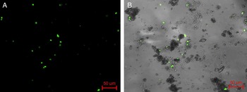
Determination of transfection rate by AMAXA© nucleofection was done analogical to the method utilized for FuGENE HD© lipofection. After 24 h transfection, mean transfection rate of all fields of view evaluated was determined at 10% (Fig. 7). Compared to FuGENE HD© lipofection there was a tendency toward lower aggregation. Regarding cell settling, there was no better adherence to the six well plates compared to lipofection. Concerning Schwann cell morphology, there was absence of prosperity indicating cell extensions in all fields of view observed in either method.
Transfection of rat Schwann cells by means of AMAXA© nucleofection. (A) Shows immunofluorescence alone and (B) shows immunofluorescence in combination with light microscopy at 20× magnification. Transfection rate was determined to be 10%. Schwann cells showed slightly lower aggregation tendency but equal failure of settling to the surface of the plates in comparison to FuGENE HD© lipofection.

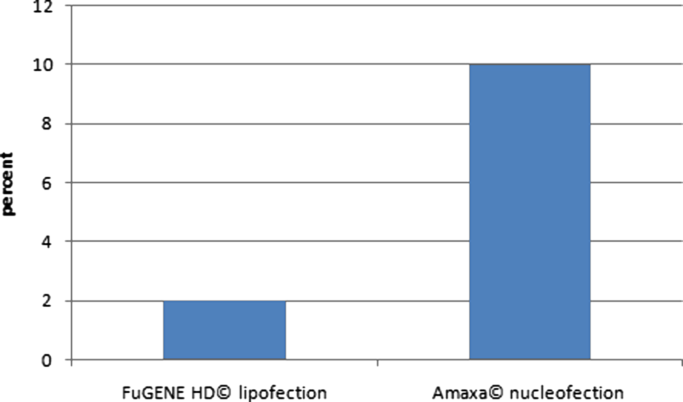
Transfection rate of FuGENE HD© lipofection was 2%, the transfection rate of AMAXA© nucleofection was 10% (Fig. 8). With both methods, Schwann cells showed pronounced aggregation what made them unfeasible for further cultivation. Cell extensions were retracted. Settling of Schwann cells on laminin and poly-l-ornithine coated six well plates was severely compromised by either method.
Transfection rates achieved with FuGENE HD© lipofection and AMAXA© nucleofection.

DISCUSSION
Genetic transfection of Schwann cells is an important scientific aim as it could enhance their regenerative potential. Especially transfection with factors such as nerve growth factor or brain-derived neurotrophic factor is of interest, as external addition has lead to promising results (Terris et al., Reference Terris, Toft, Moir, Lum and Wang2001; Chang, Reference Chang2009). However, the short half-life of these factors limits their efficiency when applied externally.
FuGENE HD© lipofection
Using FuGENE HD© lipofection, we could not achieve a transfection rate that is high enough to obtain sufficient cells for coating a conduit. Cell adhesion to the wells was also impaired so that further cultivation was compromised. Most likely, this was due to cytotoxic effects that have also been reported for other liposomes such as LipofectAMINE© (Barbu and Welsh, Reference Barbu and Welsh2007).
AMAXA© nucleofection
For AMAXA© nucleofection, transfection rates for human Schwann cells as high as 48.4% has been reported (Haastert et al., Reference Haastert, Mauritz, Chaturvedi and Grothe2007) otherwise. The same group describes the transfection rates possible for adult rat Schwann cells to be slightly lower (33%) with 79% cell viability after nucleofection (Haastert et al., Reference Haastert, Mauritz, Matthies and Grothe2006). We could not confirm these results in our experiments. Cell impairment by electric shock and toxicity seems likely with both methods so that one is reluctant to use it on rare and precious cell lines. With a transfection rate of 10% at most, laborsome procedures would be necessary to enhance culture purity, such as cotransfection with an antibiotic resistance gene and consecutive antibiotic addition to the culture. With about 12% of Schwann cell marker negative cells in our culture, it is possible that the real transfection rate of the Schwann cells is reduced by this percentage. The group of Nakayama et al. (Reference Nakayama, Sato, Shinohara, Matsubara, Yokomine and Akasaka2007) report transfection rates of 30% for porcine embryonic fibroblasts with AMAXA©. In their experiments, cell viability was also impaired to a relatively high rate (60% survival).
Despite high transfection efficiency of viral vectors (75%; Muller et al., Reference Muller, Katus and Bekeredjian2007), their application in humans is questionable. Cytopathic effects of common adenoviral or lentiviral vectors are unlikely (Shy et al., Reference Shy, Tani, Shi, Whyatt, Chbihi and Scherer1995), but disease transmission or cancerogenicity remains a problem. Therefore, non-viral transfection should be preferred for clinical application. Viral transfection should be constrained to experimental settings where high-efficiency transfection is decisive.
CONCLUSION
• Non-viral transfection of rat SC with FuGENE HD© lipofection and AMAXA© nucleofection is basically possible.
• Transfection rate is higher for nucleofection than for lipofection.
• Cell viability is compromised by either method.
• If higher transfection efficiency is required, viral transfection methods are to be considered.
ACKNOWLEDGEMENTS
This work was supported by a grant of the Fortune Programme (grant no. 1559-0-0) provided by the Eberhard-Karls University of Tübingen.
Statement of interest
The authors disclose that there is no financial or other conflict of interest within this work.