Introduction
Cystic echinococcosis (CE) caused by the metacestode of the dog tapeworm, Echinococcus granulosus, is a global zoonotic infection, which is of veterinary, medical and economic importance. Adult stages of the parasite infect canids, while metacestodes develop in several species of wild and domestic ungulates (Thompson & Lymbery, Reference Thompson and Lymbery1988; Thompson, Reference Thomson2008).
Geographically distinct strains or intraspecific variants of E. granulosus have been described in different intermediate host species as genotypes G1–G10 (Thompson & McManus, Reference Thompson and McManus2002). Comprehensive molecular genetic and phylogenetic analyses of Echinococcus populations have revealed that some strains are likely to be distinct species and a re-establishment of their formal taxonomic status has been proposed (Thompson, Reference Thomson2008). According to recent taxonomic studies, E. granulosus was an oversimplified species, and four or five cryptic species were intermixed therein. Reviewing the names of synonyms and subspecies, the species has been split into E. granulosus sensu stricto (G1–G3), E. equinus (G4), E. ortleppi (G5) and E. canadensis (G6–G10) (Nakao et al., Reference Nakao, Yanagida, Okamoto, Knapp, Nkouawa, Sako and Ito2010). Cystic echinococcosis in horses is not very common and is assumed to be caused specifically by E. equinus, previously known as horse strain (G4) (Thompson & McManus, Reference Thompson and McManus2002).
Echinococcus equinus, previously known as E. granulosus equinus, was described for the first time in the UK by Williams & Sweatman (Reference Williams and Sweatman1963). It has a classical morphology, can develop and mature in foxes (Howkins et al., Reference Howkins, Gemmel and Smyth1965) and its prepatent period extends to approximately 70 days (Cook, Reference Cook1989). However, it was not possible to infect horses experimentally (obtaining viable cysts) with E. granulosus sensu strict (s.s.) in a successful manner (Williams & Sweatman, Reference Williams and Sweatman1963; Howkins et al., Reference Howkins, Gemmel and Smyth1965).
The aim of this study was to provide molecular characterization of metacestode material obtained from a horse in Turkey with polymerase chain reaction (PCR) amplification of mitochondrial 12S rRNA (mt-12S rRNA) and partial sequencing of the mitochondrial cytochrome c oxidase subunit 1 (mt-CO1) genes.
Materials and methods
A museum sample of a hydatid cyst, which was obtained in 2008 from the liver of a horse after autopsy and preserved in 70% ethanol, was used for the genetic analysis. Genomic DNA extraction was done from cyst walls (germinal and/or laminar layers) using DNeasy Tissue Kit (Qiagen, Hilden, Germany), following the manufacturer's instructions. Before the extraction, cut tissues were washed at least five times with phosphate-buffered saline (PBS). The primer pairs E.g.ss1.for (5′-GTATTTTGTAAAGTTGTTCTA-3′) and E.g.ss1.rev (5′-CTAAATCACATCATCTTA CAAT-3′) were used to amplify partial mt-12S rRNA of E. granulosus sensu stricto (Dinkel et al., Reference Dinkel, Njoroge, Zimmermann, Walz, Zeyhle, Elmahdi, Mackenstedt and Romig2004). In addition, the primer pairs JB3 (5′-TTTTTTGGGCATCCTGAGGTTTAT-3′) and JB4.5 (5′-TAAAGAAAGAACATAATGAAAATG-3′) were used to amplify the partial mt-CO1 gene for sequence analysis (Bowles et al., Reference Bowles, Blair and McManus1992).
The PCR conditions were same for the two primer pairs. The reaction was carried out in a final volume of 50 μl, containing 25.75 μl DNase-, RNase-free sterile distilled water (Biobasic Inc., Ontario, Canada), 5 μl 10 × PCR buffer, 5 μl 25 mm MgCl2, 4 μl 1 mm deoxynucleotide triphosphate (dNTP) mix, 2.5 μl of each primer (50 pmol), 5 μl of template DNA (100–200 ng) and 0.25 μl of Taq DNA polymerase (1.25 IU) (MBI Fermentas, Lithuania). The PCR conditions were: 5 min at 95°C (initial denaturation), 35 cycles of 1 min at 95°C, 1 min at 50°C and 1 min at 72°C and, finally, 5 min at 72°C (final extension). The PCR products were separated on agarose gels (1.5%), stained with ethidium bromide, visualized and photographed on an ultraviolet (UV) transilluminator.
The 446 bp region of the mt-CO1 gene of the horse isolate was sent to a commercial company (Refgen, Ankara, Turkey) for sequencing. Analysis, multiple sequence alignment and phylogenetic tree construction of the obtained sequence of the mt-CO1 gene were performed with CLC Main Workbench software (Knudsen et al., Reference Knudsen, Knudsen, Flensborg, Sandmann, Heltzen, Andersen, Dickenson, Bardram, Steffensen, Mønsted, Lauritzen, Forsberg, Thanbichler, Bendtsen, Görlitz, Rasmussen, Tordrup, Værum, Ravn, Hachenberg, Fisker, Dekker, Schultz, Hein and Sinding2007). The phylogenetic tree was built using the neighbour-joining method (Saitou & Nei, Reference Saitou and Nei1987). The reliability of the inferred tree was evaluated by bootstrap analysis on 1000 replicates. GenBank accession numbers of the reference strains of E. granulosus for mt-CO1 used in the phylogenetic analysis are as follows: JF828337 (G1), M84662 (G2), DQ856466 (G3), M84664 (G4), M84665 (G5), M84666 (G6), M84667 (G7), AB235848 (G8) and AF525457 (G10). Taenia asiatica (AB465211) was used as an outgroup.
Results and discussion
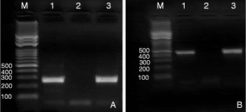
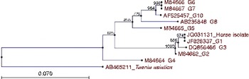
The mt-12S rRNA-PCR with the E.g.ss1.for and E.g.ss1.rev primers yielded 254 bp of amplification product. According to the detected pattern, the horse isolate was identified as E. granulosus s.s. (G1–G3 complex), and mt-CO1-PCR yielded a 446 bp sized amplification product. Amplification products are shown in fig. 1A and B. Sequence of the horse isolate (GenBank accession no. JQ031131) was entirely identical with the common sheep strain (G1) (JF828337). The phylogenetic tree of the sequence with the published sequence results is presented in fig. 2.
PCR products of mt-12S rRNA (A) and mt-CO1 genes (B). Lanes: M, marker (100 bp); 1, positive control; 2, negative control; 3, horse isolate.

(colour online) Phylogenetic relationships of E. granulosus horse isolate from this study (JQ031131) and the sequence references from GenBank (JF828337, M84662, DQ856466, M84664, M84665, M84666, M84667, AB235848, AF525457, AB465211).

Genotypic differences in the species of E. granulosus affect the life-cycle pattern, host specificity, development rate, pathogenicity, antigenicity and sensitivity to chemotherapeutic agents, transmission dynamics, epidemiology and control of cystic echinococcosis (Thompson & Lymbery, Reference Thompson and Lymbery1988; Thompson, Reference Thomson2008).
Varcasia et al. (Reference Varcasia, Garippa, Pipia, Scala, Brianti, Giannetto, Battelli, Poglayen and Micagni2008) studied 2231 horses from various Italian regions and found hydatid cysts in six horses from Sardinia, Sicily and Tuscany. Partial sequencing results of ND1, CO1 and 12S genes corresponded with E. granulosus s.s. (in two horses) and E. equinus (in three horses). Studies of equine hydatidosis are very limited in Turkey. Up to 1352 horses were necropsied in the Pathology Department of the Veterinary Faculty of Ankara University, from 1933 to 1961 and hydatid cysts were diagnosed in two animals (0.15%) (Alibasoglu & Yalciner, Reference Alibasoglu and Yalciner1965). Oge et al. (Reference Oge, Kircali, Yildirim and Oge2004), studied 80 equines slaughtered for feeding carnivores in Ankara Zoo and detected hydatid cyst infection in only 2 out of 35 (5.7%) donkeys among the equines. One of them had six cysts in the liver and the other had six cysts in the spleen, and these cysts were found to be fertile. Balkaya & Simsek (Reference Balkaya and Simsek2001) collected serum samples from 250 equines, consisting of 206 donkeys and 44 horses from various regions of Erzurum province and the vicinity. They analysed these sera by indirect enzyme-linked immunosorbent assay (ELISA) and anti-E. granulosus antibodies were detected in 78 of 250 equines (31.2%). The prevalence rate was 20.4% for horses and 33.5% for donkeys.
The parasite is usually unable to develop into fertile cysts and only small caseous cysts can be observed in infected animals (Ponce Gordo & Cuesta Bandera, 1998). In this study, we could not detect any protoscoleces in the cyst fluids and by the examination of germinal layers under a light microscope. Azlaf & Dakkak (Reference Azlaf and Dakkak2006) performed post-mortem inspection of 455 equines, including 325 horses, 60 mules and 70 donkeys, and detected a CE prevalence of 17.8%. The authors (Azlaf & Dakkak, Reference Azlaf and Dakkak2006) stated that most of the cysts collected from equines were calcified. Furthermore, DNA sequencing of a non-calcified cyst, but very small and not fertile, showed an mt-CO1 sequence identical to that of the sheep strain (G1), as in the present study.
In Turkey, different molecular techniques such as PCR, PCR-restriction fragment length polymorphism (RFLP), random amplification of polymorphic DNA (RAPD)-PCR, single-strand conformation polymorphism (SSCP) and DNA sequencing have been used to detect different strains of E. granulosus. Studies on cattle, sheep, goats, cross-breed mountain goats, mouflons, camels, dogs and humans revealed that the predominant strain is G1, both in humans and in animals, and also that G1/G3, G3, G6, G7 strains exist in Turkey (Utuk et al., Reference Utuk, Simsek, Koroglu and McManus2008; Simsek & Eroksuz, Reference Simsek and Eroksuz2009; Šnábel et al., Reference Šnábel, Altintas, D'Amelio, Nakao, Romig, Yolasigmaz, Gunes, Turk, Busi, Hüttner, Ševcová, Ito, Altintas and Dubinský2009; Utuk & Piskin, Reference Utuk and Piskin2010; Simsek et al., Reference Simsek, Balkaya, Ciftci and Utuk2011a, Reference Simsek, Kaplan and Ozercanb; Utuk et al., 2012).
In the present study, we used the mt-12S rRNA gene as a target for PCR and partial mt-CO1 gene for sequencing. At the end of the study, the horse isolate was detected as E. granulosus s.s. (G1–G3). This is the first study of the molecular characterization of the horse isolate of E. granulosus in Turkey. We consider that further molecular studies of equine hydatidosis are necessary to define the strains of E. granulosus and to develop a clear understanding of the epidemiology and control of the disease.
Acknowledgement
The authors declare no conflict of interests.