Introduction
Cardiovascular diseases are of a multifactorial origin and can be programmed during prenatal and perinatal development. Unfavourable conditions during prenatal development can have negative consequences on the postnatal development of several organs and affect health in adulthood.Reference Giussani and Davidge1–Reference Andraweera, Gatford and Care3Prenatal hypoxia (PH) is one example of such a condition. During early stages, the foetus can adapt to a reduced oxygen supply and increase blood distribution to critically important organs, such as the brain and heart, while conversely reducing renal and gastrointestinal perfusion.Reference Giussani, Camm and Niu4 In a poor intrauterine environment, the foetus develops long-lasting adaptations in cardiovascular and endocrine functions.Reference Verschuren, Morton and Abdalvand5 If PH is present for a prolonged time and during critical periods of development, it can significantly affect the function of the vasculature, heart and kidney, as well as several localReference Alma, De Groot and De Menezes6 and central regulatory mechanisms.Reference Tain, Huang and Hsu7
The timing of critical periods, which determine changes in sensitivity of organs to negative stimuli, can differ among stimuli. However, the final days of pregnancy are particularly important.Reference Svitok, Molcan and Stebelova8,Reference Van den Bergh, van den Heuvel and Lahti9 In our previous study, we found increased blood pressure (BP) and activation of the sympathetic nervous system in adult male rats exposed prenatally to two 4-h periods of 10.5% of oxygen on prenatal days 19 and 20.Reference Svitok, Molcan and Stebelova8 PH can contribute to the development of hypertension and higher susceptibility of the cardiovascular system to different environmental conditions, which occur later in development or in adulthood.Reference Shah, Matsumura and Quon10,Reference Zeman and Okuliarova11
After birth, organisms are exposed to various environmental conditions, including regular light (L) and dark (D) cycles. Rotation of the light/dark regimen is an important synchronisation factor for central circadian oscillator localised in the suprachiasmatic nucleus of the hypothalamus. These clocks generate circadian rhythms of behavioural and physiological processes, including the daily variability of BP and heart rate (HR).Reference Smolensky, Hermida and Portaluppi12,Reference Black, D’Souza and Wang13 Circadian clock outputs are transmitted via the endocrine and autonomic nervous system to the rest of body and entrain their functions to the actual environmental LD cycle.Reference Coomans, Ramkisoensing and Meijer14 Circadian clocks allow organisms to predict regular changes during the 24-h cycle and help to cope more efficiently with stressors.Reference Molcan, Vesela and Zeman15,Reference Chellappa, Vujovic, Williams and Scheer16 Disruption of circadian rhythms can be induced by several environmental variables, including artificial light at night (ALAN). Low ALAN levels (<10 lx) can disturb the circadian control of different physiological processes, including the cardiovascular system in men.Reference Obayashi, Saeki, Iwamoto, Ikada and Kurumatani17 Moreover, low ALAN levels (1–2 lx) differentially affect the circadian control of BP and HR in normotensive ratsReference Molcan, Sutovska and Okuliarova18 in comparison with spontaneously hypertensive rats,Reference Rumanova, Okuliarova, Molcan, Sutovska and Zeman19 which have an activated sympathetic nervous system.
PH increases BP and the activity of the sympathetic nervous system, through which central biological clock outputs are mediated and can be disturbed by ALAN. Therefore, in our present study, we explored the effects of ALAN on circadian rhythms in HR and BP in male offspring exposed to PH. We evaluated: (1) stability of BP and HR changes after PH; (2) circadian variability of BP and HR after 2 and 5 weeks of exposure to ALAN (1–2 lx); and (3) response of BP and HR to norepinephrine in PH offspring.
Materials and methods
Animals
In our study, we examined mature male Wistar rats from nulliparous female Wistar/DV rats (n = 16). We induced PH on prenatal day 20 by 12-h exposure of pregnant females (n = 8) to 10.5% oxygen in nitrogen. After the hypoxic insult, mothers were returned to their home cages. Control pregnant females (n = 8) were placed in the hypoxic chamber on day 20 of gestation for 12 h without inducing hypoxia. Animals were allowed to spontaneously deliver offspring and pups were assigned to the groups after weaning (21 days postpartum) using a single pup per litter to minimise genetic bias as previously described.Reference Svitok, Molcan and Stebelova8,Reference Ujhazy, Dubovicky and Navarova20 We maintained animals individually in plastic cages with food and water available ad libitum. The room temperature (21°C ± 2°C), humidity (55% ± 10%), and regular 12-h L (150 lx):12-h D (0 lx) conditions were automatically controlled. The experiment was approved by the Ethical Committee for the Care and Use of Laboratory Animals at the Comenius University in Bratislava, Slovak Republic and the State Veterinary Authority of Slovak Republic.
Experimental design
Using telemetry, we evaluated daily rhythms of systolic BP and HR in adult, 21- and 27-week-old PH (n = 7, 370 ± 8 g) or control (n = 6, 384 ± 8 g) male offspring. During this period, rats were exposed to regular 12L:12D conditions (Fig. 1). In 28–32-week-old control and PH rats, we determined the response of daily rhythms of BP and HR to ALAN: 12-h L (150 lx) and 12-h of ALAN phases (dim D; 1–2 lx). We evaluated the consequences of ALAN on circadian rhythms of BP and HR via telemetry after 2 and 5 weeks of ALAN exposure (Fig. 1). In 34-week-old rats, we administered norepinephrine and analysed BP and HR response in the control and PH group (Fig. 1).
Experimental design. LD, regular 12-h light:12-h dark regimen; PH, prenatal hypoxia; ALAN, artificial light (1–2 lx) during the dark phase of the day; NE, norepinephrine.

Telemetry
We continuously measured BP and HR via telemetry (HD-S10; Data Sciences International, MN, USA). We applied telemetry transmitters to the abdominal aorta.Reference Svitok, Molcan and Stebelova8,Reference Molcan, Sutovska and Okuliarova18 After implantation, we treated rats with tramadol (15 mg/kg; SC; Tramal, Stada, Bad Vilbel, Germany) and placed them in a heated room. We allowed the animals to recover for 2 weeks after surgery.
Norepinephrine administration
Norepinephrine (200 µg/kg; SC; arterenol bitartrate hydrate; Calbiochem, Germany) was administered subcutaneously 3 h before the lights were turned off and 3 h before the lights were turned on as we did in our previous experiments. Consequences of norepinephrine and vehicle on BP and HR we analysed previouslyReference Molcan, Vesela and Zeman15 and therefore this procedure was not repeated in this experiment. During the dark phase, norepinephrine was administered under red light and the procedure lasted maximum 1 min.
Data analysis
We evaluated systolic BP and HR data as 12-h averages separately for the L and D or dim D. We analysed 24-h variability as the difference (delta) between the L and D or dim D. Due to data normalisation, we expressed delta in % (in relation to LD week). We used Chronos-Fit software to analyse parameters of circadian rhythm (%rhythm, amplitude, acrophase and mesor) of BP and HR.Reference Zuther, Gorbey and Lemmer21 %rhythm is a chronobiological term for the coefficient of determination, that is, the squared coefficient of correlation times 100 (%rhythm = r 2·100). It represents the percentage of variation in the data that is explained by the fitted model. The amplitude is half the range of oscillation of the wave. The acrophase is the peak time of the cosine wave fitted to the data. The mesor is the rhythmically adjusted mean as the mean level of rhythm calculated by cosinor method.Reference Zuther, Gorbey and Lemmer21
We expressed systolic BP and HR responses to norepinephrine as the difference between the stimulated and basal values. We defined the basal values separately for each rat as the value from the 2-h segment before norepinephrine administration. We calculated the area under the curve from a 90-min norepinephrine response.
We tested the normality of data distribution with the Shapiro–Wilk test and subsequently analysed the data with two-tailed heteroscedastic Student’s t-test (parameters of the circadian rhythms). Stability of the PH model (Fig. 2) and the response of BP and HR to norepinephrine (Fig. 3) was evaluated by two-way analysis of variance (ANOVA) with repeated measures (factors: group, LD phase) followed by Tukey’s test. Exposure of rats to ALAN (Fig. 4) was evaluated by two-way ANOVA with repeated measures (factors: week, LD phase) followed by Tukey’s test. BP and HR delta response to ALAN exposure was evaluated by one-way repeated measures ANOVA (factor:week) followed by Tukey’s test. Differences were considered statistically significant at p < 0.05. Statistical evaluations were performed using the statistical package R, version 3.6.3 (R Foundation for Statistical Computing, Vienna, Austria). Data are presented and visualised as the arithmetic mean ± standard error of the mean (SEM; Figs. 4a–f, 3a and b; Excel Office 365, Microsoft, Redmond, USA) or as box plots with individual data points (Figs. 2, 4g–j, 3c–f; R; package: ggplot2). The box representing the range from the first to third quartiles; the band near the middle of the box is the median, and the lines above and below the box indicate the locations of the minimum and maximum value.
Effect of prenatal hypoxia on blood pressure and heart rate in mature male offspring. *p < 0.05 Data are visualised as box plots with individual data points.

(a) Blood pressure and (b) heart rate responses on norepinephrine administration. Responses of (c) blood pressure and (d) heart rate on norepinephrine analysed as the area under the curve (AUC). Maximal time in minutes of (e) blood pressure, and (f) heart rate responses on norepinephrine. *p < 0.05, **p < 0.01. Data are visualised as the arithmetic mean ± standard error of the mean (a, b) and normalised to the value before norepinephrine administration or as box plots with individual data points (c–f).
Notes: dashed grey line, prenatal hypoxia mature male offspring in the light phase of the day; dashed black line, prenatal hypoxia mature male offspring in the dark phase of the day; grey line, control rats in the light phase; black line, control rats in the dark phase of the day; light grey box plots, light phase of the day; dark grey box plots, dark phase of the day.

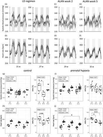
Effect of artificial light at night (ALAN) on blood pressure and heart rate in control and prenatal hypoxia affected rats. Original data from telemetry: (a) blood pressure and (b) heart rate in 21- and 27-week-old (21 week, 27 week) rats during the LD regimen (L light phase 150 lx, D dark phase 0 lx). (c) Blood pressure and (d) heart rate in 29-week-old (29 week) rats during the second week of the ALAN regimen (A2). (e) Blood pressure and (f) heart rate in 32-week-old (32 week) rats during the fifth week of ALAN (A5). The dashed black line indicates prenatal hypoxia mature male offspring, while the grey line represents control rats. Grey and white vertical bars represent the D and L phase, respectively. Data are visualised as the arithmetic mean ± standard error of the mean. Mean values of (g) blood pressure and (h) heart rate and their differences between L and D phases of days in % (delta) in control rats. Mean values of (i) blood pressure and (j) heart rate and their differences between L and D phases of days in % (delta) in prenatal hypoxia mature male offspring. Data are visualised as box plots with individual data points. *p < 0.05, ***p < 0.001.

Results
Stability of the PH model
Blood pressure
In 21-week-old males, BP was significantly higher in PH compared to control rats (134 ± 2 mmHg vs. 127 ± 2 mmHg; p = 0.0504) during the D phase. During the L phase, BP was marginally higher in PH compared to control rats (125 ± 1 mmHg; 120 ± 2 mmHg; p = 0.0849). This pattern of BP was also preserved after 6 weeks (Fig. 2). As expected, in both PH (p < 0.001) and control (p < 0.01) rats, BP was higher during the D compared to the L phase. PH did not affect parameters of the circadian rhythm of BP (%rhythm, acrophase, amplitude, and mesor; Table 1).
Parameters (mean ± standard error of the mean [SEM]) of the circadian rhythm in blood pressure after 2 and 5 weeks of artificial light at night (ALAN) exposure

*p < 0.05 vs. LD; **p < 0.01 vs. LD; ***p < 0.05 control vs. prenatal hypoxia in the same week.
Heart rate
In 21-week-old males, PH did not affect HR. We did not observe differences between control and PH males (Fig. 2). In both PH (p < 0.001) and control (p < 0.001) rats, HR was significantly higher during the D compared to the L phase of the day. PH did not affect the parameters of the circadian rhythm of HR (Table 2).
Parameters (mean ± standard error of the mean [SEM]) of the circadian rhythm in heart rate after 2 and 5 weeks of artificial light at night (ALAN) exposure

*p < 0.05 vs. LD; **p < 0.01 vs. LD; ***p < 0.05 control vs. prenatal hypoxia in the same week.
Exposure of PH male offspring to ALAN
Blood pressure
In control rats, BP was affected by both LD phase (p < 0.001) and ALAN (p = 0.035; Fig. 4g). Delta, 24-h variability, was affected by ALAN in control (p = 0.033; Fig. 4g), but not in PH (p = 0.227; Fig. 4i) rats. In control rats, the difference between L and dim D decreased significantly after 2 weeks of ALAN (p = 0.021; −60.5% ± 11.8%) and was recovered after 5 weeks of ALAN (−16.7% ± 24.8%; p = 0.667) compared to control LD conditions (Fig. 4g). In both control and PH rats, ALAN did not affect BP during the L phase of the day (Fig. 4g, i).
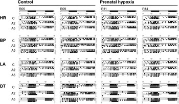
After 2 weeks of ALAN, the circadian rhythm of BP was disrupted in control rats: only two of six rats exhibited significant 24-h variability. After 5 weeks of ALAN, the circadian rhythm of BP was restored in five of six rats; however, %rhythm was still significantly lower (p = 0.029) compared to the LD regimen. In PH rats, the circadian rhythm was disrupted after 5 weeks of ALAN; %rhythm was decreased (p = 0.042; Table 1). As demonstrated with the actograms (Fig. 5), daily rhythmicity was still present for all recorded parameters after 2 and 5 weeks of ALAN in control and PH males.
Representative individual actograms of heart rate (HR), systolic blood pressure (BP), locomotor activity (LA) and body temperature (BT) in freely moving control and mature offspring exposed to prenatal hypoxia during regular 12-h light:12-h dark (C) and second (A2) and fifth (A5) week of the artificial light at night exposure. Actograms were made from raw data by Chronos-Fit. Data for each week is from a 3-day measurement.

Heart rate
In both control (Fig. 4h) and PH (Fig. 4j) rats, HR was affected by LD phase and ALAN.
The LD difference decreased by 26.4% ± 7.8% in control (p = 0.086) and by 15.8% ± 3.8% in PH (p = 0.049) rats after 2 weeks of ALAN. After 5 weeks of ALAN, the LD difference was restored in control (p = 0.721 vs. LD regimen, Fig. 4h) rats, while in PH rats it was continuously decreased (−28.7% ± 4.3%; p < 0.001, Fig. 4j) compared to the LD regimen.
Two weeks of ALAN suppressed %rhythm (p = 0.011), amplitude (p = 0.004), and mesor (p = 0.008) in control but not PH males. After 5 weeks of ALAN, the %rhythm was significantly decreased in control (p = 0.004) and PH (p = 0.016) rats (Table 2).
Discussion
Hypoxic conditions during embryonic life can contribute to hypertension in adulthood and change the response of offspring to environmental challenges. In the current study, we found that: (1) half-day hypoxia on prenatal day 20 consistently increased BP in mature (21 and 27 weeks old) male offspring; (2) PH rats were less sensitive to ALAN compared to control rats after 2-week exposure, but after 5-week exposure, the circadian control of HR and BP variability was equally suppressed in both groups in comparison with LD; and (3) BP and HR responses to norepinephrine administration were not different between PH and control rats.
PH can affect the development of the foetus and control mechanisms that act postnatally.Reference Tain, Huang and Hsu7 The extent of functional changes caused by negative prenatal conditions depends on their intensity and time when they act.Reference Schulz22 Our results showed that 12-h hypoxia during prenatal day 20 resulted in higher BP but not HR compared to control adult male rats. It is necessary to mention that changes in BP and HR caused by 12-h prenatal hypoxia were stable and did not differ between 21- and 27-week-old rats. These results are consistent with our previous data demonstrating that even shorter (4 h) durations of hypoxic conditions during prenatal days 19 and 20 can increase BP in adulthood.Reference Svitok, Molcan and Stebelova8 In contrast, the same level of hypoxia (10.5%) applied in the middle of the embryonic development (days 8–14) did not increase BP in adult male rats (our unpublished data). PH can affect different regulatory areas in the brain and alter the expression of adrenergic receptors in the heart and arteries.Reference Eckhart, Zhu, Arendshorst and Faber23–Reference Moretta, Papamatheakis and Morris25 These changes can lead to a modified function of the autonomic nervous system and increase sympathetic nervous system activity.Reference Svitok, Molcan and Stebelova8 Long-lasting PH can affect both the central and peripheral control mechanisms mediated by transcriptomic and epigenomic mechanisms.Reference Chen, Zhang and Wang26 In our experiment, we exposed animals to PH for 12 h, and even such short exposure can change endothelial mechanisms responsible for higher BP in adult rats.Reference Chen, Qi and Fan27 PH leads to endothelial dysfunction in systemic resistance arteries and affects the acute regulation of BP,Reference Verschuren, Morton and Abdalvand5,Reference Walton, Bielefeldt-Ohmann and Singh28 while chronic regulation of BP is mediated by the kidney. Prenatal adverse condition reduces foetal kidney growth and reduces the number of nephronsReference Verschuren, Morton and Abdalvand5 and glomeruli,Reference Svitok, Husková and Červenková29 an alteration that is often associated with later development of hypertension.Reference Verschuren, Morton and Abdalvand5 It leads in the kidney to upregulation of renin–angiotensin–aldosterone system.Reference Walton, Bielefeldt-Ohmann and Singh28
While daily rhythms of recorded parameters persisted after ALAN exposure, the treatment suppressed circadian variability (amplitude, %rhythm) of BP and HR in both PH and control rats. We analysed the stability of circadian rhythms by Cosinor analysis and as the difference between the L and D or dim D phase. In the control group, ALAN suppressed considerably 24-h variability of BP and HR after 2 weeks of exposure. After 5 weeks of ALAN, 24 h variability of BP and HR was restored. We identified a different pattern in the PH group: we did not find such pronounced suppression of circadian variability of BP and HR after 2 weeks of ALAN as in the control group. However, the light–dark variability of both parameters continuously decreased after 5 weeks of ALAN in PH rats, and there was no difference between the groups at that time. These results are in line with our previous study,Reference Molcan, Sutovska and Okuliarova18 in which ALAN of the same intensity suppressed circadian variability of BP and HR in rats together with changes of spontaneous baroreflex sensitivity. On the other hand, in spontaneously hypertensive rats,Reference Rumanova, Okuliarova, Molcan, Sutovska and Zeman19 the consequences of ALAN resembled those in PH rats found in this study. These data show that even low-intensity ALAN can act as a mild circadian disruptor and, consequently, interfere with circadian rhythms in cardiovascular parameters. It is necessary to mention that disruption of 24-h oscillations of cardiovascular parameters, especially BP, is associated with cardiovascular risk in humans and is used as a prognostic factor.Reference Smolensky, Hermida and Portaluppi12 Our data suggest that PH can change regulatory mechanisms that control the cardiovascular system and responses to environmental stimuli. The nature of these changes is still unknown, but the autonomic nervous system can be involved because PH and spontaneously hypertensive rats have up-regulated activity of the sympathetic nervous system.Reference Svitok, Molcan and Stebelova8,Reference Yang, Wang and Ding30
Light is received by the retina and intrinsically photosensitive ganglion cells transmit the information to the central circadian oscillator localised in the suprachiasmatic nucleus of the hypothalamus. From there, the entrained rhythmic signals are passed to the paraventricular nucleus (PVN) of the hypothalamus, which controls the endocrine and sympathetic system.Reference Thosar, Butler and Shea31 Among the hormonal signals, the circadian rhythm of melatonin synthesis in the pineal gland can be of primary importance because it represents a direct output of the central circadian clocks and melatonin has a pleiotropic action. The circadian rhythm of plasma melatonin concentrations was eliminated in our previous study by the same low light illuminanceReference Molcan, Sutovska and Okuliarova18 as we applied in the present experiment. Melatonin can influence the brain structures involved in the BP control and reactivity of peripheral vessels.Reference Nishi, Almeida and Amaral32
In addition to its role in the control of the endocrine system, the PVN is involved in the regulation of the autonomic nervous system.Reference Coote33 Morphological and electrophysiological studies have shown that PVN is reciprocally connected to regions of the brain that are involved in cardiovascular regulation. Importantly, the PVN projects to both the intermediolateral cell column of the spinal cord and the rostral ventrolateral medulla, regions critical in the control of the sympathetic nervous system and the cardiovascular system.Reference Pyner and Coote34
Gestational intermittent hypoxia,Reference Fan, Wang and Hao35 as well as perinatal hypoxia (E19 to PD14),Reference Raff, Jacobson and Cullinan36 can upregulate sympathetic outflow to the heart and vessels. Moreover, hypoxia increases expression of ɑ1-adrenergic receptors in arteries,Reference Eckhart, Zhu, Arendshorst and Faber23 but not β1-adrenergic receptors in the heartReference Hutchinson, Brew and Vu24 and chronic PH causes postnatal desensitization of β-adrenoceptors in the heart.Reference Lindgren and Altimiras37 Therefore, PH in our study could increase BP but not HR. The centrally increased sympathetic activity can at least partially explain the increased resistance of PH animals to ALAN. We hypothesise that the activated sympathetic nervous system in PH rats can stabilise the outflow to the rostral ventrolateral medulla and protect against disturbing effects of ALAN after 2 weeks of ALAN in comparison with control animals.
In the last part of our study, we tested the significance of ɑ-adrenergic signalling on vessels in rats by evaluating the response of BP to norepinephrine administration. The response to norepinephrine was phase-dependent; both control and PH groups showed higher response during the L in comparison with the D phase of the day. These data are in line with our previous studies.Reference Svitok, Molcan and Stebelova8,Reference Molcan, Vesela and Zeman15 The absence of differences in the BP response to norepinephrine administration between PH and control rats does not exclude the participation of the sympathetic nervous system in their different response to ALAN at the central level because peripherally administered norepinephrine is unable to penetrate the blood-brain barrier.Reference Kostrzewa38 Furthermore, the expression of adrenergic receptors in the heart and blood vesselsReference Eckhart, Zhu, Arendshorst and Faber23–Reference Moretta, Papamatheakis and Morris25 can be different and should be further studied.
Conclusions
Prenatal hypoxia increased BP in mature male rats and permanently affected regulatory mechanisms controlling the cardiovascular system. Daily rhythms of recorded parameters were dumped after ALAN exposure in both groups and the response of BP to ALAN was more pronounced in the control compared to PH rats after 2 weeks of exposure. The higher resistance of PH animals to ALAN can be explained at least partially by centrally increased sympathetic activity and supressed melatonin rhythmicity.
Acknowledgements
We would like to thank Proof Reading Servis.com, the UK for the professionally proofread of the manuscript.
Financial Support
The study was supported by the Slovak Research and Development Agency (APVV-17-0178) and the Scientific Grant Agency of the Ministry of Education of the Slovak Republic (VEGA 1/0492/19, VEGA 2/0154/20).
Conflicts of Interest
The authors declare that there is no conflict of interest.
Ethical Standards
The experiment was approved by the Ethical Committee for the Care and Use of Laboratory Animals at the Comenius University in Bratislava, Slovak Republic and the State Veterinary Authority of Slovak Republic.