Introduction
POU family proteins are important transcription factors conserved from Caenorhabditis elegans to vertebrates (Ryan & Rosenfeld Reference Ryan and Rosenfeld1997). POU-V genes regulate the maintenance of cell pluripotency and differentiation (Cao et al., Reference Cao, Knöchel, Donow, Miethe, Kaufmann and Knöchel2004, Reference Cao, Siegel, Donow, Knöchel, Yuan and Knöchel2007, Reference Cao, Siegel, Oswald and Knöchel2008; Reim et al., Reference Reim, Mizoguchi, Stainier, Kikuchi and Brand2004; Reim & Brand, Reference Reim and Brand2006). For example, zebrafish Pou2 regulates early neurogenesis (Iwafuchi-Doi et al., Reference Iwafuchi-Doi, Yoshida, Onichtchouk, Leichsenring, Driever, Takemoto, Uchikawa, Kamachi and Kondoh2011), axis formation (Reim & Brand, Reference Reim and Brand2006), and endoderm formation (Reim et al., Reference Reim, Mizoguchi, Stainier, Kikuchi and Brand2004). Mouse Oct3/4 (Pou5f1) is expressed during early embryogenesis and is required for development of primordial germ cells (Okamoto et al., Reference Okamoto, Okazawa, Okuda, Sakai, Muramatsu and Hamada1990; Rosner et al., Reference Rosner, Vigano, Ozato, Timmons, Poirier, Rigby and Staudt1990; Palmieri et al., Reference Palmieri, Peter, Hess and Schöler1994). Mouse Sprm1 (Pou5f2) is expressed during spermatogenesis (Pearse et al., Reference Pearse, Drolet, Kalla, Hooshmand, Bermingham and Rosenfeld1997). In early embryogenesis, Oct3/4 expression is regulated by Oct3/4 and Sox2 and their binding sites exist in tandem in the Oct3/4 promoter region (Okumura-Nakanishi et al., Reference Okumura-Nakanishi, Saito, Niwa and Ishikawa2005).
In Xenopus, the gene encoding POU-V is duplicated twice, and the three resultant genes are each used in a different manner (Frank & Harland, Reference Frank and Harland1992; Hinkley et al., Reference Hinkley, Martin, Leibham and Perry1992; Whitfield et al., Reference Whitfield, Heasman and Wylie1993). Oct60, a Xenopus POU-V transcription factor, is maternally expressed and its mRNA levels decrease starting at the blastula stage (Whitfield et al., Reference Whitfield, Heasman and Wylie1993). Other POU-V genes are zygotically expressed starting from the blastula stage. Oct60 overexpression suppresses the Activin/Nodal, BMP and Wnt signaling pathways in the early embryo (Cao et al., Reference Cao, Knöchel, Donow, Miethe, Kaufmann and Knöchel2004, Reference Cao, Siegel and Knöchel2006, Reference Cao, Siegel, Oswald and Knöchel2008), suggesting that maternally expressed Oct60 suppresses cell differentiation in early embryogenesis.
In this report, we analyzed molecular mechanisms of Oct60 transcription. Ectopic expression of Oct60 in epidermal cells was experimentally induced by culturing the cells with oocyte extracts, but not with a control of physiological saline. To analyse the transcriptional regulation of Oct60, we isolated the 2523 bp flanking the 5’ region of Oct60. Reporter analysis using this 5’-flanking region showed oocyte-specific gene expression. In a comparative analysis between Oct60 of Xenopus laevis and Xenopus tropicalis, we found four conserved regions in the 5’-flanking region. A deletion assay showed that enhancer activity exists in the third conserved region (CR3), where an Octamer-Sox binding motif was recognized. A deletion construct lacking the Octamer- and/or Sox-binding motif showed that both of the binding motifs are required for normal expression of Oct60 in oocytes. Co-injection of Oct60 mRNA activated the promoter in an Octamer-Sox motif-dependent manner. Furthermore, ChIP analysis demonstrated the direct binding of Oct60 to the Octamer motif. A conserved role for the Octamer-Sox motif in the gene encoding Oct60 in Xenopus will be discussed in comparison with the mammalian ES-related genes.
Materials and methods
Isolation and analysis of the Oct60 promoter
POU class V genes in the X. tropicalis genome were analyzed with X. tropicalis genome assembly 4.1 (http://genome.jgi-psf.org/Xentr4/Xentr4.home.html). Oct91, Oct25, and Oct60 are positioned in tandem in the X. tropicalis genome (Morrison & Brickman, Reference Morrison and Brickman2006). The sequence between the Oct25 and Oct60 cDNA coding regions was defined as the XtOct60 promoter (−2859 bp). To isolate the X. laevis Oct60 promoter, nested PCR was performed to amplify the sequence between the Oct25 and Oct60 transcript coding regions. Three nested PCR reactions were performed with the following primers: forward primer 5′-ATTTTATGCTTCCAGGATGTAAGCG-3′, backward primer 1 5′-CCTAAACCAGCAACTGCCTTGGG-3′, backward primer 2 5′-TCAGAATGACCAAGGCGCTTCCC-3′, and backward primer 3 5′-AACTCTTCCAACCCAAGGCCTGG-3′. Thermal cycling condition were composed of denaturation at 94°C for 2 min, 25 cycles at 94°C for 15 s and 68°C for 3 min, using KOD-Plus (TOYOBO). The isolated X. laevis Oct60 5′-flanking region (−2523 bp) was subcloned into the EcoRV site of pBSKII+. To identify the sequence, the flanking region was digested by PstI/KpnI. The obtained five fragments were subcloned in pBSKII+ and sequences were checked by ABI3100 and Big dye ver3.1 using T3 primer 5′- AGCGCGCAATTAACCCTCACTAAAGGGAAC-3′, and T7 primer 5′- TCTGGATCTACGTAATACGACTCACTATAG -3′. The −2523-bp Oct60 promoter was then subcloned into Venus/pCS2 (a gift from Dr Miyawaki, Laboratory for Cell Function Dynamics, Advanced Technology Development Group Brain Science Institute, RIKEN, Japan) at the SalI/EcoRI sites (pOct60-Venus) and was used for oocyte extract treatment. Sequence comparison was performed with EMBOSS Pairwise Alignment Algorithms (http://www.ebi.ac.uk/Tools/emboss/align/). Prospective transcription factor binding motifs were analysed by TFSearch (http://www.cbrc.jp/research/db/TFSEARCH.html).
Oocyte extract treatment
Ovaries were isolated from adult females and treated with 1× Ca2+ and Mg2+-free MBS (1× CMF-MBS) Modified Barth's Saline (MBS) containing 0.2% collagenase type IV (SIGMA) for 2 h at room temperature. Dumont stage-IV and -V oocytes (Dumont Reference Dumont1972) were isolated and washed with 1× CMF-MBS with 0.05 mg/ml kanamycin. After washing with 1× CMF-MBS containing a protease inhibitor cocktail (SIGMA), the oocytes were centrifuged overnight in the same medium at 18,000 g at 4°C. The middle layer of the supernatant was collected and centrifuged at 100,000 g at 4°C for 1 h. The supernatant was filtered with a 0.45 μM filter (Millipore) and used as oocyte extract. In this procedure, 700 μl of oocyte extract can be obtained from about 900 oocytes. Larval epidermal sheets were isolated from stages 53–57 tadpoles anesthetized using MS222 (Sankyo). Larval epidermal sheets were washed with 70% ethanol and dissociated into single cells by pipetting with 1× CMF-MBS containing 2 mM EDTA. To permeabilize the cells, 8 U/μl streptolysin O (SLO) was added to the cells suspension and incubated for 15 min. After stopping the SLO treatment by adding 0.5% BSA, cell suspension was diluted with 1000 cells/μl; 50 μl of the cell suspension was added to 100 μl of egg extract or 1× CMF-MBS. After 15 min of incubation, the cells were resealed by adding 150 μl of 1× MBS and incubating for 1–3 days. The pOct60-Venus construct (5 μg) was incubated in 100 μl oocyte extract containing 250 μM rNTP and 1× transcription buffer (Wako) for 2–24 h at 14°C. Total RNA was isolated using RNAiso PLUS (TAKARA). Synthesis of cDNA was performed as reported previously (Ito et al., Reference Ito, Katada, Miyatani and Kinoshita2007). RT-PCR was performed using the following primers: Oct60 forward primer 5′-GTTCTTCCATGTCCCTGTTA-3′ and Oct60 backward primer 5′-ATGGATCCTCCTCAAGGGTCATT-3′, Venus forward primer 5′-ACGTAAACGGCCACAAGTTCAGCGT-3′ and Venus backward primer 5′-TTGTAGTTGCCGTCGTCCTT-3′, Xenopus larval keratin (XLK) forward primer 5′-TCAGCATCTCTGTCATCAGC-3′ and XLK backward primer 5′-TGTAGCTGGACTTGCTGGAA-3′, and histone H4 forward primer 5′-ATCCATGGCGGTAACTGTCTTCCT-3′ and histone H4 backward primer 5′-CGGGATAACATTCAGGGTATCACT-3′. Thermal cycling condition were composed of denaturation at 94°C for 2 min, 30 cycles (histone H4) or 36 cycles (XLK) or 38 cycles (Oct60) at 94°C for 30 s, 55°C for 30 s and 72°C for 30 s, using Ex Taq (TAKARA).
Luciferase assay
Injection of plasmid and mRNA was performed following a previous report (Stewart et al., Reference Stewart, Tomita, Shi and Wong2006). Stage IV–V oocytes were dissected from ovaries with forceps into 1.3× MBS. The Oct60 promoter construct was injected in germinal vesicles with a pRL-TK construct (Promega) as an internal control. The mRNAs were synthesized from the construct encoding Oct60-glucocorticoid receptor fusion protein, and injected into the cytoplasm. Injected oocytes were incubated for 20 h at 16°C supplemented with 20 μM dexamethasone for inducing nuclear localization of Oct60 protein. Luciferase activity was measured using the Dual-Luciferase Reporter Assay System (Promega). Activity of the reporter construct was normalized with pRL-TK activity. N-terminal deletion constructs were subcloned in the XhoI/HindIII site of a pGL3-basic vector (Promega) using the following primers: −2523-bp forward primer 5′-CCGCTCGAGTCCTTGTCTTTTGTCTTCTTATGTTTTATG-3′, −1686-bp forward primer 5′-CCGCTCGAGTGTGTGATTGTAGAAGTGTG-3′, −1328-bp forward primer 5′-C C G C T C G A G G A A T A G G C C C A A T A T G T G T G - 3′, −763-bp forward primer 5′-CCGCTCGAGTAACATAACCCTGTCCCATG-3′, and backward primer 5′-C C C A A G C T T C T C C A G C A C T T G C T C A G G C C - 3′. Thermal cycling condition were composed of denaturation at 94°C for 2 min, 30 cycles at 94°C for 10 s, 55°C for 5 s and 72°C for each times (–2523-bp promoter: 150 s, –1686-bp promoter: 100 s, –1328-bp promoter: 80 s and –763-bp promoter: 50 s), 72°C for 5 min, using PrimeStar (TAKARA). Deletion constructs of the Sox motif and/or Octamer motif were made by PCR and self-ligation using the Oct60 5′-flanking region (−2523 bp)/pGL3-basic (pOct60-luc) as a template and using the following primers: forward primer 5′-TCACACCCAGCCCAGAGAATTTTC-3′, Octamer deletion backward primer 5′-CATTGTTGAAAAGTGTGAAACCACAACATGAC-3′, Sox deletion forward primer 5′-ATGCAAAGTCACACCCAGCCCAGA-3′, Sox deletion backward primer 5′AAAAGTGTGAAACCACAACATGACACTGCA-3′, Octamer and Sox deletion forward primer 5′-TCACACCCAGCCCAGAGAATTTTC-3′, and Octamer and Sox deletion backward primer 5′-AAAAGTGTGAAACCACAACATGACACTGCA-3′. Thermal cycling conditions were composed of an initial denaturation step at 98°C for 1 min, 30 cycles at 98°C for 10 s, 55°C for 15 s and 72°C for 7.5 min, using PrimeStar (TAKARA). The XDelat1 promoter was subcloned in the KpnI/XhoI site of the pGL3-basic vector (Promega) using the following primers: forward primer 5′-CAGGGTACCCTGCAGCAGCTTGTAGTTCG-3′ and reverse primer 5′-AACCTCGAGGTTGTCTGATATGCGATTGC-3’. Thermal cycling condition were composed of an initial denaturation step at 98°C for 1 min, 30 cycles at 98°C for 10 s, 55°C for 15 s and 72°C for 7.5 min, using PrimeStar (TAKARA). All construct sequences were checked by ABI3100 and Big dye ver3.1 standard protocols.
ChIP assays
ChIP assays were performed as reported previously (Stewart et al., Reference Stewart, Tomita, Shi and Wong2006). In the same manner as for the luciferase assays, −2523-pOct60-luc plasmid and Oct60-GR-HA mRNA were co-injected into stage V oocytes. Oocytes were incubated in 1.3× MBS containing 20 μM dexamethasone for 20 h at 16°C. The oocytes were fixed with 1% w/v formaldehyde, sonicated with a Sonifier 250 (BRANSON), and immunoprecipitated with anti-HA antibody (Santa Cruz Biotechnology). PCR was performed with the following primers: forward primer 5′-ATATAAAGGCACAAAGTTCCAGGTGAG-3′ and backward primer 5′-ATATAAAGGCACAAAGTTCCAGGTGAG-3′. Thermal cycling condition were composed of denaturation at 94°C for 2 min, 40 cycles at 94°C for 30 s, 55°C for 30 s, 72°C for 30 s, using Ex Taq (TAKARA).
Results
Oct60 expression was induced in larval epidermal cells by treatment with oocyte extracts
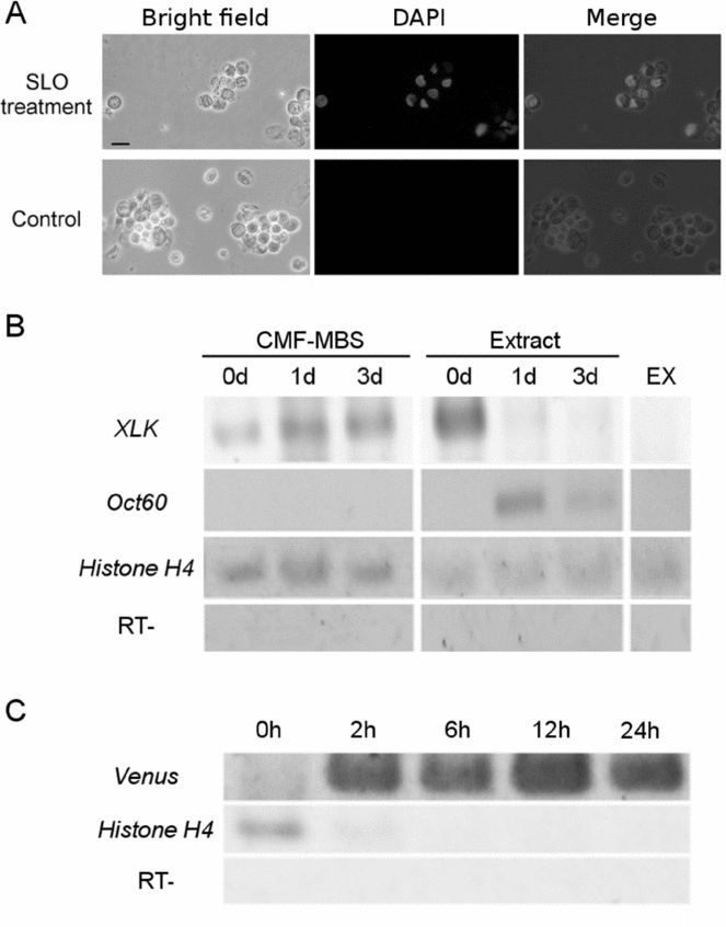
In Xenopus, somatic nuclei were reprogrammed by transferring them into oocytes (Gurdon Reference Gurdon2006). To analyse whether Oct60 expression was activated in oocytes, epidermal cells that do not express endogenous Oct60 were isolated from stage 53–57 tadpoles. The cells were treated with SLO to permeabilize the cell membrane (Fig. 1A). After penetration with the oocyte extract, the permeabilized cells were resealed and cultured in 1× MBS for 1 or 3 days at 16 °C. After culture, gene expression was analysed using RT-PCR. In the oocyte extract-treated cells, keratin expression decreased after 1 day, whereas epidermal cells cultured in MBS maintained their keratin expression for 3 days. Oct60 expression was activated between 1–3 days of incubation (Fig. 1B). In order to analyse the molecular mechanism of Oct60 expression in oocytes, the 5′-flanking region of the gene encoding Oct60 was isolated and conjugated with Venus gene (pOct60-Venus). The Oct60 promoter-conjugated Venus construct was incubated with the oocyte extract, and gene expression was examined using RT-PCR (Fig. 1C). The early response of reporter suggested that the oocyte extract contains the transcription factor that binds and activates the Oct60 promoter directly.
Oct60 expression induced by treatment of oocyte extract. (A) Effect of SLO on the permeabilization of the cell membrane. Epidermal cells were isolated from stage 57–63 tadpoles and treated with SLO. Nuclear staining with DAPI was recognized only on the SLO-treated cells. (B) Gene expression of the epidermal cells treated with oocyte extract. The cells were incubated in oocyte extract or MBS for 3 days, and gene expression was examined by RT-PCR. Oct60 expression was induced within 1 day of treatment with oocyte extract, while Xenopus larval keratin (XLK) expression was suppressed by treatment with oocyte extract. Oocyte extract without permeabilized cells (EX) was used as a negative control. (C) Activation of the Oct60 promoter induced by the oocyte extract. Isolated 5′-flanking region of the Oct60 gene was conjugated with Venus (pOct60-Venus) and incubated in oocyte extract. Venus transcripts were detected 2 h after the treatment.

Oct60 promoter constructs were activated in oocytes
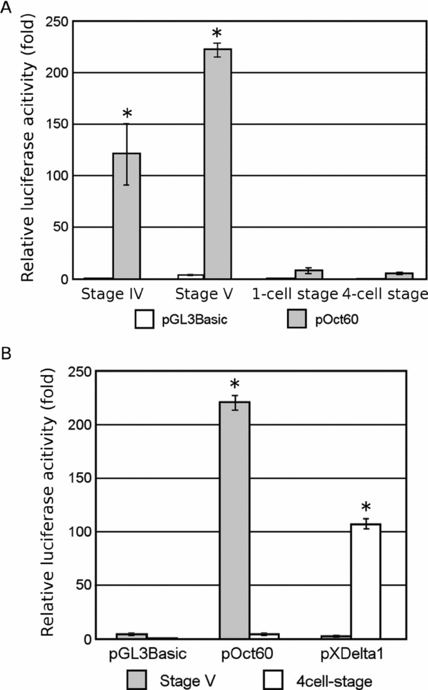
The results suggest that the oocyte extract contains activating factors for the Oct60 promoter. In order to clarify the period of early development during which activation of the Oct60 promoter occurs, Oct60 promoter activity was analyzed from oocyte maturation to early embryogenesis. The pOct60-luc construct was injected into stage IV–V oocytes or 1-cell stage embryos, and luciferase activity was measured after 20 h of incubation. As shown in Fig. 2A, pOct60-luc activity was strongly activated during oogenesis, whereas pOct60-luc activity was highly repressed after fertilization. To compare with the promoter activity, the same promoter assay was performed using an X-Delta-1 promoter that showed zygotically expression. pXDelta1-luc was activated in the early embryo but not in oocytes, which was different from the oocyte-specific activation of pOct60-luc (Fig. 2B). These results show that the oocytes contain factors that specifically activate the Oct60 promoter.
Oocyte-specific activation of the Oct60 promoter. A −2523-pOct60-luc construct was injected into stage IV–V oocytes and 1-cell- or 4-cell-stage embryos. After incubation for 20 h at 16°C, the injected oocytes or embryos were harvested for luciferase assays. (A) Luciferase activity during oogenesis and early development. Luciferase activity was high in oocytes but low in the embryos. (B) Comparison of promoter specificity. A pGL3-basic promoter was used as a negative control. Oct60 promoter was activated in oocytes but not in the 4-cell-stage embryos. However, activation of XDelta1 promoter occurred in the 4-cell-stage embryos, and not in the oocytes. Luciferase activity of pGL3-basic in stage V oocytes is indicated as 1-fold. Error bars represent mean standard error, n = 3. Asterisks indicate that values differ from control at *P < 0.05; Student's t-test was used.
Deletion of Octamer-Sox sequence decreased activity of the Oct60 promoter
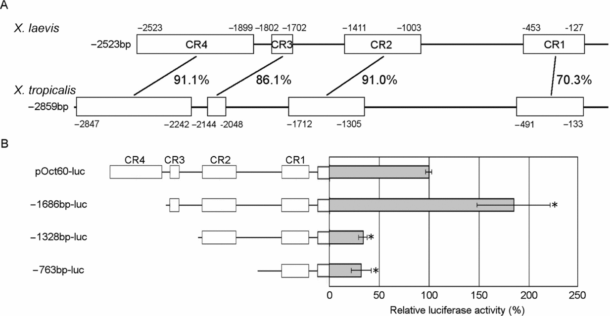
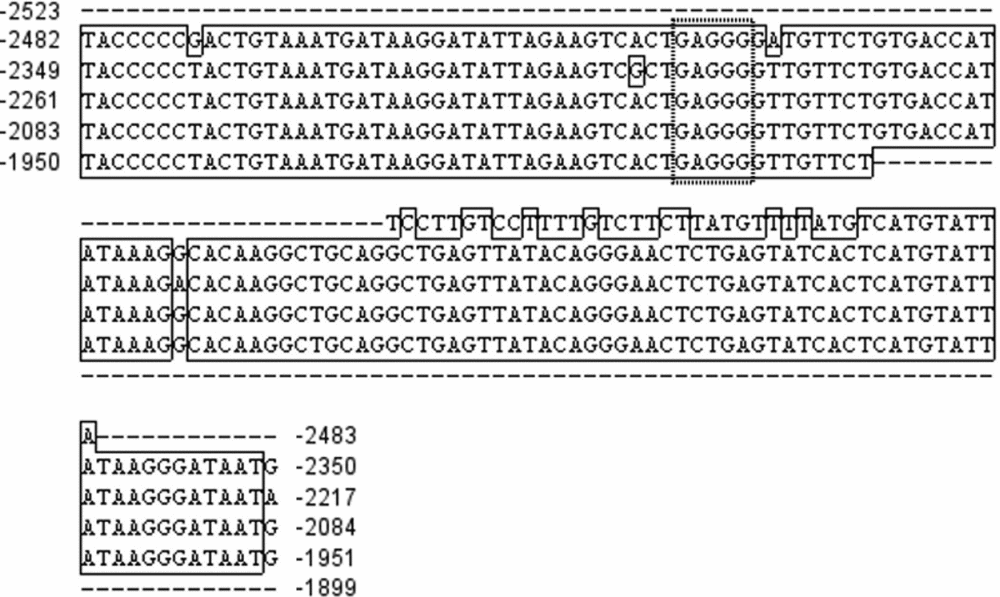
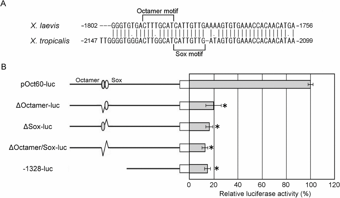
To identify the important region of Oct60 promoter activity, we compared X. laevis and X. tropicalis promoter sequences and analysed the importance of conserved regions by luciferase activity (Fig. 3). Four conserved regions were found and named: CR4, CR3, CR2, and CR1 (Fig. 3A). CR4 and CR2 were tandem repeat sequences (Fig. 4 and data not shown). In X. laevis and X. tropicalis genomes, these repeats were found scattered in the insulator regions, promoter regions, introns, and untranslated regions of mRNA. The CR2 sequences exist in the Oct25 promoter (data not shown). The CR3 and CR1 regions showed 86.1% and 70.3% homology, respectively. The function of these conserved regions was analyzed by using several deletion constructs. In the CR4 deletion construct, the luciferase activity was about twice as high as that of the full-length promoter (Fig. 3B). On the other hand, luciferase activity decreased to 34.0% in the CR3 deletion construct. The CR2 deletion did not remarkably affect the luciferase activity (Fig. 3B). These results suggest that CR3 plays an important role in Oct60 promoter activity. In order to know the role of CR3, the region was searched for prospective transcriptional binding motifs. Comparative analysis of the Oct60 promoters between X. laevis and X. tropicalis showed that Octamer and Sox binding motifs are conserved in the 5′-end of the CR3 region (Fig. 4A). To analyze the importance of these motifs, luciferase activity was measured using the constructs in which the Octamer and/or Sox binding motifs were deleted. In all the deleted constructs, the luciferase activity decreased to approximately 20% the activity of the full-length construct (Fig. 4B). These results indicate that the Octamer and Sox motifs may have an essential role in the function of the Oct60 promoter.
Promoter activity of 5′-flanking region in the Oct60 gene. (A) Comparison of 5′-flanking region in Oct60 gene between X. laevis and X. tropicalis. White boxes labelled CR4, CR3, CR2, and CR1 are highly conserved regions. Percentages represent homology. (B) Luciferase assays using the deletion construct of Oct60. Deletion of CR4 showed upregulation of the promoter, while CR3 deletion caused remarkable downregulation. Full-length promoter activity is indicated as 100%. Error bars mean standard error, n = 3. Asterisks indicate that values differ from the control at *P < 0.05; Student's t-test was used.
Alignment of tandem repeat sequences in the CR4 region of the XlOct60 promoter. CR4 region is five repeats of 133 bp. CTCF binding motifs were conserved in the repeats. Dotted box means CTCF binding motif.
Oct60 protein could bind to Octamer-Sox sequence and activates transcription
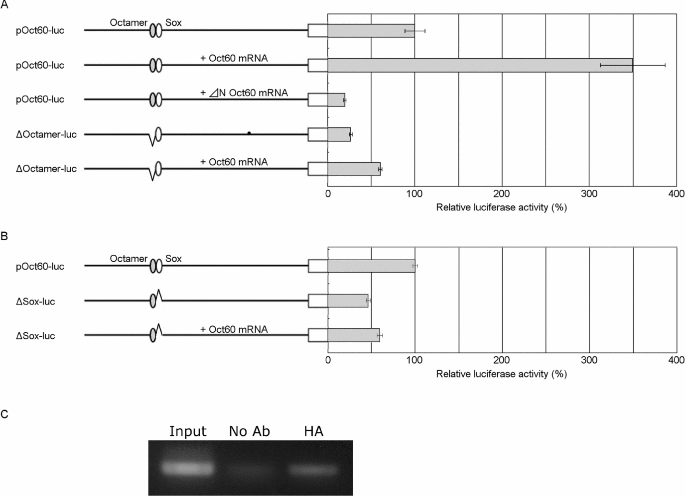
To analyse whether Oct60 protein activates the Oct60 promoter itself, we performed luciferase assays using oocytes injected with Oct60 mRNA. In comparison with the oocytes injected with pOct60-luc alone, the luciferase activity was increased 3.5-fold in the oocytes co-injected with Oct60 mRNA (Fig. 5A). To confirm the role of Oct60, an N-terminal deletion construct of Oct60 (ΔN Oct60) mRNA was coinjected with pOct60-luc into oocytes, and the luciferase activity was measured. The luciferase activity decreased to 20% of the control oocyte injected with pOct60-luc alone. These results suggest that Oct60 protein activates the Oct60 promoter in vivo.
Positive feedback loop of Oct60 expression. Effect of Oct60 protein on the expression of −2523-pOct60-luc. (A, B) pOct60-luc or DNA binding domain deleted construct was coinjected with Oct60 mRNA or ΔN Oct60 mRNA into stage V oocytes. After 20 h of incubation, luciferase activity was measured. Luciferase activity of reporter constructs was normalized by pRL-TK activity. Full-length promoter activity without Oct60 mRNA is indicated as 100%. Left bar, Oct60 promoter; grey ellipse, Octamer binding motif; and white ellipse, Sox binding motif. Error bars mean standard error, n = 5. Asterisks indicate that values differ from the control at *P < 0.05; Student's t-test was used. (C) ChIP assay of the Oct60 protein. Stage V oocytes were coinjected with −2523-pOct60-luc and Oct60-GR-HA mRNA. After 20 h of incubation with 20 μM dexamethasone, ChIP assay was performed using anti-HA antibodies. Oct60 promoter was coprecipitation with the Oct60-GR-HA protein.
In addition, the Octamer motif-deleted construct showed a remarkable decrease in luciferase activity, regardless of co-injection with Oct60 mRNA, suggesting that Oct60 activates the Oct60 promoter via the Octamer motif. Sox binding motif-deleted construct did not activate by coinjection with Oct60 mRNA (Fig. 5B). This result showed that Oct60 promoter self-transcriptional activation of Oct60 is dependent of Sox binding motif. Finally, to confirm that Oct60 protein binds the Octamer motif directly, ChIP assays were performed using oocytes that were injected with Oct60-GR-HA mRNA and Oct60 promoter constructs. Immunoprecipitation was performed using anti-HA antibodies. Figure 5C shows the binding of Oct60 protein to the Oct60 promoter. These results suggest that Oct60 activates Oct60 promoter by binding the Octamer motif.
Discussion
The present study reveals that the Oct60 promoter is activated in oocytes but the activity is remarkably reduced after fertilization. It has been known that Oct60 mRNA exists maternally in the egg and early stage embryo, but its level decreases after gastrulation (Whitfield et al., Reference Whitfield, Heasman and Wylie1993). The maternal expression of Oct60 is converse of the zygotic expression of the other POU subclass V genes Oct25 and Oct91 (Frank & Harland, Reference Frank and Harland1992; Hinkley et al., Reference Hinkley, Martin, Leibham and Perry1992). Oocyte-specific activation of the Oct60 promoter in the present study suggests that the −2523-bp 5′-flanking region of the Oct60 gene plays a key role in the maternal expression of Oct60.
In the treatment of permeabilized cells with oocyte extract, Oct60 expression was induced temporarily at 1 day after the treatment and quickly decreased. Oct60 promoter construct was rapidly activated and maintained for long time. These results are consistent with the previous report using Xenopus oocytes (Simonsson & Gurdon, Reference Simonsson and Gurdon2004). Their report has demonstrated that gene activation of Oct3/4 from plasmid DNA occurs faster than that of whole nuclei. This time lag must come from the remodeling of nuclear proteins. In the reprogramming study using Xenopus oocytes, gene expression of Oct3/4 from somatic nuclei could be detected for more than 6 days (Byrne et al., Reference Byrne, Simonsson, Western and Gurdon2003). In the present experiment, however, gene activation of Oct60 decreased at 3 days after the treatment. This difference might be caused by the difference of the experimental system. In this study, the nuclei of epidermal cells are separated from the egg extract after resealing with calcium-containing culture medium. This finding is quite different from a previous study using Xenopus oocytes which are able to activate continuously the gene expression of somatic nuclei within the germinal vesicles.
Between X. laevis and X. tropicalis, four conserved regions were found in the Oct60 promoter region. Among the four conserved regions, CR4 and CR2 were composed of simple sequence repeats. In the present study, a CR4 deletion caused the enhancement of Oct60 promoter activity suggesting the existence of a suppressor of the CR4 region. The CR4 region is five repeats of 133 bp and includes five CTCF binding domains (Fig. 6). CTCF binding domains are well known as insulators in vivo. But, plasmid containing CTCF domains sometimes act as transcriptional repressors in vitro (Phillips & Corces Reference Phillips and Corces2009). Therefore, the CR4 region probably plays a role as an insulator between the Oct25 and Oct60 promoters. CR4-like repeat sequences are found widely in the genome, suggesting that the CR4 sequence may act as an insulator and/or as a transcriptional repressor in various genome regions.
Role of Octamer-Sox tandem motif in Oct60 promoter. (A) CR3 region of the Oct60 promoter. The sequence of Octamer-Sox binding motif in CR3 region is conserved between X. laevis and X. tropicalis. (B) Luciferase assay of pOct60-luc. Luciferase activities were compared among promoters with deletions in the Octamer (grey circle) and/or Sox (white circle) binding motif. Luciferase activity was downregulated by deletion of the Octamer- and/or Sox-binding site. Full-length promoter activity is indicated as 100%. Error bars mean standard error, n = 3. Asterisks indicate that values differ from the control at *P < 0.05; Student's t-test was used.
The Octamer-Sox tandem motif is conserved in the promoter region of ES cell-related genes such as Oct3/4, Sox2, Nanog, Utf1, Fbx15, and Fgf4 (Nishimoto et al., Reference Nishimoto, Fukushima, Okuda and Muramatsu1999; Tokuzawa et al., Reference Tokuzawa, Kaiho, Maruyama, Takahashi, Mitsui, Maeda, Niwa and Yamanaka2003; Chew et al., Reference Chew, Loh, Zhang, Chen, Tam, Yeap, Li, Ang, Lim, Robson and Ng2005; Okumura-Nakanishi et al., Reference Okumura-Nakanishi, Saito, Niwa and Ishikawa2005; Rodda et al., Reference Rodda, Chew, Lim, Loh, Wang, Ng and Robson2005). It has been reported that Oct3/4 transcription is regulated with a positive feedback loop via the Octamer-Sox tandem motif (Okumura-Nakanishi et al., Reference Okumura-Nakanishi, Saito, Niwa and Ishikawa2005). The present study showed that the Octamer-Sox tandem motif is conserved in Oct60 promoter region, and that the positive feedback loop may also be conserved in the Oct60 promoter. In the Sox2 promoter, Sox2 activates transcription through the Sox binding motif synergistically with Oct3/4 (Tomioka et al., Reference Tomioka, Nishimoto, Miyagi, Katayanagi, Fukui, Niwa, Muramatsu and Okuda2002). Oct3/4 promoter activity depended on both of Octamer and Sox binding motif (Okumura-Nakanishi et al., Reference Okumura-Nakanishi, Saito, Niwa and Ishikawa2005). These finding are consistent with the present results that deletion of the Sox binding motif caused downregulation of the Oct60 promoter with or without Oct60 overexpression in oocytes. Therefore, we hypothesized that activation of Oct60 promoter is dependent on Oct60-Sox heterodimer. As it has been shown that the Oct3/4–Sox2 heterodimer forms the effective transcriptional complex on the Nanog promoter (Rodda et al., Reference Rodda, Chew, Lim, Loh, Wang, Ng and Robson2005). It is possible that Oct60 proteins are not able to bind the Octamer motif without Sox protein. If this is the case, Sox protein is necessary for the maternal expression of Xenopus Oct60. As Sox2 is not expressed in oocytes (Yuan et al., Reference Yuan, Corbi, Basilico and Dailey1995), other Sox family genes may activate the Oct60 promoter.