Introduction
Kleinsasser et al. are credited with the first description of salivary duct carcinoma, in 1968; they selected this term because of the tumour's resemblance to ductal carcinoma of the breast.Reference Kleinsasser, Klein and Hubner1 The use of this term has reportedly confused pathologists and clinicians, even though the majority of salivary duct carcinoma arises from the ductal system.Reference Ellis, Auclair, Gnepp and Goode2 Review of the literature has resulted in some reported cases being reclassified as other tumours (specifically epithelial-myoepithelial carcinoma or polymorphous low-grade adenocarcinoma) which were not considered or reported as specific categories in the original series, making the incidence of salivary duct carcinoma difficult to estimate. Since the introduction of the term salivary duct carcinoma, it has been used in a generic sense to describe any primary adenocarcinoma demonstrating focal ductal differentiation.Reference Seifert, Batsakis and Brocheriou3 The term should be reserved only for tumours that histologically resemble ductal carcinoma of the breast.Reference Gnepp, Henley, Simpson, Eveson and Gnepp4
Barnes et al. Reference Barnes, Rao, Krause, Contis, Schwartz and Scalaogna5 have suggested that, following the initial reporting and definition of salivary duct carcinoma, this tumour type was slow to be recognised, and was thus either not recognised, not correctly identified or confused with other salivary pathologies. Early reported series were retrospective, sourced mainly from tertiary centres in Europe and the US with access to large case series.Reference Jaehne, Roeser, Jaekel, Schepers, Albert and Löning6 More recent reports on salivary duct carcinoma have indicated that the incidence is low – some have reported that this tumour constitutes less than 2 per cent of all salivary tumours,Reference Delgado, Klimstra and Albores Saavedra7 while others have suggested that the true incidence is likely to be greater.Reference Gnepp, Henley, Simpson, Eveson and Gnepp4
Salivary gland malignancies are recognised as a heterogeneous disease group. It is important that pathologists and clinicians use a classification system that has been researched and agreed.Reference Eveson, Watkinson, McGurk and Renehan8 In the latest World Health Organization (WHO) classification,Reference Barnes, Eveson, Reichart and Sidransky9 low-grade cribriform cystadenocarcinoma, which had previously been considered a low-grade form of salivary duct carcinoma, was reclassified as a separate, distinct entity.Reference Delgado, Klimstra and Albores Saavedra7, Reference Laco, Podhola and Dolezalova10–Reference Brandwein-Gensler, Skalova, Nagao, Barnes, Eveson, Reichart and Sidransky12
The survival of salivary duct carcinoma patients is poor, with most dying within three years.Reference Gnepp, Henley, Simpson, Eveson and Gnepp4, Reference Jaehne, Roeser, Jaekel, Schepers, Albert and Löning6, Reference Lewis, McKinney, Weiland, Ferreiro and Olsen13, Reference Simpson and Di Palma14 The five-year overall survival of stage I, II, III and IV disease is reported as 42, 40, 30 and 23 per cent, respectively.Reference Jaehne, Roeser, Jaekel, Schepers, Albert and Löning6
The WHO has now introduced a more stringent definition and description of salivary duct carcinoma.Reference Barnes, Eveson, Reichart and Sidransky9 The clinical disease diagnosed using this new definition is likely to behave in a more aggressive fashion, compared with disease encountered under earlier diagnostic systems. Considering this change, a review of salivary duct carcinoma was felt to be needed.
Pathology
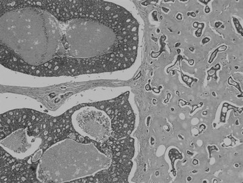
Salivary ductal carcinoma closely resembles ductal carcinoma of the breast, both macroscopically and microscopically, due to its abundance of very firm, fibrous, often ill-defined matrix. Comedo-type necrosis and calcification are seen microscopically (Figure 1), with cystic changes on macroscopic inspection.Reference Lewis, McKinney, Weiland, Ferreiro and Olsen13
Photomicrograph of salivary duct carcinoma, showing cribriform ductal component with comedo necrosis on the left and the invasive desmoplastic carcinoma on the right. (hematoxylin and eosin, ×4)

The majority (80 per cent) of salivary duct carcinoma arises de novo, although in approximately 20 per cent of cases it arises within a pre-existing pleomorphic adenoma.Reference Seifert, Batsakis and Brocheriou3 The true incidence of salivary duct carcinoma involvement with pleomorphic adenoma may be higher, but this is difficult to estimate as many cases can overrun the pleomorphic adenoma, making the latter difficult to recognise.Reference Hosal, Fan, Barnes and Myers15 Carcinoma ex-pleomorphic salivary adenoma is defined as ‘a carcinoma that arises in association with a [pleomorphic salivary adenoma]’,Reference Lewis, Olsen and Sebo16 and an antecedent or coexistent pleomorphic salivary adenoma must be identified to make this diagnosis. The clinical findings that indicate a greater likelihood of malignant transformation include an origin in the submandibular gland, longer duration of tumour, older patient age (compared with a mean of 61 years) and larger tumour size (compared with a mean of 4 cm).Reference Lewis, Olsen and Sebo16 Typically, carcinoma ex-pleomorphic salivary adenoma is widely invasive and associated with presentation at an advanced stage, and has a very poor five-year survival. Carcinoma ex-pleomorphic salivary adenoma with salivary duct carcinoma tends to have a better prognosis than salivary duct carcinoma alone, with a reported five-year survival of 65 per cent. Survival is better if the tumour proportion is less than 50 per cent, if capsular invasion beyond the mixed tumour extends less than 15 mm, and if cervical nodal disease is absent.Reference Olsen and Lewis17, Reference Zbaren, Zbaren, Caversaccio and Stuffer18
A pure, in situ form of salivary duct carcinoma has been described; this is considered rare and is classified as intraductal carcinoma, but its existence is controversial.Reference Simpson, Desai and Di Palma19, Reference Cheuk and Chan20 Pure, in situ salivary duct carcinoma has not been recognised as an entity by the WHO classification system.Reference Seifert, Batsakis and Brocheriou3
The proportion of intraductal and extraductal growth in reported specimens is variable and has prognostic significance. Although the majority of salivary duct carcinoma behaves in a high-grade fashion, cases which have a predominant intraductal component of greater than 90 per cent and which are minimally invasive (i.e. less than 8 mm) are reported to have a good prognosis. The intraductal component may represent the pre-invasive phase of invasive salivary duct carcinoma, which by itself is non-metastasising and associated with an excellent prognosis.Reference Simpson and Di Palma14, Reference Simpson, Desai and Di Palma19, Reference Cheuk and Chan20
Macroscopically, salivary duct carcinoma is un-encapsulated, poorly circumscribed and typically multi-nodular. Comedo-type necrosis, calcification and cystic change may also be seen. Reported tumour sizes range between 1 and 10 cm.Reference Gnepp, Henley, Simpson, Eveson and Gnepp4
Microscopically, salivary duct carcinoma arises from the larger intralobular and extralobular ducts, and is characterised by intraductal and infiltrating components similar to the intraductal and infiltrative ductal carcinoma of the breast. Comedo necrosis is evident within the ducts, with ‘Roman bridge formation’, and evidence of an intraductal cribriform pattern is typically seen. Perineural invasion has been reported in 60 per cent of cases, and around 30 per cent show lymphovascular invasion.Reference Seifert, Batsakis and Brocheriou3, Reference Gnepp, Henley, Simpson, Eveson and Gnepp4, Reference McHugh, Vissher and Barnes21
Most cases of salivary duct carcinoma are diagnosed on a haematoxylin and eosin slide preparation. Special stains are at present mainly confirmatory, although some immunohistochemical markers may in future be considered of therapeutic importance.Reference Gnepp, Henley, Simpson, Eveson and Gnepp4
Morphological variants
Several histological variants of salivary duct carcinoma have been described in the literature. However, such variants should not pose a diagnostic problem because a component of classical salivary duct carcinoma is always present, at least focally.Reference Cheuk and Chan20
Mucin-rich variant
This histological type demonstrates areas of mucinous or colloid carcinoma in which clusters of carcinoma cells, with and without cytoplasmic mucin, appear to float in mucin pools. Four patients have been reported, all treated by surgery and post-operative radiotherapy, but only one was alive at 36 months.Reference Simpson, Prasad, Lewis, Skalova and David22
Invasive micropapillary variant
This variant is highly aggressive and is characterised by morule-like tumour cell clusters without a fibrovascular core, surrounded by morphologically clear spaces.Reference Nagao, Gaffey, Visscher, Kay, Minato and Serizawa23, Reference Yamamoto, Uryu, Segawa and Tsuneyoshi24 This tumour variant expresses human epidermal growth factor receptor-2/neu, and some specimens express human epidermal growth factor receptor-2/neu gene amplification, which is considered to contribute to the observed aggressive behaviour and poor prognosis.Reference Nagao, Gaffey, Visscher, Kay, Minato and Serizawa23, Reference Yamamoto, Uryu, Segawa and Tsuneyoshi24
Sarcomatoid variant
Only five cases of this variant have been reported.Reference Delgado, Vuitch and Albores-Saavedra25, Reference Padberg, Sasse, Huber and Pfaltz26 Histopathologically, while specimens show components of typical salivary duct carcinoma, there are also adnexal sarcomatoid components comprising anaplastic spindle cells, bizarre multinucleated giant cells, rhabdoid cells and, rarely, osteosarcomatous cells. Conceptually, these tumours are considered a variant, de-differentiated form of salivary duct carcinoma,Reference Padberg, Sasse, Huber and Pfaltz26 and can be expected to behave as typical salivary duct carcinoma.Reference Delgado, Vuitch and Albores-Saavedra25
Diagnosis using fine needle aspiration cytology
The poor prognosis and aggressive behaviour of salivary duct carcinoma would suggest that earlier diagnosis may increase the chances of early intervention and thus improved outcomes. The cytological literature relating to salivary duct carcinoma is confined to single cases or small case series.Reference Khurana, Pitman, Powers, Korourian, Bardales and Stanley27, Reference Garcia-Bonafe, Catala, Tarragona and Tallada28 Cribriform, trabecular, acinar and papillary formations can be seen.Reference Gilcrease, Guzman-Paz, Froberg and Pambuccian29 The individual cells are large, monomorphic to pleomorphic, and polygonal to cuboidal, with abundant, finely granular cytoplasm. Squamoid and oncocyte-like appearances are also seen. The nuclei usually exhibit marked pleomorphism, with granular chromatin and small to prominent nucleoli.
The presence of definitive or apparent cribriforming cell groups should alert the pathologist to a possible diagnosis of salivary duct carcinoma.Reference Garcia-Bonafe, Catala, Tarragona and Tallada28, Reference Gilcrease, Guzman-Paz, Froberg and Pambuccian29 Such cytological suspicion may make it possible to investigate patients for evidence of more extensive disease, by magnetic resonance imaging or positron emission tomography scanning, even in the absence of palpable cervical lymphadenopathy.Reference Khurana, Pitman, Powers, Korourian, Bardales and Stanley27
Molecular and biomarker analysis
Molecular and biomarker analysis of salivary duct carcinoma detects a high level of androgen receptor expression (85–95 per cent); in contrast, most breast ductal carcinoma is androgen receptor negative. In addition, salivary duct carcinoma is usually positive for carcino-embryonic antigenReference Hoang, Callander, Sola Gallego, Huang, Sneige and Luna30, Reference Moriki, Ueta, Takahashi, Mitani and Ichien31 but almost uniformly negative for oestrogen and progesterone receptors.Reference Etges, Pinto, Kowalski, Soares and Araujo32 Studies have shown that peroxisome proliferator activated receptor gamma (located in the cytoplasm) is frequently expressed in salivary duct carcinoma at high levels, and may provide a potential molecular target for treatment using agonist ligands.Reference Vadlamudi, Balasenthil, Sahin, Keis, Weber and Kumar33, Reference Mukunyadzi, Ai, Portilla, Barnes and Fan34
Immunohistochemistry and genetic abnormalities
Human epidermal growth factor receptor-2 is a protein found on the surface of breast ductal carcinoma cells. The gene encoding this receptor is known as human epidermal growth factor receptor-2/neu (also known as HER-2/neu or c-erbB-2), and is amplified in more than 20 per cent of invasive breast cancers; it is known to be a negative prognostic indicator.Reference Etges, Pinto, Kowalski, Soares and Araujo32, Reference Skálová, Stàrek, Vanecek, Kucerova, Plank and Szepe35
Because breast ductal carcinoma and salivary duct carcinoma are similar, over-expression of the human epidermal growth factor receptor-2/neu proto-oncogene has also been studied. The incidence of human epidermal growth factor receptor-2 expression in salivary duct carcinoma is 20–25 per cent.Reference Jaehne, Roeser, Jaekel, Schepers, Albert and Löning6, Reference Williams, Roberts, Kies, Mao, Weber and El-Naggar36 Expression of human epidermal growth factor receptor-2/neu has been linked to early local disease recurrence, distant metastasis and poorer survival.Reference Jaehne, Roeser, Jaekel, Schepers, Albert and Löning6, Reference Etges, Pinto, Kowalski, Soares and Araujo32 However, gene amplification, as detected by fluorescence in situ hybridisation, is not always consistent and may indicate a possible independent role for this marker.Reference Cornolti, Ungari, Morassi, Facchetti, Rossi and Lombardi37–Reference Ettl, Schwarz, Kleinsasser, Hartmann, Reichert and Driemel39
Differential diagnosis
BarnesReference Barnes40 has reviewed metastases to the major salivary glands, and found that only approximately 15 per cent are from sites below the clavicle (especially lung, kidney, prostate and breast). Metastatic prostate carcinoma can be a diagnostic dilemma, as some salivary duct carcinomas themselves secrete prostate-specific antigen.Reference Fan, Wang and Barnes41 The differential diagnosis for high-grade salivary duct carcinoma includes the papillary cystic and microcystic variants of acinic cell carcinoma, metastatic squamous cell carcinoma, metastatic breast cancer, melanoma, mucoepidermoid carcinoma, and oncocytic carcinoma.
Based on nuclear findings, it may be possible to distinguish salivary duct carcinoma from other high-grade salivary gland carcinomas, by immunostaining for androgen receptor, gross cystic disease fluid protein-15 and p63.Reference Kawahara, Harada, Akiba and Kage42, Reference Moriki, Ueta, Takahashi, Mitani and Ichien43
Clinical features
Men are more likely (by a factor of three) to receive a diagnosis of salivary duct carcinoma than women, and both sexes present in the fifth to sixth decade.Reference Seifert, Batsakis and Brocheriou3, Reference Jaehne, Roeser, Jaekel, Schepers, Albert and Löning6, Reference Brandwein-Gensler, Skalova, Nagao, Barnes, Eveson, Reichart and Sidransky12, Reference Hosal, Fan, Barnes and Myers15
More than a quarter of patients will present with facial nerve dysfunction or paralysis, and more than a third have palpable cervical lymphadenopathy, highlighting the aggressive behaviour of this tumour.Reference Jaehne, Roeser, Jaekel, Schepers, Albert and Löning6, Reference Hosal, Fan, Barnes and Myers15
Due to the tumour's rapid increase in size, approximately two-thirds of patients will present with tumour stage three or four tumours. Of patients who present with node (N) stage zero disease, 50 per cent will be discovered to be histologically pN+ following neck dissection.Reference Hosal, Fan, Barnes and Myers15
The parotid gland is the most common site of salivary duct carcinoma, accounting for 88 per cent of reported cases.Reference Hosal, Fan, Barnes and Myers15 The submandibular gland is an uncommon site and accounts for approximately 8 per cent of cases.Reference Hosal, Fan, Barnes and Myers15 There has only been a single case report arising from the sublingual gland.Reference Sartorius, Gille, Bédrossian-Pfingsten and Kemp44
The involvement of minor oral salivary gland sites has been reported, such as the palate, retromolar trigone and tongue; 20 such cases had been reported to 2003.Reference Huh, Heo, Lee and Choi45 There are three reported cases of salivary duct carcinoma occurring in the larynx.Reference Jeong, Son, Ko and Kim46, Reference Goel, Agrawal and Srivastava47
Current management
Major salivary glands
Patients with salivary duct carcinoma have a high incidence of loco-regional recurrence and distant metastasis, resulting in dismal survival rates. Thus, aggressive treatment should be pursued. Tumours arising in the parotid should be managed with total parotidectomy. Radical parotidectomy should only be performed in patients presenting with facial nerve paralysis. Delgado et al. Reference Delgado, Vuitch and Albores-Saavedra25 recommend total parotidectomy with facial nerve preservation for smaller tumours (not defined). In contrast, Colmenero Ruiz et al. Reference Colmenero Ruiz, Patron Romero and Martin Perez48 argue for radical surgery with facial nerve sacrifice even in smaller tumours, citing the tumour's aggressive behaviour. Due to rapid tumour growth, the overlying skin can often be involved, and needs to be resected en bloc with the tumour.
In cases of submandibular gland involvement, there are often micro- and macroscopic extra-glandular tumour extensions, so wide local excision and resection of adjacent structures (e.g. the digastric muscle, mandible and skin) may be indicated.
Sublingual and minor salivary glands
In cases involving the sublingual and minor salivary glands, an adequate biopsy is essential prior to definitive surgery. This allows accurate diagnosis and enables elective evaluation of tumour spread, before embarking on major resection of the primary site. An elective neck dissection should also be considered in all such cases.
Neck management
Salivary duct carcinoma metastasises to the neck in 65 per cent of patients.Reference Hosal, Fan, Barnes and Myers15 Thus, an elective selective or modified neck dissection should be performed in all cases presenting with N0 or N+ disease when possible.
Role of radiotherapy
In the literature, the majority of long-term survivors of salivary duct carcinoma had received post-operative radiotherapy. However, the small patient numbers and retrospective nature of the data make it difficult to assess the effectiveness of this policy.Reference Hosal, Fan, Barnes and Myers15
Prognostic indicators
Few prognostic indicators have emerged from clinicopathological studies of salivary duct carcinoma, probably because of the overwhelmingly aggressive nature of the disease.
Small tumour size, absence of perineural or lymphatic invasion, lack of cervical metastases, and tumour diploidy have failed to predict the few patients with long-term disease-free survival. Expression of p53 protein is associated with a high risk of recurrence and metastasis.Reference Jaehne, Roeser, Jaekel, Schepers, Albert and Löning6 The prognostic significance of human epidermal growth factor receptor-2/neu expression remains uncertain at present, but there seems to be a better clinical correlation with protein expression on immunocytochemistry than with gene amplification on fluorescence in situ hybridisation.Reference Skálová, Stàrek, Vanecek, Kucerova, Plank and Szepe35, Reference Cornolti, Ungari, Morassi, Facchetti, Rossi and Lombardi37
The treatment and clinical outcomes for the mucin-rich and sarcomatoid variants are similar to those for conventional salivary duct carcinoma, whereas the micropapillary variant has thus far been reported to be more aggressive.Reference Nagao, Gaffey, Visscher, Kay, Minato and Serizawa23
Potential role of adjuvant therapy
In breast cancer, studies have shown that determination of the human epidermal growth factor receptor-2 gene amplification status improves the identification of likely responders to targeted therapy with trastuzumab (Herceptin, Genentech, San Francisco, California, USA).Reference Simpson, Desai and Di Palma19, Reference Wolff, Hammond, Schwartz, Hagerty, Allred and Cote49, Reference Ligibel and Winer50 Clinical trials have demonstrated a resultant prolongation of survival.
Some early publications have reported promising results of a good response of metastatic salivary duct carcinoma to trastuzumab.Reference Prat, Paraera, Reyes, Peralta, Cedres and Andreu51–Reference Nabili, Tan, Bhuta, Sercarz and Head53
Docetaxel (Taxotere; Sanofi-Aventis, Guildford, UK), an anti-mitotic drug that interferes with cell division, may also be more effective when used in combination with trastuzumab, due to a synergistic interaction.Reference Nashed and Casasola54 This combination is well established in advanced breast cancer treatment, and up to two-thirds of patients positive for human epidermal growth factor receptor-2 show a good tumour response.
Immunoreactivity for androgen receptor is almost 100 per cent in salivary ductal carcinoma cases, suggesting that a treatment approach using an anti-androgen drug should be investigated.Reference Cornolti, Ungari, Morassi, Facchetti, Rossi and Lombardi37 A complete remission was achieved in a single patient with recurrent, androgen receptor positive disease, using an anti-androgen blockade drug, triptorelin, given monthly.Reference Moriki, Ueta, Takahashi, Mitani and Ichien43 Therefore, although salivary duct carcinoma resembles breast ductal carcinoma, it appears to carry an immunophenotype closer to prostate carcinoma than to breast carcinoma.Reference Locati, Quattrone, Marchiano, Cantu and Licitra55 Salivary duct carcinoma has almost no oestrogen or progesterone receptor immunoreactivity, in contrast to its androgen receptor immunostaining, suggesting that its carcinogenesis process may resemble that of prostate cancer, and involve androgen receptor, prostate-specific antigen, tumour growth factor alpha and epidermal growth factor receptor.Reference Fan, Melhem, Hosal, Grandis and Barnes56 Exploitation of anti-androgen therapy seems to be the corollary of these observations.Reference Locati, Perrone, Losa, Mela, Casieri and Orsenigo57
Clearly, more research is needed in order to target the biological aspects of salivary duct carcinoma as, despite aggressive surgical resection and post-operative adjuvant radiotherapy, the overall patient survival rate remains dismal.
At present, chemotherapy has only been used for clinically demonstrable disseminated salivary gland cancers, administered on the basis of optimism rather than evidence of a likely, predictable response.Reference Hamakawa, Nakashiro, Sumida, Shintani, Myers and Takes58, Reference Milano, Longo, Basile, Iaffaioli and Capongigro59 One argument is to consider adjuvant therapy following surgery, perhaps initially in combination with radiotherapy.
Conclusion
Salivary duct carcinoma is one of the most aggressive of the salivary gland carcinomas, and phenotypically resembles high-grade ductal breast carcinoma. The parotid gland is the most common site. Clinically, tumours are typically characterised by a rapidly growing parotid mass with pain, the presence of cervical metastases, and early facial nerve involvement. Following conventional treatment, comprising surgery with or without radiotherapy, the local recurrence rate is high, associated with both cervical nodal involvement and distant metastasis, resulting in a poor prognosis.
Treatment needs to be more aggressive, including total surgical removal of the primary site together with a neck dissection. Post-operative radiotherapy should be recommended in all cases.
Traditionally, chemotherapy has only been offered for clinically recurrent, disseminated disease. However, evidence suggests that improved disease control could be achieved by employing adjuvant chemotherapy in combination with post-operative radiotherapy.
Advances in immunocytochemistry have identified tyrosine kinase receptors and hormone receptors in salivary duct carcinoma. Certain specific histotypes carry promising immunophenotypic profiles, which should be exploited to identify patients suitable for innovative treatment options, administered within prospective clinical trials.