Introduction
Onchocerciasis, commonly known as river blindness, remains a major health problem for numerous countries in tropical Africa, America and the Arabian Peninsula. This filarial disease is caused by Onchocerca volvulus and transmitted by the black flies of the genus Similium, which breeds in fast-flowing rivers. Adult female O. volvulus, which can produce up to 1500 microfilariae daily, are known to survive for more than 10 years in the definitive host (Schulz-Key & Karam, Reference Schulz-Key and Karam1986; Plasier et al., Reference Plasier, van Oortmarssen, Remme and Habbema1991). According to World Health Organization statistics, about 120 million individuals are at risk of the disease and over 18 million infected, with more than 500,000 visually impaired (WHO, 1995). About 90% of affected persons live in Africa. Onchocerciasis is characterized in humans by debilitating skin pathology and can ultimately lead to blindness (OMS, 2011). People with this disease are usually stigmatized in their communities as they may, unfortunately, lose their productivity. Thus, the impact of onchocerciasis is not only on individuals but also on the productivity and the economy of the endemic country. The disability-adjusted life year (DALY) rates from onchocerciasis are calculated to be 884 per 100,000 inhabitants.
Over several decades, much progress has been made towards developing a number of programmes, including the African programme for onchocerciasis, and strategies to control the disease. Control is based on the elimination of vectors and a chemotherapeutic approach. The vector-control approach is based on the killing of Similium larvae by regular rotatory spraying with organophosphate larvicides in areas under control (OMS, 2011). Although the procedure of spraying the larvicides was monitored, this approach has been shown to be quite unsafe for several non-targeted insect species, and severely compromised the biodiversity of the concerned areas. Some treated areas were re-invaded by blackflies coming from surrounding areas with a relatively low larvicide pressure. In addition to the use of larvicides, drugs such as diethylcarbamazine (DEC) and suramin were used between the seventeenth and the twentieth centuries as first-choice drugs for the treatment of the disease. However, since 1987 ivermectin, a derivative of avermectin (Crump & Omura, Reference Crump and Ōmura2011) has been used as the main antifilarial drug for human onchocerciasis treatment. It has been found to contain a potent microfilaricidal substance and, so far, remains the most effective, available and efficient drug against onchocerciasis. Specifically, in Cameroon, this drug was reported to induce some severe inflammatory reactions in people co-infected with loasis and onchocerciasis (Tanya et al., Reference Tanya, Wandji, Kamgno, Achukwi and Enyong2013). Ivermectin is also used as long-term treatment until the destruction of adult worms, which can live up to 15 years (Duke, Reference Duke1968). Furthermore, resistance of the parasites to the drug was observed several years ago in some communities in Ghana and Sudan where mass treatment was undertaken (Ali et al., Reference Ali, Mukhtar, Baraka, Homeida, Kheir and Mackenzie2002; Awadzi et al., Reference Awadzi, Boakye, Edwards, Opoku, Attah, Osei-Atweneboana, Lazdins-Helds, Ardrey, Addy, Quartey, Ahmed, Boatin and Soumbey-Alley2004; Osei-Atweneboana et al., Reference Osei-Atweneboana, Eng, Boakye, Gyapong and Prichard2007). Another recent approach involves treatment with antibiotics such as tetracycline or doxycycline (Townson et al., Reference Townson, Tagboto, McGarry, Egerton and Taylor2006), which are targeted to Wolbachia. This approach is motivated by the presence of Wolbachia endobacteria as an obligatory endosymbiont in some worms. Langworthy et al. (Reference Langworthy, Renz, Mackenstedt, Henkle-Dührsen, Bronsvoort, de, Tanya, Donnelly and Trees2000) reported the efficiency of oxytetracyline on macro- and microfilariae in cattle after 9 months of treatment. The chemotherapy based on the use of antibiotics was shown to have limitations because of the absence of the endobacteria in some nematodes, such as Loa loa (Ghedin et al., Reference Ghedin, Hailemariam, DePase, Zhang, Oksov, Unnasch and Lustigman2009). The increasing reliance of onchocerciasis control on oxytetracycline, tetracycline or doxycycline could lead to the emergence of antibiotic resistance (Büttner et al., Reference Büttner, Wanji, Bazzocchi, Bain and Fischer2003). Briefly, the length of treatment is still a stumbling block (Pfarr & Hoerauf, Reference Pfarr and Hoerauf2006), as there is still no effective drug that can kill the macrofilariae. The ideal solution could be a macrofilaricidal drug that acts in a short time period, without serious side-effects, but with a high specificity and adaptability of the treatment, and adequate access. The bovine parasite O. ochengi is the closest relative of O. volvulus and both parasites are transmitted by the same blackfly, namely Similium damnosum, making it an appropriate model for trials on onchocerciasis.
Nematodes, and all helminths, are known to release products that enable them to invade, subsist and persist inside a host by modulating the immune system response. Proteases, protease inhibitors, antioxidants and orthologues of host cytokines and their receptors (Vermeire et al., Reference Vermeire, Cho, Lolis, Bucala and Cappello2008; Soblik, Reference Soblik2009; Ajonina-Ekoti et al., Reference Ajonina-Ekoti, Ndjonka, Manchang, Wilbertz, Younis, Boursou, Kurosinski, Eberle, Lüersen, Perbandt, Breloer, Brattig and Liebau2012) are reported to contribute to the establishment of the infection in various kinds of nematodes. Such molecules could be collected in situ as excretory–secretory (ES) products in worm culture (McSorley et al., Reference McSorley, Hewitson and Maizels2013). Due to their similarity, the excretory–secretory products (ES products) of O. ochengi could contain several proteins similar to those of the human parasite O. volvulus, and certainly some specific or homologous proteins, since immunization has been reported using irradiated O. volvulus L3 in cattle (Achukwi et al., Reference Achukwi, Harnett, Enyong and Renz2007). In the immune reaction against host invasion, the proteins involved are an interesting area of study and discovery of new immunological approaches for immune system consolidation. Studying those proteins could certainly improve understanding of, and the therapeutic approach against, onchocerciasis, boost host immunity against infestations of parasites and even overcome the disease. The purpose of the present work was to investigate and compare ES products from adults of O. ochengi as an excellent parasite model for O. volvulus, and to examine the immune recognition of proteins by cattle and human host antibodies.
Materials and methods
Culture, harvesting and analysis of ES products
Skin pieces from the umbilical area of cattle (mostly females) containing palpable nodules were collected in the slaughterhouse of Ngaoundéré. Samples were taken directly to the laboratory within 2 h. Skin pieces were then washed thoroughly with distilled water, and then with 70% ethanol and left to dry for 10 min. If not stated otherwise, all chemicals were purchased from Sigma-Aldrich (Deisenhofen, Germany).
Nodules were dissected and isolated from the skin using a scalpel blade, and put directly in phosphate-buffered saline (PBS). Isolated nodules were dissected under a binocular microscope. Females were isolated by nodule digestion with 5% collagenase for 10 h at 37°C and cleaned using sterile PBS supplemented with 100 U/ml penicillin and 100 μg/ml streptomycin. Males were essentially collected by nodule dissection. Isolated males were washed three times in sterile PBS and both genders were washed twice in the RPMI 1640 culture medium supplemented with penicillin 100 U/ml, streptomycin 100 μg/ml, l-glutamine and 0.08 mg/ml NaHCO3, as described by Cho-Ngwa et al. (Reference Cho-Ngwa, Daggfeldt, Titanji and Grönvik2005) and modified by Ndjonka et al. (Reference Ndjonka, Agyare, Lüersen, Djafsia, Achukwi, Nukenine, Hensel and Liebau2011). The culture was performed in 24-well culture plates at 37°C in a CO2 incubator with a maximum of 25 males and 1–3 females (depending on the worm size) separately per 2 ml of culture medium. Females are about ten times bigger than males. After each 48 h, culture media were changed and old media were stored at − 70°C in 10% trichloroacetic acid (TCA) for subsequent analysis. The culture was continued until a maximal time of 8 days, beyond which worms were usually not alive. Dead adult worms were kept in RNA stabilization solution (RNAlater; Qiagen, Hilden, Germany) and stored at − 70°C. A general total of 938 ES products was collected and refers to 938 separate worm cultures. Recombinant proteins were obtained from cloning, expression and purification of templates originating from O. volvulus.
Stored culture media were defrosted, collected and centrifuged in 2-ml Eppendorf tubes at high speed (5000 × g for 15 min). The supernatant was discarded and the pellet collected in 8 μl medium for the next steps. Pellets were mixed with 2 μl of 10 × concentrated loading buffer and heated at 95°C for 5 min. Samples were then put on ice for 2 min, spun down (quick spin), loaded on to 10% sodium dodecyl sulphate (SDS) polyacrylamide gels and stained with 0.1% Coomassie brilliant blue (Carl Roth, Karlsruhe, Germany). The destaining operation was performed with acetic acid–methanol solution and the gel was washed using distilled water. The stained gel was scanned using a Canon scanner (Canon CanoScan LIDE 220; Canon, Krefeld, Germany).
Enzyme-linked immunosorbent assay (ELISA)
Semi-quantitative analysis of the serum IgG antibody level was performed by ELISA to find antibody end-points, as described previously by Mpagi et al. (Reference Mpagi, Büttner, Tischendorf, Erttmann and Brattig2000) with modifications. Onchocerca volvulus and O. ochengi extracts were used as positive controls. The antigens used were coated at 200 ng/well (using a 96-well polystyrene plate (Nunc, Roskilde, Denmark)) in carbonate buffer (pH 9.6) and incubated overnight at 4°C. The incubation was made with ES products, extracts of O. ochengi and O. volvulus, and, simultaneously, some expressed proteins, such as macrophage migration inhibitory factor (MIF) from O. volvulus (Ajonina-Ekoti et al., Reference Ajonina-Ekoti, Ndjonka, Manchang, Wilbertz, Younis, Boursou, Kurosinski, Eberle, Lüersen, Perbandt, Breloer, Brattig and Liebau2012) and from Strongyloides ratti as control samples. Twenty-four hours later the plate was washed four times with PBS +0.05% Tween 20. After removing unbound proteins, the plate was blocked with 200 μl of 5% bovine serum albumin (BSA) per well for 2 h at 37°C. Excess BSA was removed by washing four times, and the plate was incubated for another 2 h at 37°C with human sera (1/2000, 1/8000 and 1/32,000) and with cattle sera (1/2000, 1/10,000 and 1/50,000) diluted in 200 μl PBS +0.5% BSA. The non-specifically bound antibodies were washed out and the plate was incubated with 100 μl of 1/1000 horseradish peroxidase-conjugated goat anti-human IgG (Roche, Mannheim, Germany) and goat anti-cow IgG (Sigma, St. Louis, Missouri, USA) for human and cattle sera, respectively, for an hour at 37°C. The anti-human IgG excess was removed by another washing step. Detection was performed using 100 μl of tetramethyl benzidine substrate (TMB) in the dark for 5 min and stopping the reaction with 0.02% H2SO4 at room temperature. The optical density (OD) was measured at 405 nm using an ELISA reader (Dynatech, Denkendorf, Germany).
Data analysis
The Kruskal–Wallis test was used for intra-group comparison, followed by Dunn's multiple comparison test to compare the reactivity of immunoglobulins to each protein or mixture of proteins. OD values were transformed using a regression formula, y= ax+b where x= index, y= ODs, a= the slope calculated with the standard dilution of 1/500, 1/1000 and 1/2000, and b= y 0. OD values were presented as mean ± standard error, with values < 0.1 being discarded, and P < 0.05 was considered significant.
Results and discussion
Harvesting and analysis of ES products
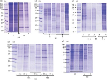
Nine hundred and thirty-eight separate worm cultures, generated from adult females and males of O. ochengi, were collected in 2 ml of culture medium and analysed using the testing system mentioned above. After centrifugation in 2-ml Eppendorf tubes, ES products and total extracts of the adult stages were separated by SDS-PAGE to compare the pattern of protein bands. Many worm cultures were found without pellets and no visible protein bands on SDS-PAGE. Only 156 samples or supernatants loaded on the SDS gels showed visible bands. Lanes with no visible protein bands on the SDS gels were discarded. Gels confirm the presence of huge amounts of proteins of various molecular masses in the whole worm extracts, which appeared denser for females than for males (fig. 1a). Further, worm somatic extracts displayed a wider repartition of protein bands (from 250 to 10 kDa) with higher band frequency compared to ES products (250 to 10 kDa) (fig. 1a and b). Compositions of ES as well as extracted products comprised similar and differing proteins. The ES production seems to be a function of time of culture and number of cultured worms in a medium (fig. 1c and d). The main observation is that the increase in protein amounts is not strongly correlated with the time of culture but rather with the number of cultured parasites. Concentrated samples showed a similar pattern as non-concentrated ES products (fig. 1e). Proteins in female extracts displayed prominent bands at < 10–15, 23, 27, 30, 37–40, 70 and ≥ 100 kDa, while prominent protein bands in male extracts occurred at 15, 27, 30, 37–48 and 70 kDa. Major differences between proteins in females versus males included dominant bands at < 15, 23, 40 and high molecular mass >100 kDa, while males showed a more prominent band at 15 kDa. Bands of ES products displayed by females showed prominent bands at low molecular mass between 10 and 15 kDa and between 20 and 70 kDa, while male ES products showed marked protein bands at 23 and 30–50 kDa (fig. 1b). However, marked variability occurred in the protein patterns from various experiments of ES production (fig. 1c and d). Compared to the worm extract (fig. 1a) ES products displayed fewer bands for the products of four female worms at 48 h and 23 males at the same time point (fig. 1c and d). Band patterns displayed at the same time point for the same number of worms are not strictly identical. For 23 males at 48 h culture, for both lanes, the intensity of bands is different (fig. 1d). In the first lane of these ES products of 23 males, bands are not as intense as in the second lane. For female ES products at 24 h two lanes are shown (one for products from two females and one for those from three females) which are very similar in band separation and intensity (fig. 1c).
SDS gels of Onchocerca ochengi proteins to show (a) somatic extracts (SX) of male (m) and female (f) worms (about 36 μg); (b) excretory–secretory (ES) products released from males and females; (c) ES products from females and (d) from males; (e) protein patterns from somatic extracts (SX) and pooled male (m) and female (f) ES products.

To confirm that ES products originated from the physiological activity of the worm and not from leaking body material, worms were checked under the microscope for integrity from one end to the other. Wounded worms usually show a discontinuous flow of fluid from their bodies and are relatively less motile than unwounded worms. Trichloroacetic acid (TCA), which allows the highest protein yield (Moreno & Geary, Reference Moreno and Geary2008), was used for sample conservation. Collecting samples at different time points displayed an important variability of product release by the worms (fig. 1c and d). The same observation has been made for Echinococcus granulosus ES product collection, where the time of culture did not significantly influence the amount of collected proteins (Virginio et al., Reference Virginio, Monteiro, Drumond, de Carvalho, Vargas, Zaha and Ferreira2012). It is much more a matter of physiological differences. The age, the origin, the parasitological load and the medical care provided to the infected individual probably influences the release of proteins from adult worms. In any case, there is no direct relationship between the time of culture and the amount of collected proteins (Virginio et al., Reference Virginio, Monteiro, Drumond, de Carvalho, Vargas, Zaha and Ferreira2012).
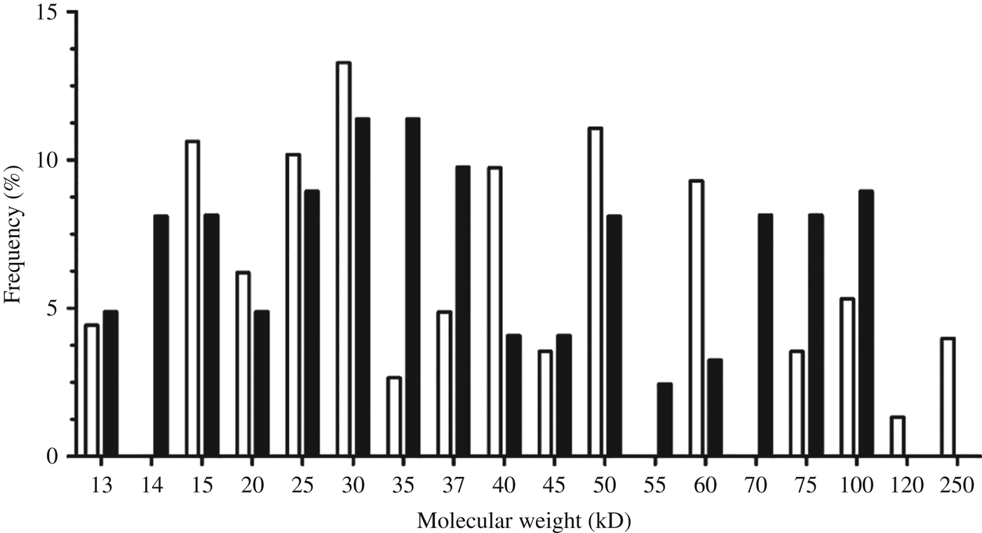
Male and female ES product protein bands were distributed within a scale of 250 to 10 kDa (fig. 2). Bands were widely and frequently distributed within the scale, but it was noticed that most of the bands were localized between 15 and 100 kDa. Bands of male ES products were highly represented at 15, 25, 30, 40, 50 and 60 kDa, with percentage values ranking between 8 and 14% of the total counted bands. SDS gels from ES products of females displayed highly represented protein bands at approximately 15, 25, 30, 35, 37, 50, 70, 75 and 100 kDa.
The frequency (%) of protein bands from ES products of female (black bars) and male (white bars) Onchocerca ochengi.

Immune recognition of proteins
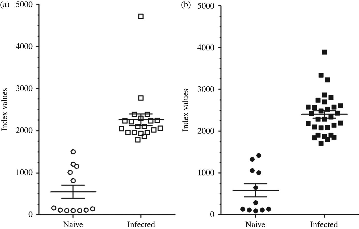
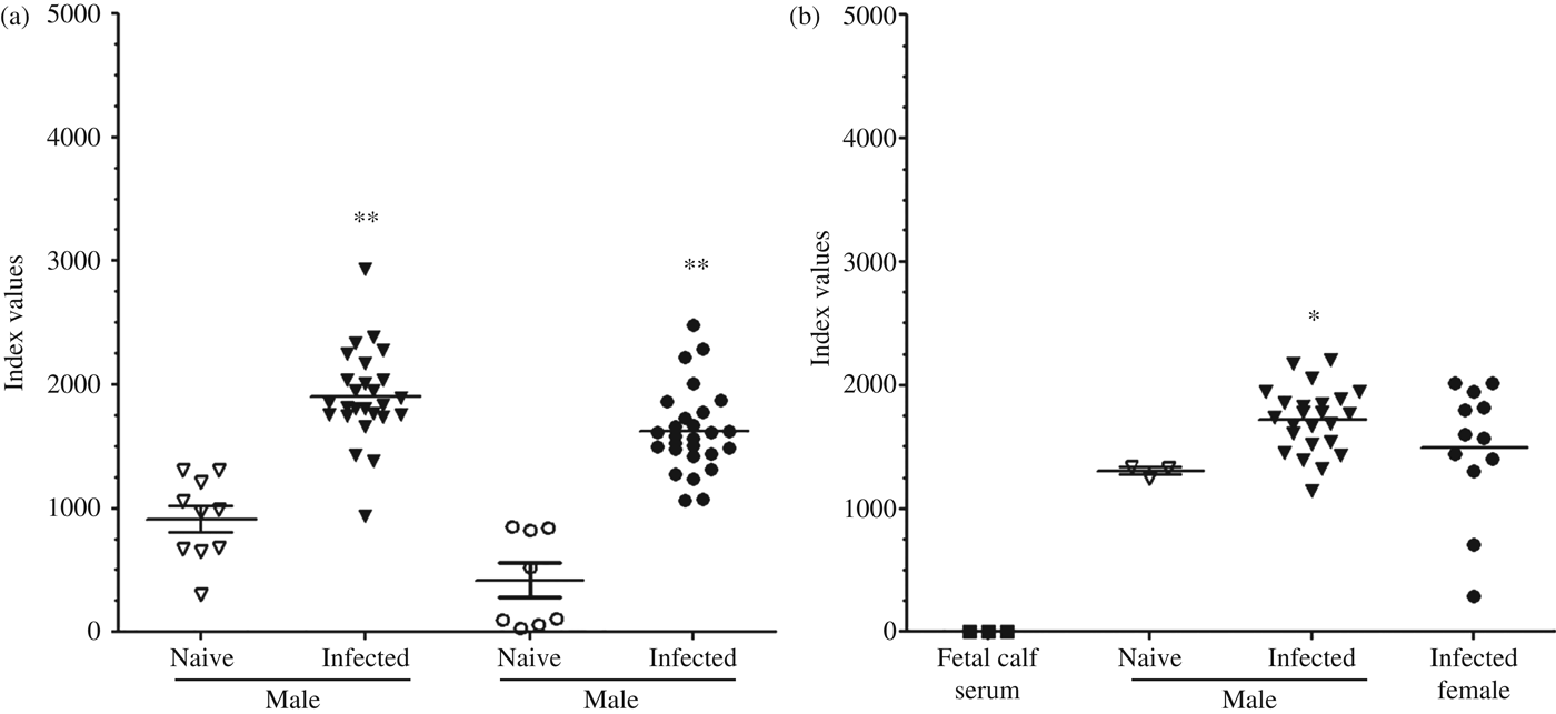
ES products were tested for recognition by antibodies from cattle and humans infected with Onchocerca spp. The analysis involved 13–24 sera from cattle 4 months post-infection with O. ochengi as well as a maximum of 36 sera from humans with onchocerciasis, infected with O. volvulus. For negative controls, sera from naïve, non-exposed cows and Europeans were included. The reactivity of O. volvulus and O. ochengi extracts and O. ochengi ES products to sera from humans with a generalized form of onchocerciasis is demonstrated (figs 3 and 4). The recognition of antigen is observed for both species, showing a cross-reaction between infected human sera and O. volvulus or O. ochengi ES products as antigens, and a relatively weak recognition by naïve or non-infected sera was equally observed for both species. Index values for the antigens ranged between 96 and 4714 for O. ochengi and O. volvulus somatic extract against naïve and infected human sera, respectively (fig. 3a and b). The reactivity of cow IgG to O. ochengi proteins was also observed (fig. 4). The recognition was effective for the antigens used, except for the fetal calf serum, which did not show any reactivity with infected cow sera. Evaluated index values were scaled from 284 to 2201, corresponding respectively to the minimum index value of recognition of ES products of O. ochengi females by sera of infected cattle (fig. 4b) and the maximum index value of recognition of ES products of O. ochengi males by the same sera (fig. 4b).
Immune recognition, expressed as index values of somatic extracts of (a) Onchocerca volvulus and (b) Onchocerca ochengi, by both naïve and infected human sera.

Immune recognition, expressed as index values of O. ochengi male and female ES products, by (a) human sera and (b) cattle sera. *P< 0.05, **P< 0.001.

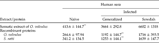
Means of index values of the recognition of recombinant proteins by human sera from the hyperactive, or sowdah, form and hyporeactive, or generalized, form of onchocerciasis are shown (table 1). All antigens showed an affinity for human sera. The recognition of antigen was very high with sera from the sowdah form and relatively low with sera from the generalized form of onchocerciasis. The immune recognition of proteins in the whole-worm extract and ES products of O. ochengi compared to recombinant proteins from human (O. volvulus) and rodents (S. ratti) parasite worms was demonstrated by an indirect ELISA, but with different specificity (figs 3, 4 and table 1). IgG in sera from humans infected with O. volvulus in contrast to healthy individuals strongly recognized not only O. volvulus extract but also O. ochengi extracted proteins and, less pronounced, ES products from O. ochengi males and females (figs 3 and 4). However, some isolated index values were observed, such as the highest values 3900 and 4714 for the recognition of O. ochengi and O. volvulus somatic extract, respectively, by sera from infected humans (fig. 3); and the lowest values 28 and 299 for the recognition of O. ochengi female and male ES products, respectively, by naïve human sera (fig. 4a). Globally, the recognition of extracts from O. volvulus and O. ochengi is higher than the recognition of female ES products, but not male ES products (figs 3 and 4). Compared to the negative control represented by the naïve sera (non-infected sera), immune recognition of the antigens by infected sera is strong. A comparison of index values of antigen groups by the Kruskal–Wallis test brought out significant differences between the medians of index values among the eight groups represented by O. ochengi extracts and O. ochengi ES products. Nevertheless, a comparison using Dunn's test of index values of ES products by gender underlined a non-significant recognition of male ES compared to female ES products. Thus, the recognition of supernatant from males and females is similar, but compared to the positive controls (O. ochengi and O. volvulus somatic extract by sera from infected humans), male ES products are slightly more reactive than female (fig. 4).
Immune recognition recorded as mean index value (± standard error) of somatic extracts and recombinant proteins (macrophage migration inhibitory factor) of Onchocerca volvulus and Strongyloides ratti using human sera from generalized and sowdah forms of onchocerciasis; level of significance given as *P < 0.05 and **P < 0.001.

Recombinant proteins from two parasitic nematodes O. volvulus and S. ratti were recognized by human IgG (table 1). Sera from the sowdah form recognized the O. volvulus extract more strongly, but not significantly, than those from the generalized form. The recognition of recombinant proteins was no stronger than the recognition of O. volvulus extract (mean of index value = 3664 ± 292.8) by sera from the generalized form of onchocerciasis or the sowdah form (6602 ± 1318) (table 1). Elsewhere, none of the recombinant proteins presented a significantly higher affinity with infected human sera from the generalized form compared to O. volvulus extract proteins (table 1). Nevertheless, no differences have been observed in the reactivity of recombinant proteins to both categories of sera and between the reactivity of recombinant proteins and O. volvulus extract to sera from the sowdah form of onchocerciasis, although O. volvulus MIF showed the highest index value of the recombinant proteins (not significant).
Cattle sera recognized O. ochengi proteins. Indeed, the infected sera revealed affinities with O. ochengi extract (result not shown) and ES products. Nevertheless, naïve cow sera showed a weak recognition of O. ochengi ES products (fig. 4b).
Proteins of O. ochengi (somatic extract and ES products) showed immunogenicity against onchocerciasis-infected human and cow sera. The similar reactivity of both O. volvulus and O. ochengi is evidence of the reported phyletic compliance between both parasites (Morales-Hojas et al., Reference Morales-Hojas, Cheke and Post2007). The observed immune reactivity in both genders could arise from the recognition of Wolbachia endobacteria, which are reported to be involved in onchocerciasis pathogenesis (Hise et al., Reference Hise, Gilette-Ferguson and Pearlman2004). Specifically, the peptidoglycan of the bacterial wall is the main factor triggering the immune reaction against the host (Baldo et al., Reference Baldo, Desjardins, Russell, Stahlhut and Werren2010).
Using ELISA, O. ochengi proteins were less specifically recognized in cattle compared with human sera, suggesting that epitopes in infected human sera correspond more closely to human than to cattle IgG (fig. 4). On the other hand, weaker infections in 4-month-old calves may neither have shown a strong immune reaction nor antibody specificity to O. ochengi. The intriguing considerable recognition of the O. ochengi ES products and extract (result not shown) by naïve cattle sera could originate from a cross-recognition of some element(s) of proteins existing in the extract and the ES products (fig. 4b). Livestock are targeted by several kinds of nematode infections that initiate the production of antibodies targeting proteins that are common, or not, from one parasite to the other. The recognition of a mixture of proteins by the IgG induced by a different nematode could explain that fact. Furthermore, the observed reactivity is the result of a cumulative effect of more specific Ig subtypes that react differently to the same antigen (Dafa'alla et al., Reference Dafa'alla, Ghalib, Abdelmageed and Williams1992), showing the difference of reactivity in function of the Ig subclass ratio in sera.
Another significant consideration is the reactivity of the cow immune system compared to the human immune system. The cattle parasite seems to be much more in equilibrium with its host (tolerance of the infection by cattle) than the human one, which gives rise to more severe pathogenesis. In addition, cattle immunoglobulin (IgG1) is reported to be transferred maternally through the colostrum (Manchang et al., Reference Manchang, Ajonina-Ekoti, Ndjonka, Eisenbarth, Achukwi, Brattig, Liebau and Breloer2015), which could induce the inhibition of establishment of O. ochengi L3 in the host. It is not possible after this immune recognition to define which antigen, either in ES products or in whole extract, is most antigenic against each of the sera used, since they are a mixture of proteins (Maizels et al., Reference Maizels, Hewitson, Murray, Harcus, Dayer, Filbey, Grainger, McSorley, Reynolds and Smith2011).
The presence of different kinds of proteins in ES products is the main factor triggering the immune recognition. According to these types of proteins, the recognition is therefore different in strength. The cytokine or cytokine receptor-like proteins, like MIF, are an interesting group that could be present in our ES products. They are commonly found in numerous ES products of nematodes (Hewitson et al., Reference Hewitson, Harcus, Curwen, Dowle, Atmadja, Ashton, Wilson and Maizels2008) and are reported to mimic human cytokine MIF which is an ubiquitous molecule reported to contribute in numerous physiological processes (Falcone et al., Reference Falcone, Loke, Zang, MacDonald, Maizels and Allen2001; Vermeire et al., Reference Vermeire, Cho, Lolis, Bucala and Cappello2008).
The higher reactivity of proteins with sowdah sera could be due to the high amount of IgG and presence of subtypes IgG1, IgG2 and IgG3. Sera of the sowdah form compared to the generalized form could induce a cumulative effect of the individual affinity of immunglobulin subtypes (Brattig et al., Reference Brattig, Krawietz, Abakar, Erttmann, Kruppa and Massougbodji1994, Mpagi et al., Reference Mpagi, Büttner, Tischendorf, Erttmann and Brattig2000). That fact is demonstrated by the weak microfilarial load in biopsies of sowdah patients (Dafa'alla et al., Reference Dafa'alla, Ghalib, Abdelmageed and Williams1992). The weak difference in the reactivity of recombinant proteins to the two types of sera could arise from the affinity of IgG4 reported to be important in the generalized form of onchocerciasis (Brattig et al., Reference Brattig, Krawietz, Abakar, Erttmann, Kruppa and Massougbodji1994).
Excretory and secretory products are reported to dampen the immune response against worm invasions, having a key role in increasing the longevity of the parasite in the host body. They induce immune suppression by inducing apoptosis of T cells (Hartmann et al., Reference Hartmann, Brenz, Manchang, Ajonina-Ekoti, Brattig, Liebau and Breloer2013) and play an important role in evasion of the host immune response and immunopathology (Franck et al., Reference Franck, Wisnewki, Brandt, Carter, Jenninqs and Selkirk1999). An immunity induced to these proteins could prevent the establishment of onchocerciasis infection or infestation by nematodes globally.
Moreover, further investigations of ES products, such as mass spectrometry analysis, must be initiated to add to the present work on the protein. Identification of the proteins of O. ochengi ES products, their structures and functions in O. ochengi host–parasite relationship, with the purpose of effective vaccination of onchocerciasis patients, should be the ultimate aim.
In conclusion, these results alerted us to an important range of O. ochengi proteins that have to be investigated. Results strengthened and justified the closeness of O. ochengi and O. volvulus through the cross-reactivity observed. In addition, ELISA analysis confirmed the antigenicity of the whole mixture of ES products. Recombinant proteins were successfully recognized by infected human sera, which is proof of the antigenicity of recombinant proteins originating from the ES products. Ideally, the detection and isolation of sequences and subsequent in situ hybridization will create the possibility to collect more information on O. ochengi proteins that could be interesting as vaccine candidates.
Acknowledgements
This work is a part of the PhD thesis of D.B. The authors thank Dr Alfons Renz of the University Tübingen (Germany) for technical help, and Professor Charles E. Bell of the Ohio State University (USA) and Dr Willie Mushing of the Department of English, the University of Ngaoundere, for reading and correcting the manuscript.
Financial support
This research was supported by funds provided by the Deutsche Forschungsgemeinschaft (DFG) grant LI 793/5-1 to the Cameroonian–German Cooperation Project (CGCP), http://www.cameroon.uni-muenster.de.
Conflict of interest
None.
Ethical standards
Skin pieces of cattle containing palpable nodules were collected at the slaughterhouse of Ngaoundéré with permission of the Health Authorities of the Adamawa Region in Ngaoundéré, Cameroon. The taking of blood samples for research purposes was approved by the ethical committees and health authorities of Hamburg (Germany) and Ngaoundéré (Cameroon).