Introduction
Sub-optimal transfer of passive immunity from sow to piglet has been associated with increased disease prevalence in nursery pigs (Drew and Owen, Reference Drew and Owen1988). Of these diseases, post-weaning diarrhea caused by enterotoxigenic Escherichia coli (ETEC) is most common. In a recent survey, it was shown that over 60% of nurseries reporting diarrhea outbreaks tested positive for E. coli K88+ and that subsequent economic losses were considerable (Amezcua et al., Reference Amezcua, Friendship, Dewey, Gyles and Fairbrother2002). When taken in combination with another report from the UK suggesting that upwards of 20% of piglet mortality could be attributed to poor colostrum intake and unsuccessful passive immunity (Edwards, Reference Edwards2002), it seems critical that any means of bolstering passive immune transfer be investigated.
A novel way by which passive immunity may be strengthened is through dietary supplementation with conjugated linoleic acid (CLA; Bontempo et al., Reference Bontempo, Sciannimanico, Pastorelli, Rossi, Rosi and Corino2004). CLA is a group of positional and geometric isomers of linoleic acid (Banni, Reference Banni2002) with proven health benefits to both humans and livestock (Belury, Reference Belury2002). The ability of CLA to stimulate immune function has been well documented in nursery pigs (Bontempo et al., Reference Bontempo, Sciannimanico, Pastorelli, Rossi, Rosi and Corino2004; Changhua et al., Reference Changhua, Jindong, Defa, Lidan, Shiyan and Jianjun2005). It has also been shown that dietary CLA supplementation during lactation results in colostrum and milk enrichment, therefore allowing access of CLA isomers to the suckling piglet (Bee, Reference Bee2000a). Although it has been suggested that supplementing sow diets with CLA could lead to improved piglet immune status post weaning (Bontempo et al., Reference Bontempo, Sciannimanico, Pastorelli, Rossi, Rosi and Corino2004), this theory is yet to be confirmed with the use of a disease challenge model in the piglet. It thus follows that dietary CLA supplementation pre- and post weaning may be a potential means of reducing the incidence of post-weaning diarrhea.
The objectives of this study were to evaluate early post-weaning performance, gut health and immune stimulation of piglets from sows provided CLA supplementation through gestation and lactation then receiving CLA in nursery diets (ND) immediately post weaning and orally challenged with enterotoxigenic E. coli K88+.
Material and methods
Animals and diets
All experimental procedures were reviewed and approved by the University of Manitoba Animal Care Committee (Protocol # F05-015) and pigs were cared for according to the guidelines of the Canadian Council on Animal Care (CCAC) (1993). All diets were formulated to meet or exceed the nutrient recommendations for each animal category (NRC, 1998).
Fourteen Cotswold sows were randomly assigned to two treatment diets (0% CLA n = 6 or 2% CLA n = 8) beginning on day 85 of gestation. Diets were fed as gestation rations from day 85 through day 112 and as lactation rations (LRs) from day 112 until weaning at day 17 of lactation. Details of sow management and feeding regimes have been reported elsewhere (Patterson, 2006).
At weaning (17 ± 1 day of age), 78 mixed-sex piglets were randomly selected (total of 39 piglets per sow treatment group), balanced for BW and housed in groups of two or three per pen (1.2 m × 1.5 m), each with plastic-coated woven metal flooring in an environmentally controlled room. Initial room temperature was 31°C and was decreased by 1.5°C each week. Two factors, LR and ND, were arranged in a 2 × 2 factorial to provide four treatment groups, which were randomly assigned to replicate pens as follows: (1) 0%-0% [(0% CLA in LR; 0% CLA in ND), 17 piglets, six pens], (2) 0%-2% [(0% CLA in LR; 2% CLA in ND), 17 piglets, six pens], (3) 2%-0% [(2% CLA in LR; 0% CLA in ND), 23 piglets, eight pens] and (4) 2%-2% [(2% CLA in LR; 2% CLA in ND), 21 piglets, seven pens].
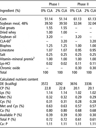
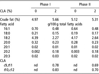
Piglets received diets formulated to meet or exceed NRC (1998) requirements and were given ad libitum access to feed, with free access to water, for 3 weeks in two phases (Phase I: day 17 to day 22; Phase II: day 23 to day 36; Table 1). Piglet diets were supplemented with either a commercial source of CLA (CLA-60, 60% methyl esters 1 : 1 ratio of c9,t11 and t10,c12, BASF Corporation, Mississauga, ON, Canada) or soybean oil (chosen so that diets would be balanced for linoleic acid; Table 2). Piglets’ feed intake and BW were recorded weekly.
Composition and calculated nutrients of piglet diets

1CLA-60 containing 28% c9-t11 and 28% t10-c12 isomers as guaranteed minimums.
2Provided per kilogram of complete diet: vitamin A, 8225 IU; vitamin D3, 1000 IU; vitamin E, 10.9 IU; vitamin B12, 0.115 mg; vitamin K, 1.1 mg; Niacin, 36.8 mg; Choline chlorine, 781.2 mg; Biotin, 0.25 mg, Folic acid, 0.75 mg, Mn (as MnO), 55 mg; Zn (as ZnO), 50 mg, Fe (as FeSO4·H2O), 80 mg, Cu (as CuO), 5 mg; Se (as NaSeO3), 0.1 mg, I (as Ca(IO3)2), 0.28 mg.
Fatty acid composition of piglet diets1

1Piglet diets separated based on inclusion of 0% or 2% CLA.
nd = not detected.
Bacterial culture, oral challenge and health status
After 11 ± 2 days on the ND, all piglets were given an oral challenge with a strain of E. coli expressing the K88+ (F4) fimbria obtained from the Animal Health Center, Veterinary Services Branch, Manitoba Agri-Food and Rural Initiatives (Winnipeg, MB, Canada). Cultures were grown overnight in Luria-Bertani (LB) broth (BD Bioscience, Mississauga, ON, Canada) at 37°C from stock cultures stored at −20°C. DNA was extracted and polymerase chain reaction (PCR) applied to confirm the expression of F4 fimbria and heat liable (LT) toxin. Briefly, DNA was extracted by centrifuging cell cultures at 3000 r.p.m. for 10 min (IEC CentraGP8, Needham, MA, USA) and discarding supernatant. Cells were then transferred to sterile microcentrifuge tubes (Fisher, Fairlawn, NJ, USA), washed with 400 μl of Tris EDTA buffer (TE), centrifuged and the supernatant discarded. Cells were re-suspended in 400 μl of TE buffer and heated at 95°C for 15 min to rupture the bacterial cell membrane. Into heated samples, 400 μl of phenol chloroform was added and vortexed thoroughly for 30 s. The mixture was cooled to −70°C for 15 min and then centrifuged at 13 000 r.p.m. (Microlite, Thermo IEC, Waltham, MA, USA). After centrifugation, 300 μl of supernatant was collected into clean 1.5 ml microcentrifuge tubes. Phenol chloroform treatment was repeated by adding an equal volume of phenol chloroform to the supernatant, vortexing and centrifugation as described above.
PCR was performed using a thermocycler (Techne genius, Duxford, Cambridge, UK) with the following 36-cycle program: 94°C for 1 min, 72°C for 1 min plus 5 min at 68°C for final elongation. The following K88+ primers designed in-house for this study: 5′-GCACATGCCTGGATGACTGGTG-3′ forward, 5′-CGTCCGCAGAAGTAACCCCACCT-3′ reverse. The primers used for LT were: 5′-CCGTGCTGACTCTAGACCCCCA-3′ forward, 5′-CCTGCTAATCTGTAACCATCCTCTGC-3′ reverse according to Kotlowski et al. (2006). Amplification products were then electrophoresed with a 2% agarose gel and viewed following UV exposure. Samples were considered positive when a distinctive band was produced that corresponded to a migration pattern consistent with a standard DNA fragment.
At 28 ± 2 days of age, each piglet was gavaged with 6 ml of cell suspension containing 109 cfu/ml of ETEC K88+ from a syringe attached to a polyethylene tube to ensure ingestion of inoculum. Scour severities were monitored at 8, 24, 48 and 56 h post challenge based on the methods of Marquardt et al. (Reference Marquardt, Jin, Kim, Fang, Frohlich and Baidoo1999). Briefly, at each period, scour severity scoring (0 = no scours, 1 = soft feces, 2 = mild diarrhea, 3 = severe diarrhea) was performed on each pen by trained personnel with no prior knowledge of the dietary treatments.
Digesta collection and microbial enumeration
At 36 ± 2 days of age, one piglet per pen, selected based on closest individual BW to the average treatment BW, was held under general anesthesia with isoflorane for blood and tissue collection. Blood was collected via jugular puncture into 10 ml vacutainers (BD Bioscience) for serum and plasma, the latter containing lithium heparin. A 5 cm segment of the terminal ileum was collected and immediately placed on ice for microbial enumeration. Piglets were then euthanized via an intra-cardiac injection of sodium pentobarbital (110 mg/kg BW). Spleen, duodenum, jejunum and ileum were removed, flushed with 0.9% saline and blotted dry with adsorbent paper before weighing and determining the length of the small intestine. Another 5 cm segment of the ileum was collected from each pig and fixed in saline buffered formalin (Fisher Scientific Canada, Ottawa, ON, Canada) for histological evaluation. Fresh samples of cecal digesta were collected and frozen at −80°C until analyzed for indices of gut health.
Mucosal adhered lactic acid bacteria (LAB) and total coliforms (TC) were enumerated from terminal ileum samples following a modified method of Krause et al. (Reference Krause, Easter, White and Mackie1995). Each sample was cut longitudinally and flushed with distilled water to remove non-adhered material, and then the epithelial mucosa and sub-mucosa were removed from the gut wall using a blunt sterile knife handle. Scrapings were weighed and 1 : 10 serial dilutions (10−2, 10−3, 10−4, 10−5 and 10−6) made with sterile peptone buffered water (BD Bioscience). LAB were cultured in duplicate on sterile De Man, Rogosa, Sharpe (MRS) agar and TC on sterile LB agar (BD Bioscience) by dispensing ten 10 μl-drops onto appropriate plates. TC plates were incubated for 24 h and LAB for 48 h at 37°C. Morphologically distinct colonies were counted on each plate and the highest dilution used for calculating cfu/g.
Chemical analyses
Total lipids in plasma were extracted using 2 : 1 chloroform : methanol, based on a procedure modified from Folch et al. (Reference Folch, Lees and Stanley1956). To each sample 100 μl of heptadecanoic acid (17:0; Sigma-Aldrich, Oakville, ON, Canada) was added to serve as an internal standard. Lipid extracts were methylated using 0.5 M methanolic acid and the reaction was carried out at 80°C for 1 h. Fatty acid methyl esters were determined using a Hewlett Packard (Hewlett Packard Canada, Mississauga, ON, Canada) HP 5890A gas chromatogram equipped with a flame ionization detector and separated on a HP88 100 m × 0.25 mm × 0.2 μm fused-silica column (Agilent Technologies Inc., Mississauga, ON, Canada). Oven temperatures were as follows: initial temperature 70°C for 1 min; raised to 180°C at 8°C/min; raised to 195°C at 1°C/min and held for 10 min; raised to 220°C at 1.2°C/min and held for 5 min; total run time 65.58 min. The injection temperature was 220°C and the detection temperature was at 290°C. Individual isomers were identified via comparisons to known standard retention times. Crude fat was determined via hexane extraction using a VELP SER 148 solvent extractor (VELP Scientifica, Plainview, NY, USA).
Plasma samples were analyzed for urea N using a Nova Stat profile M blood gas and electrolyte analyzer (Nova Biomedical Corporation, Waltham, MA, USA). Volatile fatty acids (VFA) and ammonia-N were extracted at room temperature by adding 50 ml of 0.1N HCl to 5 g of digesta followed by continuous overnight (~16 h) shaking (Newbrunswick Scientific, Edison, NJ, USA). After shaking, 10 ml of liquid was removed and frozen at −20°C until analyzed for ammonia-N. One ml of 25% metaphosphoric acid was added to remaining extracts, for VFA analysis, and mixed thoroughly prior to freezing.
VFA samples were prepared from cecal digesta using a modified method of Nyachoti et al. (Reference Nyachoti, Omogbenigun, Rademacher and Blank2006). Briefly, after thawing frozen extracts, 0.4 ml of NaOH and 0.64 ml of 0.3 M oxalic acid were added to each sample, mixed thoroughly and then centrifuged for 20 min at 3000 r.p.m. Approximately 2 ml of supernatant was then added to clean GC vials and analyzed using a Varian model 3400 gas chromatogram (Varian, Walnut Creek, CA, USA) equipped with a Carbopack B-DA 4% CARBOWAX 80/120 20 M column.
Cecal digesta samples were analyzed for ammonia-N using the method of Novozamsky et al. (Reference Novozamsky, Van Eck, Showenburg and Walinga1974). Briefly, 1.5 ml of reagent one, composed of 100 ml alkaline phenolate, 200 ml 0.05% sodium nitroprusside and 10 ml 4% Na2 EDTA plus 2.5 ml of reagent two, composed of 400 ml phosphate buffer and 100 ml 10% NaOCl were added to 50 μl of digesta extract and shielded from light to prevent UV interference. Following 30 min of incubation at room temperature, absorbencies were read at 630 nm and concentrations determined from a standard curve regression with a range of 2.5 to 20 mg/l.
Immunoglobulins A and G titer evaluation
Serum immunoglobulin (Ig) titers were measured using a quantitative commercial ELISA kit (E100-104, Bethyl Laboratories Inc., Montgomery, TX, USA). Both capture and detection antibodies were goat anti-pig IgA and IgG. Detection antibody was conjugated to horseradish peroxidase whose reaction with an alkaline phosphatase substrate formed a product after 5 min incubation at room temperature. Samples were read at 450 nm with a Bio-Rad 3550 Microplate Reader (Bio-Rad Laboratories, Hercules, CA, USA).
Histology
Histological samples were prepared as per the procedures of Owusu-Asiedu et al. (Reference Owusu-Asiedu, Nyachoti and Marquardt2003b). Briefly, 5 μm cross-sections of paraffin-embedded samples were stained with hematoxylin and eosin and mounted in duplicate. Villous height (VH) and crypt depth (CD) were measured on 10 well-oriented villi per sample where the crypt–villus junction was readily distinguishable using a Nikon YS100 compound light microscope equipped with a Sony DSP 3CCD color video camera. Similarly, mucosal thickness was calculated as the ratio of CD to crypt width as per the method of Hontecillas et al. (Reference Hontecillas, Wannemeulher, Zimmerman, Hutto, Wilson, Ahn and Bassaganya-Riera2002). Images were captured and processed using Northern Eclipse Image Processing Software v. 6.0 (Empix Imaging, Inc., Mississauga, ON, Canada).
Statistical analysis
Data were subjected to ANOVA as a completely randomized design using the GLM procedure of SAS (SAS Institute, Inc., Cary, NC, USA). For nursery piglet data, pen was considered the experimental unit for all measured response criteria. Due to the unbalanced nature of the design, data were reported as least square means. Treatment means were separated using Tukey’s test with P values >0.05 ⩽ 0.10 considered to be trends and P values <0.05 considered significant.
Results
Performance
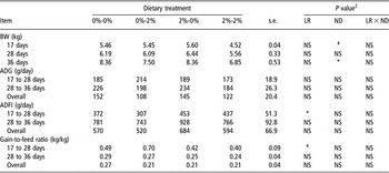
There were no significant differences in piglet BW during the nursing period (P > 0.10). At weaning there was a trend (P < 0.10) for piglets allocated to CLA-supplemented sows to have reduced BW, but by day 28, BW were similar across treatment groups (Table 3). On day 36 piglets receiving 2% CLA-supplemented ND were lighter (P < 0.05) than piglets receiving the non-supplemented diet. The main effects of sow LR, ND and their interaction had no effect on average daily gain (P > 0.05). However, piglets weaned from CLA-supplemented sows had higher (P < 0.05) average daily feed intake (ADFI) from day 17 to day 28 and tended (P < 0.10) to have reduced gain-to-feed ratios. ADFI and G : F over the entire experimental period were not affected either by the main effects of LR and ND or by their interaction (P > 0.05; Table 3).
Growth performance for piglets born to control or 2% CLA-supplemented sows fed nursery diets with or without CLA and challenged with E. coli K88+1

1Values are least square means ± pooled s.e.
2Main effects of lactation ration (LR), nursery diet (ND) or their interaction (LR × ND).
NS = not significant, ‡ = P < 0.10.
Scour scores
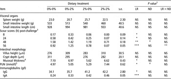
Scours were detected at 8 h post challenge in all treatment groups except the 2%-2% CLA group (Table 4). Scour severity reached a maximum level at 48 h post challenge with the 0%-0% group (control piglets) having the most severe and the 2%-2% group the least severe scours. Piglets weaned from CLA-supplemented sows had reduced (P < 0.05) scours at each post-challenge period compared with piglets weaned from control sows. At 48 h, piglets receiving 2% CLA-supplemented ND tended to have reduced scour severity (P < 0.10), a benefit further enhanced in 2%-2% piglets at 56 h as evidenced by the significant interaction (P < 0.01) between LR and ND.
Effect of CLA supplementation on indices of enteric health and immune stimulation post E. coli K88+ challenge1

1Values are least square means ± pooled s.e.
2Main effects of lactation ration (LR), nursery diet (ND) or their interaction (LR × ND); NS = not significant, ‡=P < 0.10; PUN = Plasma urea nitrogen.
3Scour scores: 0 = no scours, 1 = soft feces, 2 = mild diarrhea, 3 = severe diarrhea.
4Mucosal thickness: calculated as ratio of crypt depth to crypt width.
5Plasma urea nitrogen was measured 7 days post E. coli K88+ challenge.
Visceral organs, intestinal morphology, PUN and immunoglobulins
Spleen weight plus small intestine weight and length were not affected by dietary treatments (P > 0.05; Table 4). VH and CD were unaffected by dietary treatment (P > 0.05; Table 4). However, piglets weaned from CLA-supplemented sows had thinner (P < 0.05) gut mucosa compared with those weaned from sows fed non-supplemented diets.
Plasma urea N (PUN) levels were greater for piglets from CLA-supplemented sows than from control sows (P < 0.05; Table 4). After the E. coli K88+ challenge, serum IgA (P < 0.001) and IgG (P < 0.05) levels were greater in piglets weaned from CLA-supplemented sows compared to piglets weaned from control sows (Table 4).
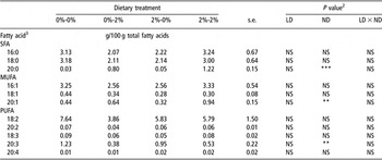
Fatty acids
Plasma saturated fatty acid, monounsaturated fatty acid and polyunsaturated fatty acid isomers remained relatively constant in all treatment groups (Table 5). However, piglets receiving 2% CLA-supplemented ND had greater circulating levels of 20:0 (P < 0.001) and 20:1 (P < 0.01) and lower concentrations of 20:3 isomers (P < 0.01) compared to piglets receiving control ND. In addition, piglets receiving 2% CLA-supplemented ND had greater (P < 0.001) concentrations of circulating c9,t11 and t10,c12 CLA isomers in comparison to piglets receiving control ND. Detectable levels of CLA isomers were only observed within the colostrum of sows receiving CLA-supplemented diets (n = 5, as samples could not be collected from three sows; data not shown) with an average level of 5.95 g c9,t11 and 5.28 g t10,c12 per 100 g of fatty acids, respectively.
Plasma fatty acid profiles post E. coli K88+ challenge for piglets born to control or 2% CLA-supplemented sows1

1nd = not detected.
2Main effects of lactation ration (LR), nursery diet (ND) or their interaction (LR × ND); NS = not significant.
3SFA = saturated fatty acids; MUFA = mono-unsaturated fatty acids; PUFA = poly-unsaturated fatty acids.
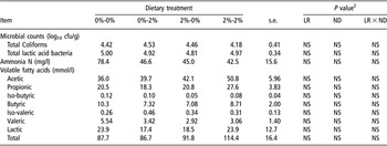
Microbial populations and fermentation products
TC and LAB populations were unaffected by dietary treatments (P > 0.05; Table 6). Ammonia-N levels were numerically greatest in 0%-0% piglets and were approximately 75.6% more than in the 0%-2%, 2%-0% and 2%-2% treatment groups, even though concentrations were not significantly different between dietary groups (P > 0.05). In general, VFA concentrations were unaffected by dietary treatment, although acetic and propionic acid concentrations were numerically higher in 2%-2% piglets than in those receiving other treatments.
Microbial enumeration and intestinal fermentation measurements in piglets born to control or CLA-supplemented sows post E. coli K88+ challenge1

1Values are least square mean ± pooled s.e.
2Main effects of lactation ration (LR), nursery diet (ND) or their interaction (LR × ND).
NS = not significant.
Discussion
Animal performance
Dietary supplementation of CLA in post-weaning diets did not affect piglet growth performance, results that are in agreement with Weber et al. (Reference Weber, Schinckel, Houseknecht and Richert2001) and Bontempo et al. (Reference Bontempo, Sciannimanico, Pastorelli, Rossi, Rosi and Corino2004). Furthermore, Bee (Reference Bee2000b) reported that piglets weaned from CLA-supplemented sows showed significantly increased weight gain, feed intake and final weight at day 70 of age. However, piglets in the study of Bee (Reference Bee2000b) were weaned at day 35 (v. day 17 in this study), presumably allowing piglets to ingest more CLA isomers prior to weaning, which may have accounted for their superior post-weaning growth performance.
Immune stimulation & indicators of gut health
Based on scour severity, piglets weaned from CLA-supplemented sows appeared to be healthier than piglets weaned from control sows during the first 56 h after oral ETEC challenge. However, it is important to note the inherent subjectivity of scour scoring. Here, although scores rarely exceeded a level of mild diarrhea, which suggests that all piglets remained relatively healthy, it is possible that piglets were sicker than the scour score indicated due to lethargy and inactivity noted during routine animal health observation performed in the period following infection.
Microbial populations within the upper gut of pigs are affected by many variables including age, diet and health status and a dynamic relationship exists between commensal and pathogenic micro-flora that are affected by these variables (Fairbrother et al., Reference Fairbrother, Nadeau and Gyles2005). In young pigs, proliferation of opportunistic pathogenic microbes may lead to the antigenic stimulation of intestinal tissue, resulting in inflammation and reduced availability of nutrients required for growth (Nyachoti et al., Reference Nyachoti, Omogbenigun, Rademacher and Blank2006). As previous studies have shown dietary CLA supplementation can lead to improved gut health in the face of an ETEC challenge, it was speculated that these improvements might be derived from an increase in Lactobacillus species populating the intestinal wall. The control and CLA-supplemented diets were formulated to contain the same protein and carbohydrate concentrations, which are known substrates for enteric microorganisms. If these constituents had been unequal across diets, it is possible that shifts in LAB or TC populations could have occurred. However, since these were kept consistent may explain why microbes populating the intestinal wall were unchanged. It should be noted that individual species within the total LAB population may have been altered but were undetected due to the assay used to measure LAB in the current experiment. Additionally, it should be keep in mind that long-chain fatty acids such as CLA are not favorable fermentation substrates for most microbes, and in some cases may even be bactericidal (Knapp and Melly, Reference Knapp and Melly1986). Nonetheless, a thorough microbial census was not performed and other micro-flora not detected may have been affected.
VFA results coincide with microbial results in that there were no significant differences observed among treatment groups. However, acetic acid and total VFA levels were numerically higher in piglets weaned from CLA-supplemented sows than those from non-supplemented sows. Also, the propionic acid level was numerically greatest in 2%-2% piglets compared to the other treatment groups. These differences may have been beneficial, in that if these piglets had reduced intestinal pH it would create an inhospitable environment for ETEC. Because both acetic and propionic acids have relatively high pKa values (4.76 and 4.88, respectively), their production could lower overall digesta pH (Partanen and Mroz, Reference Partanen and Mroz1999). However, this cannot be confirmed since digesta pH was not measured in this experiment.
Production of ammonia from amino acids (AA) and other nitrogenous compounds is a result of enteric microbial fermentation, and coliform bacteria such as E. coli have been shown to contribute to ammonia production within the upper gut of pigs (Dierick et al., Reference Dierick, Vervaeke, Decuypere and Henderickx1986). Elevated intestinal ammonia is thus a biomarker for the proliferation of potentially pathogenic bacteria. As mentioned previously, microbial enumeration performed in this study was limited and biased in favor of culturable bacteria. The fact that ammonia was elevated in 0%-0% piglets suggests the proliferation of pathogenic E. coli, which may explain the advanced incidence of scours and impaired enteric health in these treatment animals.
In addition to being used as a marker of protein and AA utilization, PUN has been used to indicate the extent of lean tissue catabolism, immune induction and acute phase protein synthesis in diseased piglets (Owusu-Asiedu et al., Reference Owusu-Asiedu, Nyachoti, Baidoo, Marquardt and Yang2003a). Although little work had been done establishing normal PUN values in the suckling piglet, elevated PUN levels could result from the catabolism of dietary AA not incorporated into muscle tissue. Because 2%-2% piglets had levels approximating normal (7.4 to 21.4 mmol/l; CCAC, 1993), an alternative explanation could be that PUN concentrations of piglets nursing control sows could be depressed, potentially as a result of reduced protein absorption.
Maternally derived colostrum and milk immunoglobulins contribute to intestinal development and serve as a primary means of disease resistance for the hypo-immune piglet (Le Dividich et al., Reference Le Dividich, Rooke and Herpin2005). These large macro-molecules, however, can only be absorbed for a brief period post partum after which absorption is no longer possible due to a shift in intestinal cell populations known as gut closure (Rooke and Bland, Reference Rooke and Bland2002). In the present study, dietary CLA had no effect on circulating immunoglobulin concentrations prior to weaning, results that do not agree with previous research showing that dietary CLA can significantly increase piglet IgG at days 2, 10 and 20 of age (Bontempo et al., Reference Bontempo, Sciannimanico, Pastorelli, Rossi, Rosi and Corino2004). The fact that immunoglobulins were not affected by dietary CLA in our study may have been due to the presence of primiparous sows, which are less efficient at mobilizing plasma fatty acids and immunoglobulins into colostrum and milk (Slevin and Wiseman, Reference Slevin and Wiseman2003) than the mostly multi-parity sows used by Bontempo et al. (Reference Bontempo, Sciannimanico, Pastorelli, Rossi, Rosi and Corino2004). Nevertheless, the sows used by Bontempo et al. (Reference Bontempo, Sciannimanico, Pastorelli, Rossi, Rosi and Corino2004) only received 0.5% CLA 8 days prior to farrowing v. 2% CLA for 30 days in the current study. This extended and elevated gestational supplementation may have contributed to a greater post-weaning carry-over immune stimulation, resulting in piglets weaned from CLA-supplemented sows having greater serum IgA and IgG concentrations at days 36 of age.
The duration for which dietary CLA must be supplied in order to stimulate immunity in nursery pigs is currently unknown. For example, 42 days of post-weaning CLA supplementation was required for lymphocyte proliferation to be affected in immune-challenged piglets (56 days of age; Bassaganya-Riera et al., Reference Bassaganya-Riera, Hontecillas-Magarzo, Bregendahl, Wannemuehler and Zimmerman2001). Whereas in immune-competent piglets suckling CLA-supplemented sows, 25 days (46 days of age) was required to increase serum IgG (Bontempo et al., Reference Bontempo, Sciannimanico, Pastorelli, Rossi, Rosi and Corino2004). The ability of young pigs to digest complex lipid nutrients such as CLA is limited during the first week post weaning, which may explain why immune stimulation is not observed in piglets less than 8 weeks of age. It is also possible that the methyl ester form of CLA included into the diets may have resulted in reduced absorption since free fatty acids are absorbed more readily than other linked forms such as triglycerides. However, our results appear to be in line with previous data indicating that immune stimulation can be accelerated when piglets are weaned from sows fed CLA-supplemented diets.
During enteric infections, secretion of IgA by intestinal lymphocytes serves as an important defense mechanism by reducing the ability of invading pathogens to attach to epithelial receptors and deactivating bacterial toxins (Mestecky et al., Reference Mestecky, Russell and Elson1999; Salmon, Reference Salmon1999). The fact that circulating IgA in pigs weaned from CLA-supplemented sows were significantly greater than controls could indicate a correlation with mucosal IgA concentrations (O’Shea et al., Reference O’Shea, Bassaganya-Riera and Mohede2004) and thus an indicator of stimulated secretory immunity in these piglets.
As E. coli K88+ colonizes the small intestine during infection, local inflammation is facilitated through the synthesis and secretion of LT (Fairbrother et al., Reference Fairbrother, Nadeau and Gyles2005). A consequence of this colonization and resulting inflammation is that local tissue damage manifest as erosions, villi atrophy and mucosal thickening. CLA has the ability to reduce inflammation through multiple means, such as the production of a less-active series of eicosaniods (Belury, Reference Belury2002). One outcome of E. coli K88+ infection is the activation of phospholipase-A by LT leading to the liberation of membrane-bound arachidonic acid and downstream synthesis of pro-inflammatory prostaglandin-E2 (PGE2; de Haan and Hirst, Reference de Haan and Hirst2004). Once CLA becomes incorporated into cellular membranes, it is metabolized in a similar fashion as linoleic acid, resulting in the formation of a conjugated isomer of arachidonic acid (Banni, Reference Banni2002). Enzymatic oxidation of this compound by cyclooxidase (COX) produces PGE4, a less-potent inflammatory agent (O’Shea et al., Reference O’Shea, Bassaganya-Riera and Mohede2004). Additionally, CLA has been shown to competitively inhibit COX activity, resulting in reduced PGE2 production and less inflammatory response initiation (Bulgarella et al., Reference Bulgarella, Patton and Bull2001). Although CLA was not detected in plasma of 2%-0% piglets, this does not preclude residual CLA remaining within the gut tissue, which accumulated during nursing. Lack of mucosal thickening and reduced scours observed in 2%-0% and 2%-2% piglets suggests synthesis of less-active prostaglandins within the intestinal tissue, which may have contributed to reduced local inflammation and superior epithelial integrity.
CLA is a known ligand for peroxisome proliferator-activated receptor gamma (PPAR-γ; O’Shea et al., Reference O’Shea, Bassaganya-Riera and Mohede2004) whose activation has been associated with reduced intestinal inflammation in pigs (Hontecillas et al., Reference Hontecillas, Wannemeulher, Zimmerman, Hutto, Wilson, Ahn and Bassaganya-Riera2002; Bassaganya-Riera and Hontecillas, Reference Bassaganya-Riera and Hontecillas2006). Although not measured in this study, PPAR-γ activation may have been responsible for reduced mucosa thickening observed in 2%-0% and 2%-2% piglets. It is also possible that PPAR-γ activation along with the reduced production of proinflammatory eicosanoids contributed to the superior enteric health observed in 2%-0% and 2%-2% piglets. However, future studies will need to be conducted to confirm this mode of action.
Conclusion
Supplementation of sow gestation and lactation diets with CLA appears to be a practical strategy to improve the enteric health and immune status of nursery piglets. The results of the present study indicate that piglets weaned from CLA-supplemented sows had superior intestinal health and immune status markers as indicated by reduced intestinal mucosal inflammation and elevated serum IgG and IgA. The fact that ND supplementation with CLA did not improve intestinal health markers may have been related to piglet age or feeding duration. In light of these beneficial findings, further studies are required to determine the specific routes by which dietary CLA supplementation is facilitating these favorable effects.
Acknowledgments
Support for this project was received from Manitoba Agri-Food Research and Development Initiative (ARDI) and Manitoba Pork Council. Special thanks to G. H. Crow for assistance with statistical analysis and the Glenlea Swine Research Unit and T. K. Cheung Centre for Animal Science Research staff members for animal care.