Introduction to biofabrication
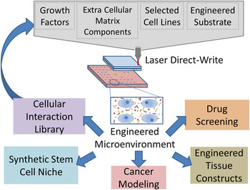
The ability to control the structure through placement of smaller individual sub-units (processing) allows for materials to be engineered with selected properties. Although, conventionally, this premise has been applied to inorganic materials, wherein the structure is built up through homogeneous layers with discrete interfaces, within the past 20 years, a quantum jump has been made into engineering biological materials in the new field of biofabrication.1 As the behavior and properties of mammalian cells are defined by their surrounding microenvironment, biofabrication focuses primarily on the creation of idealized constructs—defined as the reproducible assembly of a biomaterial structure—in vitro that mimic the in vivo environment/design. Idealized constructs and engineered cell cultures will allow a better understanding of cellular communication mechanics, thereby advancing stem cell, cancer, and tissue engineering researchReference Mironov, Trusk, Kasyanov, Little, Swaja and Markwald2–Reference Prestwich4 (Figure 1). Many of today’s state-of-the-art biofabrication techniques still utilize conventional biomaterials; biodegradable poly-L-lactic acid (PLLA)Reference Inui, Kokubu, Makino, Nagura, Toyokawa, Sakata, Kotera, Nishino, Fujioka and Kurosaka5 is widely used in tissue engineering scaffolds, and gelatin, derived from denatured collagen, is employed extensively for timed drug delivery.Reference Young, Wong, Tabata and Mikos6 Novel processing of these biomaterials using techniques such as inkjet printing,Reference Roth, Xu, Das, Gregory, Hickman and Boland7–Reference Cui, Dean, Ruggeri and Boland9 dip-pen nanolithography,Reference Huang, Patlolla, Abilez, Sharma, Rajadas, Beygui, Zarins and Cooke10, Reference Salaita, Wang and Mirkin11 vacuum deposition, and laser direct-writing allow for the creation of precise geometries over large areas. Such techniques may even be extended to create three-dimensional scaffoldsReference Nirmalanandhan, Juncosa-Melvin, Shearn, Boivin, Galloway, Gooch, Bradica and Butler12 to more closely mimic the in vivo environment.
Concept diagram of laser direct-write techniques. ECM, extracellular matrix.

Idealized construct fabrication can benefit tremendously from incorporation of pluripotent stem cells into engineered environments. Given their unlimited growth potential and undifferentiated state, stem cells are versatile and robust tools that are the focus of much of the current research in tissue engineering and regenerative medicine.Reference Keller13, Reference Watt and Hogan14 Maintenance of stem cell pluripotency (i.e., the ability to differentiate into any cell type) is dependent on the stem cell niche, or local microenvironment, which consists of extracellular matrix, neighboring cells (both stem and differentiated cells), and soluble factors (i.e., hormones, proteins, nutrients).Reference Scadden15, Reference Morrison and Spradling16 Thus, when utilizing stem cells to create idealized constructs, it is critical to culture the cells in distinct, controlled microenvironments due to the profound influence of intercellular communication on stem cell differentiation.Reference Tang, Peng and Ding17–Reference Liu and Chen19 As such, new fabrication techniques are needed that can manipulate the arrangement of stem cells and matrix within their local microenvironment with high speed, precision, and reproducibility.
One such technique to write biomaterials is inkjet printing, which is noted for its fast deposition rate and suitable resolution (10–70 μm) that has been adapted from its original purpose of printing patterns of text and figures. The cartridge of a standard inkjet printer can be sterilized and filled with either a cell suspension or biopolymer solution, such as adherence proteins or growth factors. Depositing these solutions allows for customizable patterns of live cells to be printed onto glass coverslipsReference Cui, Dean, Ruggeri and Boland9 or scaffolds such as Dermamatrix.Reference Cooper, Miller, Decesare, Usas, Lensie, Bykowski, Huard, Weiss, Losee and Campbell8 However, even using a 20-μm nozzle to deposit a cell suspension in microdroplets, inkjet printing is found to be incapable of controlling cell placement.Reference Miller, Li, Kanade, Weiss, Walker and Campbell20 The nozzle outlet diameter dramatically limits the amount of control a user has over the distance between deposited cells, and the use of a suspension of cells results in an uncontrolled number of cells being deposited per drop. Although single cells have been deposited,Reference Liberski, Delaney and Schubert21 the number of cells deposited per drop is based on statistical probability and cannot be repeated a priori. In order to study cell-cell interactions, reproducible placement of cells is necessary, and novel methods to process biomaterials are needed.
Reproducible placement can be achieved in the high-resolution movement found with atomic force microscopy (AFM), where control is obtained within fractions of a nanometer to measure nanometer-scale surface topographies. This high-resolution technology has been exploited in the dip-pen nanolithography (DPN) technique. DPN utilizes an AFM tip as a dip-pen such that material can be placed with a resolution better than 50 nm,Reference Salaita, Wang and Mirkin11 allowing for single cell attachment sites to be formed. By depositing multiple biomaterials within microns of one another, complex patterns are createdReference Huang, Patlolla, Abilez, Sharma, Rajadas, Beygui, Zarins and Cooke10 of preferentially adherent cells. Although DPN provides extensive control, samples are limited to small areas due to the experimental setup.
Although DPN and inkjet printing have their individual advantages, neither fulfill the two requirements for large-scale biofabrication: speed and precision. Although DPN provides nanoscale resolution, the time required to produce even small samples is significant. Conversely, inkjet printing, while adequate in speed, lacks the precision necessary to produce reliable detailed patterns. Laser direct-write techniques have been shown to print electronic and organic materials with high precision at speeds comparable to ink-jet techniques, making it attractive for biological printing. This article summarizes the advances laser-based processing has made in biofabrication, starting with large-area deposition of bio-organic materials and continues with laser direct writing.
Laser deposition of bio-organic films over large areas
For many large-scale applications (e.g., sensors, catalysts, or anti-fouling coatings), the soft inorganic, organic, polymer, or biomaterial thin films must maintain its structural and chemical integrity, while also being smooth, thin, dense, adherent, and of accurately and precisely predictable thickness. When these performance requirements became critical, a large-area laser-based vapor deposition technique, matrix-assisted pulsed-laser evaporation (MAPLE), was developed to deposit thin and uniform polymer films without notable decomposition. The patented process,Reference McGill and Chrisey22 developed in the late 1990s at the Naval Research Laboratory in Washington, DC, can generate high-quality polymeric, organic, and biomaterial films on many types of substrates;Reference Chrisey, Pique, McGill, Horwitz, Ringeisen, Bubb and Wu23–Reference Kokkinaki and Georgiou33 however, it cannot deposit viable cells due to the deposition configuration.
Contrasting most traditional polymer or organic thin-film fabrication techniques, MAPLE simultaneously deposits contamination-free films with monolayer thickness control in user-defined patterns. Moreover, MAPLE requires minimal amounts of material and provides enhanced film adhesion to the substrateReference Gyorgy, Perez del Pino, Sauthier and Figueras34, Reference Smausz, Megyeri, Kekesi, Vass, Gyorgy, Sima, Mihailescu and Hopp35 without damage to the substrate’s structure or chemistry and biological functionality.Reference Ringeisen, Callahan, Wu, Pique, Spargo, McGill, Bucaro, Kim, Bubb and Chrisey36 MAPLE is capable of depositing a wide variety of materials onto an equally diverse set of substrates in user-defined patterns.Reference Cristescu, Mihaiescu, Socol, Stamatin, Mihailescu and Chrisey37–Reference Frycek, Jelinek, Kocourek, Fitl, Vranata, Myslik and Vrobova52
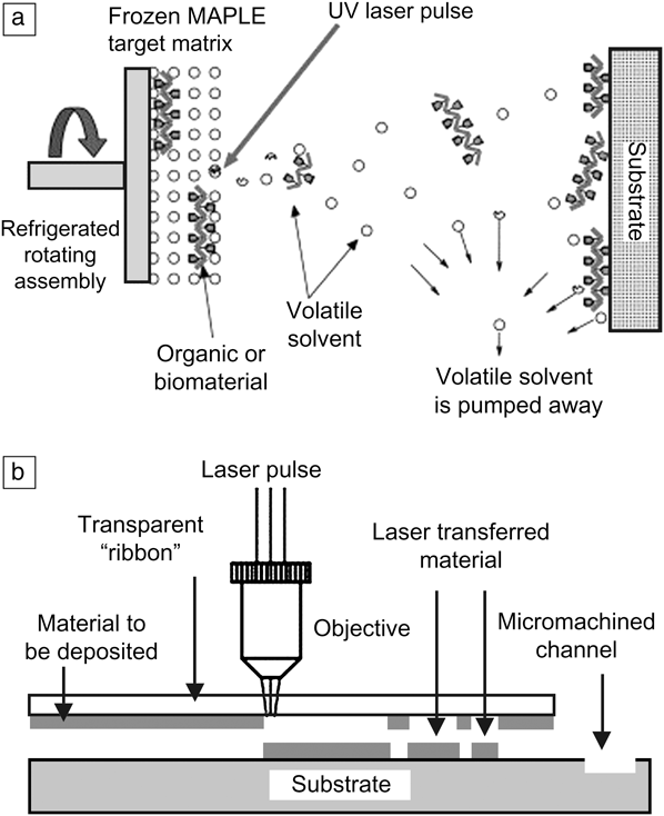
The MAPLE system is similar to pulsed laser deposition, employing a pulsed laser beam, vacuum chamber, pump system, MAPLE target mounted on a refrigerated target holder, and a substrate (Figure 2a).Reference Piqué, Wu, Ringeisen, Bubb, Melinger, McGill and Chrisey53 MAPLE uses a cryogenic composite dilute mixture of the material to be deposited with a light absorbent, high vapor-pressure solvent matrix as the target. Constant rotation of the target during the evaporation process avoids hole formation caused by localized heating. Ideally, the incident laser pulse used for MAPLE initiates two photo-thermal processes in the matrix: evaporating the frozen composite target and releasing the material of interest into the chamber. Because of the low concentration (1–5 wt%) in the composite target, the simultaneous action of the evaporation gently desorbs the delicate material. The photon energy absorbed by the solvent is converted to thermal energy that vaporizes the solvent and heats the soft material. As the surface solvent molecules are evaporated into the gas phase, molecules of the material to be deposited are exposed at the gas-target matrix interface. The molecules attain sufficient kinetic energy to be transferred in the gas phase through the collective collisions with the evaporating solvent molecules. The volatile solvent molecules, which have very low sticking coefficients, are pumped away from the deposition chamber. With careful optimization of the MAPLE deposition conditions (laser wavelength, laser fluence [energy per unit area], repetition rate, solvent type, material concentration, target temperature, background gas, vacuum chamber pressure), this process can occur without any significant degradation of the deposited material.Reference Bubb, Papantonakis, Toftmann, Horwitz, McGill, Chrisey and Haglund54–Reference Toftmann, Rodrigo, Schou and Pedrys64
Schematic of (a) the matrix-assisted pulsed laser evaporation system and (b) the matrix-assisted pulsed laser evaporation direct-write.Reference Wu, Ringeisen, Krizman, Frondoza, Pique, Spargo, McGill and Chrisey27

The MAPLE process continues layer-by-layer, depleting the target of solvent and material of interest in the same concentration as the starting matrix. So far, the substrate temperature has been shown to be an important parameter during deposition, largely affecting the morphology and other film characteristics.Reference Rodrigo, Czuba, Toftmann, Schou and Pedrys65–Reference Rotaru67 When a substrate is placed directly in the plume path, a uniform thin film, whose material properties such as chemical structure and functionality have been maintained, starts to form from the evaporated complex material molecules.
MAPLE is capable of depositing biopolymer films onto a receiving substrate without loss of chemical integrity or biological functionality; however, it does not allow for precise patterning of discrete molecular subunits in a user-generated pattern. Also, the manufacturing setup (e.g. vacuum chamber and cryogenic target) is a hostile environment that would kill live cells that are to be deposited. In order to achieve deposition of healthy, living cells, the additive deposition process of laser direct-write must be employed.
Laser direct-writing for biofabrication
Laser direct-writing combines optical imaging, speed, and high precision attributes that are essential for fabrication of larger, robust tissue constructs by utilizing a laser to induce micro-bubble formation in a cell suspension. The resulting micro-bubble collapse deposits the cell suspension onto a receiving substrate. In comparison to other laser direct-writing methods (e.g., laser-induced forward transfer [LIFT]),Reference Schiele, Corr, Huang, Raof, Xie and Chrisey68 MAPLE direct-write (MAPLE DW) utilizes a sacrificial biopolymer instead of a dynamic release layer (e.g., a thin-film metal) to interact with the laser.Reference Fogarassy, Fuchs, Kerherve, Hauchecorne and Perriere69–Reference Koch72 MAPLE DW can achieve a spatial resolution of less than 10 μm, allowing individual cells to be deposited.Reference Barron, Krizman and Ringeisen73 By accurately patterning single cells, or groups of cells, a controllable distance apart, intercellular communication methods, which regulate differentiation, can be experimentally explored to be better understood (Figure 1). Furthermore, in contrast to more traditional patterning techniques such as micro-contact printing of adhesion proteins, where cells are limited to specific cell-adhesion islands, MAPLE DW allows patterned cells to migrate and proliferate uninhibited outside of the initial pattern, creating dynamic signaling that better mimics in vivo cellular behavior and cell-cell interactions.
Engineered constructs fabricated using MAPLE DW capture the advantages of conventional scaffolds, as they may be engineered to have specific topographies, mechanical stiffness, and chemical properties that create a customizable cellular niche to direct the differentiation of stem cells. The spatial resolution gives the necessary control to create custom complex patterns of cells in culture. The speed at which samples can be prepared and deposited is significantly greater than that of DPN techniques, making MAPLE DW an appropriate method for large-scale, high-throughput construct fabrication and for studying cellular interaction.
MAPLE DW systems were first used in the rapid prototyping of electronics to deposit metals for thick and thin-film conductors and semiconductors achieving resolutions of 2 μm.Reference Fogarassy, Fuchs, Kerherve, Hauchecorne and Perriere69, Reference Chrisey, Pique, Modi, Wu, Auyeung and Wu74–Reference Fitz-Gerald, Chrisey, Pique, Auyeung, Mohdi, Young, Wu, Lakeou and Chung76 The proven reliability and precision of MAPLE DW has driven its application for the deposition and patterning of biomaterials and living cells.Reference Fernandez-Pradas, Colina, Serra, Dominguez and Morenza70, Reference Barron, Ringeisen, Kim, Spargo and Chrisey77–Reference Ringeisen, Kim, Barron, Krizman, Chrisey, Jackman, Auyeung and Spargo82 MAPLE DW is of particular interest for modification for application to biomaterial and soft matter depositions.
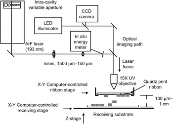
In order to use this process for live cells, several modifications must be made involving the environment during deposition. Using cryogenic targets, low pressure vacuum, and heated substrates required in traditional MAPLE would prove to be fatal to living cells. To deposit cells into user-generated patterns, a laser-transparent quartz disk (print ribbon) is coated with a sacrificial biopolymer, on which cells are then embedded. The MAPLE DW system uses a low-powered UV pulsed laser to deposit live cells from the print ribbon to a parallel receiving substrate a short (200–7000 μmReference Patz, Doraiswamy, Narayan, He, Zhong, Bellamkonda, Modi and Chrisey83) distance away (Figure 2b). The short duration (~8 ns) laser pulse volatilizes the biopolymer layer, ejecting the cells and depositing them onto the target receiving substrate or long-term growth surface. A typical MAPLE DW setup features multiple apertures, focusing objective, a charge coupled device (CCD) camera, XY control of the print ribbon, and XYZ control of the receiving substrate (Figure 3). The CCD camera and optical path co-focused with the laser on the print ribbon stage allows for the visualization and targeting of specified cells when coupled with XY print ribbon control.Reference Schiele, Chrisey and Corr84 To increase repeatability and speed of deposition, the typical MAPLE DW system is also coupled with computer-aided design/computer-aided manufacturing (CAD/CAM) control of the receiving substrate to generate user-specified, automated patterns.
Schematic of the matrix-assisted pulsed laser evaporation direct-write system adapted for biomaterial deposition.

For viable cell deposition from the print ribbon, the receiving substrate must provide a moist environment that promotes cell adhesion and long-term growth. In early work using MAPLE DW, the receiving substrate and ribbon were both coated with the biopolymer hydrogel Matrigel (56% laminin, 31% Collagen IV, 8% Entactin)85 that provided the proper conditions for growth and cushioned the impact upon deposition.Reference Lin, Huang, Huang, Tzeng and Chrisey79, Reference Patz, Doraiswamy, Narayan, He, Zhong, Bellamkonda, Modi and Chrisey83, Reference Schiele, Koppes, Corr, Ellison, Thompson, Ligon, Lippert and Chrisey86 The numerous parameters of the system, including laser beam diameter, substrate and print ribbon biopolymer choice, and laser fluence, allow for precise control of cell placement, as well as the flexibility to fine-tune the system for different cell lines to maintain optimal cell viability following deposition.
Mechanisms behind MAPLE DW
The deposition of cells via MAPLE DW can be broken into three stages: droplet formation, travel, and landing. During travel, the droplet is in free-fall, and it can be assumed that the cells contained within are not experiencing acceleration and, therefore, are not in danger of damage. During droplet formation and landing, however, the cell is exposed to considerable acceleration and stress and must be examined to understand how cell viability may be affected.
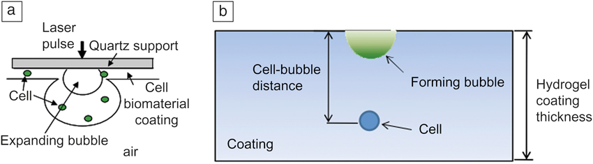
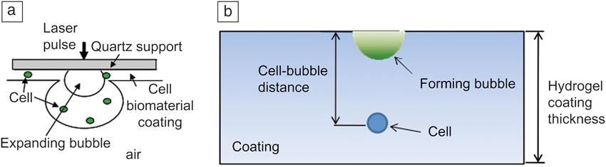
Droplet formation relies on induced bubble collapse in order to create a shockwave that will project a droplet off of the print ribbon. As shown in Figure 4, when the laser pulse interacts with the quartz ribbon and sacrificial biopolymer interface, the biopolymer is vaporized, forming a bubble that expands through the coating thickness. The pressure of the rapidly expanding bubble causes the ejection of a cell suspension droplet to the receiving substrate. Significant pressure can develop, generating a stress wave as the bubble is confined in rigid media. The cell membrane may be damaged when exposed to the resulting stress waves during bubble expansion, creating membrane permeability that allows an undesirable influx of medium or permanent rupture.Reference Lee and Doukas87 Modeling the velocity, acceleration, and stress profiles on the cell when the droplet is ejected from the print ribbon allows the user to refine system parameters (laser fluence, substrate choice) to obtain optimal cell viability.Reference Wang, Li and Huang88 The ejection velocity, which can reach up to 1000 m•s−1,Reference Young, Auyeung, Pique, Chrisey, Dlott, Young and Pique89 sets the initial velocity of impact during cell landing, contributing to the stresses on the cell and therefore the viability of cells following deposition.
(a) Schematic of bubble formation during transfer. (b) Simplified illustration used for physical modeling of bubble formation.Reference Wang, Li and Huang88
Finally, the collision of the droplet onto a hydrogel-coated receiving surface also imparts significant stresses onto the cell. Two impacts occur during the landing process: the droplet collision with the substrate and the cell collision with the substrate.Reference Wang, Huang, Grujicic and Chrisey90 By examining the stresses of several points in the cell droplet via modeling, it has been seen that the droplet significantly reduces the amount of stresses that a cell, assumed to be at the center of the droplet, experiences: 0.2 MPa on the cell versus 1 MPa on the bottom of the droplet.Reference Wang, Huang and Chrisey91 Modeling simulations can be utilized to determine a set, or sets, of system parameters that would yield cell deposition with the lowest droplet stress to achieve the highest cell viability for the unique characteristics of a given application (e.g., cell type(s), spot sizes, substrate viscoelastic properties).
Parameters for preventing cell damage
Given the large stresses and accelerations laser deposited cells may experience, the largest challenge in MAPLE DW is to preserve cell viability and to maintain unaltered growth and function following deposition. Given a growth environment that maintains moisture and provides a surface for cell adhesion, maintaining cell viability then depends on controlling the initial pressure from the bubble and ejection velocity, which determines the stresses cells experience upon landing. The material properties and thickness of the biopolymers chosen for the print ribbon and receiving substrate significantly affect cell viability.
Material selection
The optimal biopolymer for the print ribbon will allow release of cells from the quartz ribbon with a single laser pulse, while still providing a surface for cells to adhere. Matrigel has been used extensively to coat both ribbons and receiving substrates, as it allows cells to easily adhere. However, several complications are introduced with the utilization of Matrigel, including the introduction of various growth factors and extracellular matrix (ECM) components that may not be required for a particular cell type or application. Because Matrigel is derived from murine tumors, lot-to-lot variation may impart uncontrolled and undesired stimuli on the cells. Such stimuli (either growth factor or ECM components) may have severe consequences, such as the unintended induction of stem cell differentiation.Reference Vukicevic, Kleinman, Luyten, Roberts, Roche and Reddi92 Also, because of the ECM content in Matrigel, cells readily adhere to Matigel-coated print ribbons via specific cell-binding proteins; these adhesions may be disrupted during deposition, causing unnecessary damage to the cells. In order to reduce the chance of cell damage, during propulsion from Matrigel, dynamic release layers such as triazene have been employed at the ribbon-biopolymer interface to reduce the laser energy needed and thereby possible damage from the pressure-induced stress wave.Reference Doraiswamy, Narayan, Lippert, Urech, Wokaun, Nagel, Hopp, Dinescu, Modi, Auyeung and Chrisey93
As an alternative to Matrigel, gelatin has recently been shown to be an effective sacrificial biopolymer for MAPLE DW. Unlike Matrigel, gelatin contains no extraneous growth factors, mitigating a potential source of unintended differentiation. Furthermore, the thermal properties of gelatin are exploited to adhere cells on the print ribbon. By temporarily warming the gelatin in an incubator, the cells can become partially encapsulated within the gelatin layer prior to forming cell adhesions. This partial encapsulation of cells improves the protection of the cells from generated stress waves during deposition and avoids the adhesion rupture that may be seen with cells adhered to ribbons coated with Matrigel. By increasing the thickness of the biopolymer to above 40 μm, cell viability approached 100% due to distancing the cell suspensions from the bubble-induced stress wave.Reference Ringeisen, Kim, Barron, Krizman, Chrisey, Jackman, Auyeung and Spargo82 Because hydrogels exhibit fluid-like behavior, it has been found that as viscosity increases, the peak magnitude of the stress wave will increase as well.Reference Wang, Li and Huang88 Therefore, by selecting less viscous biopolymers, the stress wave generated in the print ribbon during deposition can be reduced to non-critical levels.
At the other end of the cell deposition process, the receiving substrate must act as a cushion to reduce the acceleration-induced stresses on the cell during landing to maintain moisture during transfer and to provide the proper growth environment post-transfer. The viscosity of the hydrogel is taken into account in the modeling of impact, and therefore the user can control the resulting impact stresses on the cell by tailoring the hydrogel viscosity. Increasing the thickness of the hydrogel coating on the receiving substrate diminishes the resulting stresses significantly.Reference Wang, Huang, Grujicic and Chrisey90 However, visualization and imaging post-transfer can become an issue if the receiving substrate layer is too thick or opaque. Removal of the hydrogel layer on the growth surface may be necessary, but by doing so, potential harm to the deposited layer or disruption of the intended pattern may occur. Recently, gelatin has been shown to melt away during the time it takes for deposited cells to adhere, leaving only the base substrate and cells without negatively influencing the registry or integrity of the cellular pattern.Reference Schiele, Chrisey and Corr84 This property allows removal of dissolved gelatin by changing the culture media, which is minimally invasive to deposited cells.
Laser parameters
Although mechanical damage is the largest concern with deposition of live cells through laser processing, it is also necessary to maintain biological functionality and avoid molecular alterations (e.g., DNA damage, protein cross-linking, heat-induced stress) when biofabricating tissue constructs. The use of a UV laser exposes the cells to radiation, which may cause damage to genetic material. The majority of the energy from the laser, however, is absorbed or attenuated by the biopolymer layer on the ribbon. This greatly diminishes the chance for direct exposure of cells to UV radiation.Reference Lin, Huang, Huang, Tzeng and Chrisey79 The level of energy transmitted to the cells is sufficiently low, such that DNA damage due to radiation can be considered negligible.Reference Koch72, Reference Ringeisen, Kim, Barron, Krizman, Chrisey, Jackman, Auyeung and Spargo82, Reference Schiele, Chrisey and Corr84
Although high energies can be detrimental to cell viability, varying the laser energy can be used for deposition into three-dimensional scaffolds. By adjusting the energy of the laser pulse and the viscosity of the receiving substrate, the penetration depth of cells deposited into the scaffold can be controlled.Reference Patz, Doraiswamy, Narayan, He, Zhong, Bellamkonda, Modi and Chrisey83 If the receiving scaffold is assumed to behave as a Newtonian fluid, Stokes’ law can be used to determine the resisting force a deposited cell will experience during deposition. Patz et al. used Matrigel as a receiving substrate, as it remained to act as a scaffold during incubation.Reference Patz, Doraiswamy, Narayan, He, Zhong, Bellamkonda, Modi and Chrisey83 However, as previously discussed, extraneous growth factors, which would promote undesired differentiation, prevent it from being used to study stem cell behavior. As gelatin would be ineffective due to its low melting temperature, other biomaterials for a passive three-dimensional scaffold must still be investigated.
Case studies
The successful deposition of living mammalian cells (e.g., human dermal fibroblasts, rat neural stem cells, mouse embryonic stem cells) via MAPLE DW methods has been demonstrated previously.Reference Schiele, Corr, Huang, Raof, Xie and Chrisey68, Reference Barron, Ringeisen, Kim, Spargo and Chrisey77, Reference Ringeisen, Kim, Barron, Krizman, Chrisey, Jackman, Auyeung and Spargo82, Reference Schiele, Koppes, Corr, Ellison, Thompson, Ligon, Lippert and Chrisey86, Reference Doraiswamy, Narayan, Lippert, Urech, Wokaun, Nagel, Hopp, Dinescu, Modi, Auyeung and Chrisey93, Reference Ringeisen, Kim, Young, Spargo, Auyeung, Pique, Chrisey and Wu94 Recently, advances in gelatin-based MAPLE DW have been explored to gain better control of the receiving substrate microenvironment while maintaining high cell viability. This technology has been extended for use with embryonic stem cells.
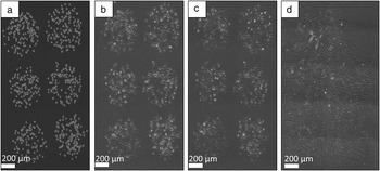
Schiele et al. demonstrated the practicality of using gelatin on both the print ribbon and receiving substrate.Reference Schiele, Chrisey and Corr84 Human dermal fibroblasts, cells isolated from skin tissue, were deposited from a gelatin-coated quartz print ribbon onto a gelatin-coated receiving substrate. Because gelatin melts at 37ºC, allowing cells to be more easily washed away during the addition of media, it was important to ensure that the pattern registry was maintained within the first hour of incubation at 37ºC following transfer. Using a custom MATLAB program, the cells were tracked for the first hour after transfer, finding that the average displacement was only 5.6 ± 2.5 μm, well within the typical diameter of a trypsinized (unattached) cell (~10 μm). This ability to specifically pattern cells into an in vitro engineered environment that allows unhindered cell proliferation and migration is a significant advancement in MAPLE DW. Human dermal fibroblasts patterned onto a gelatin-coated receiving dish were shown to grow into a monolayer over the first 24 hours post-transfer (Figure 5). The growth to a confluent monolayer from an initial pattern on a well-controlled substrate demonstrates the utility of gelatin-based MAPLE DW for tissue engineering applications and biofabrication.
Phase microscopy images of human dermal fibroblasts (a) 0 min, (b) 30 min, (c) 45 min, and (d) 24 hr post-transfer.Reference Schiele, Koppes, Corr, Ellison, Thompson, Ligon, Lippert and Chrisey86

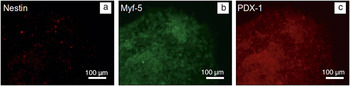
When using MAPLE DW to create patterns of stem cells, maintenance of pluripotency, the ability to differentiate into any adult cell type, is essential in addition to high viability. Stem cell pluripotency must be maintained following transfer to allow for directed differentiation later in culture. Again, despite its previous applications,Reference Ringeisen, Kim, Barron, Krizman, Chrisey, Jackman, Auyeung and Spargo82, Reference Patz, Doraiswamy, Narayan, He, Zhong, Bellamkonda, Modi and Chrisey83, Reference Schiele, Koppes, Corr, Ellison, Thompson, Ligon, Lippert and Chrisey86, Reference Doraiswamy, Narayan, Lippert, Urech, Wokaun, Nagel, Hopp, Dinescu, Modi, Auyeung and Chrisey93 the extraneous growth factors present in Matrigel greatly limits, or precludes, its use with stem cells due to the risk of uncontrolled or undesired differentiation. Raof et al. applied gelatin-based MAPLE DW for the deposition of mouse embryonic stem (mES) cells, demonstrating spatially precise depositions of viable stem cells, with maintained pluripotency.Reference Raof, Schiele, Xie, Chrisey and Corr81 Three hours following deposition, the mES cells maintained the expression of the Oct4 (a canonical marker for undifferentiated embryonic stem cells). After one week of incubation following deposition, cells were stained for markers representing the three germ layers: ectoderm (e.g., epidermal tissue, nervous system tissue), mesoderm (e.g., muscle, fat, bone, cartilage, tendon, blood), and endoderm (e.g., lungs, stomach lining, tissue of the gastrointestinal tract). The presence of all three biomarkers demonstrates that deposited cells can differentiate into cells of all three germ layers, thereby establishing the long-term maintenance of pluripotency following deposition (Figure 6). With this nearly unlimited differentiation potential, and in combination with engineered microenvironments, stem cell fate may be more easily directed through patterns designed to better mimic in vivo stem cell niches.
Mouse embryonic stem cells one week after deposition onto a gelatin receiving substrate show differentiation potential by biomarker expression into (a) ectoderm, (b) mesoderm, and (c) endoderm. Biomarker expression demonstrating long-term maintenance of pluripotency.

Summary
The ability to precisely control (±5 μm) the placement of single, selected cells to form an idealized construct is necessary for the further investigation into cellular communication phenomena, stem cell differentiation, and tissue engineering (Figure 1). Laser direct-write processes have been shown to provide such precision in the electronics industry, and their application to printing live mammalian cells has proven successful, with consistently >90% cell viability. Cell tracking has shown that following deposition, the pattern registry is maintained, confirming the precision of matrix-assisted pulsed laser evaporation direct-write (MAPLE DW). By controlling the receiving substrate coating, idealized microenvironments may be created for the deposition of multiple cell types. Altering the substrate properties may allow for improved control of stem cell differentiation and provide a more complete understanding of the mechanisms that drive differentiation and the associated contributing factors. The short time for preparing a sample and depositing cells, combined with the high precision and computer-aided design/computer-aided manufacturing control of deposition, makes MAPLE DW an efficient means to study cellular interactions. Further advances involving printing in three-dimensions can lead to a greater understanding of intercellular communication and factors that influence stem cell fate decisions so that in vitro idealized constructs may be created to better mimic in vivo conditions for applications in tissue engineering and regenerative medicine.