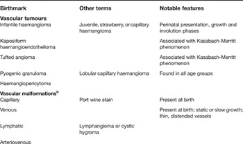
Infantile haemangiomas are vascular tumours that exhibit a generally predictable life cycle consisting of rapid growth in the first months of life followed by a variable period of involution spanning months to years. Occasionally, these tumours can cause significant cosmetic deformity or even induce life-threatening complications. There is no uniformly safe and effective treatment option for these problematic tumours. A better understanding of the pathophysiology of these lesions, and vascular anomalies in general, is needed in order to develop appropriate therapies. Progress in this field has been hampered by lack of a clear and accurate nomenclature to distinguish between different types of vascular lesions as well as lack of an animal model that faithfully mimics the human condition. Fundamental information such as incidence, commonly estimated at 10%, is based on older studies that may have included lesions other than infantile haemangiomas. In 1982, Mulliken and Glowacki proposed a classification scheme that has been generally accepted and also promoted the use of common terminology in this field (Ref. Reference Mulliken and Glowacki1). The International Society for the Study of Vascular Anomalies (http://www.issva.org) has adopted this nomenclature and has raised awareness within the medical community about the important distinctions between various lesions, as well as advancing our understanding of the clinical and biological characteristics of vascular anomalies (Table 1). Infantile haemangiomas possess interesting and complex characteristics that hold relevance to the fields of vascular biology, stem cell biology, oncology, immunology, dermatology and others.
Classification and features of vascular birthmarksa

a The table is adapted from Ref. Reference Hand and Frieden108 [Hand, J.L. and Frieden, I.J. (2002) Vascular birthmarks of infancy: resolving nosologic confusion. Am J Med Genet 108 (4), 257-264] (Copyright © 2002 Wiley-Liss, Inc.), with permission of Wiley-Liss, Inc., a subsidiary of John Wiley & Sons, Inc.
b Well-defined combined malformations: capillary–lymphatic–venous (so-called CLVM – includes most cases of Klippel Trenaunay); capillary–venous (includes many milder cases of Klippel Trenaunay); capillary–venous with arteriovenous shunting and/or fistulae (Parkes–Weber syndrome); cutis marmorata telangiectatica congenita; lymphatic–venous.
Clinical characteristics
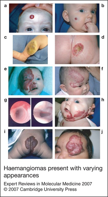
Infantile haemangiomas are at least three times more likely to develop in females than in males, and premature infants are at significantly higher risk (Refs Reference Finn, Glowacki and Mulliken2, Reference Amir3). Those of Caucasian heritage appear more likely to develop haemangiomas, as are infants whose mothers suffered from placental abnormalities during gestation or delivery (Ref. Reference Haggstrom4). Haemangiomas vary both in age of onset and clinical characteristics (Fig. 1). The classic presentation is that of a completely normal examination at birth with subsequent development of a red spot on the head or neck during the first few weeks to months of life. However, a small fraction of haemangiomas are ‘fully formed’ at birth. These congenital lesions have been shown to differ from infantile haemangioma and thus likely represent a distinct vascular tumour. Infantile haemangiomas may initially exhibit a ‘precursor lesion’, which appears as a blanched area of pallor, sometimes with superimposed telangectases. A bright red patch subsequently develops within this area, followed by rapid growth of a papular or nodular mass over the next weeks to months.
Haemangiomas present with varying appearances. (a) Superficial focal haemangioma; (b) combined haemangioma. These types of lesions may leave a parchment-like scar but will not cause significant deformity. (c) Congenital haemangioma. This lesion involuted during the first year of life. (d) Ulcerated haemangioma. This location is associated with significant pain and risk of infection. (e) Large ulcerated facial haemangioma obstructing the visual axis. This case is a medical emergency necessitating intervention. (f) Segmental haemangioma associated with PHACES (‘posterior fossa cranial anomalies, haemangioma, arterial and cardiac defects, eye and sternal anomalies’). (g) Subglottic haemangioma associated with (h) ‘beard-like’ haemangioma distribution. (i) Lumbosacral haemangioma associated with tethered cord. (j) Large haemangioma obstructing vision and displacing eyelid and orbit; this requires immediate intervention. Images were obtained from patients being treated at The Children's Hospital and Health Center (San Diego, CA, USA) and were reproduced with consent.

The clinical significance of haemangiomas varies greatly, and is often linked to their location, size and type (Ref. Reference Haggstrom5). Although they can present anywhere on the body, they have a predilection for the head and neck regions, where they have the potential to obstruct the visual axis or interfere with breathing and eating. Genital lesions are at higher risk for erosion and pain, as are any lesions that develop in an area where friction may occur. Lesions located in the lumbosacral area may be a marker for spinal dysraphism, and haemangiomas that involve the ‘beard’ area (neck, chin, pre-auricular area) may indicate the presence of airway haemangiomas (Ref. Reference Orlow, Isakoff and Blei6). In each of these circumstances, appropriate evaluations to rule out such possibilities should be performed.
Haemangiomas are generally categorised based on certain morphological characteristics, and antiquated terms such as ‘strawberry’ and ‘cavernous’ have been supplanted by more accurate terms such as ‘superficial’, ‘deep’ or ‘mixed’. Superficial lesions usually consist of red lobulated papules, plaques or nodules that appear on the surface of the skin. Deep lesions are usually less defined, located beneath the skin in subcutaneous fat, and may possess a blue tint. A ‘combined’ lesion will possess both deep and superficial qualities. Others have further subdivided lesions as localised, segmental, indeterminate and multifocal (Ref. Reference Chiller, Passaro and Frieden7). Segmental haemangiomas are generally larger, plaque-like lesions (Refs Reference Waner8, Reference Haggstrom9), and are more likely to be associated with underlying syndromes such as PHACES (Refs Reference Frieden, Reese and Cohen10, Reference Metry11), a constellation of findings including posterior fossa cranial anomalies, haemangioma, arterial and cardiac defects, eye and sternal anomalies. Segmental morphology is also more likely to portend complications and need for active intervention and therapy.
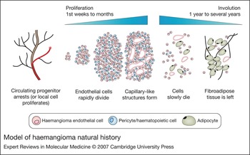
The majority of haemangiomas progress through a sequential life cycle that begins with a rapid proliferative phase lasting approximately 6 months, followed by continued slower growth during the first year of life (Fig. 2). The proliferative phase is followed by what appears clinically to be a more quiescent or ‘plateau’ phase where cellular proliferation and apoptosis offset each other. When cell death outpaces proliferation, this marks the transition to a longer period of involution that can last months to years: 10% of haemangiomas have completely regressed at 1 year of age, 50% by 5 years of age, 70% by 7 years, and so on. Following involution, some patients are left with an area of redundant cutaneous and/or fibrofatty tissue, while others manifest a central area of thin, parchment-like tissue, sometimes with superimposed telangectasia.
Model of haemangioma natural history. Recent evidence suggests that haemangiomas originate from the clonal expansion of a progenitor cell. The progenitor cells may be of placental origin or blood-borne endothelial progenitor cells. Alternatively, local cells under the influence of unusual conditions may serve as progenitors. The haemangioma progenitor cell may embolise or extravasate and begin dividing under appropriate conditions. The growing tumours comprise haemangioma endothelial cells (red) as well as nonendothelial cells, including haematopoietic cells and pericytes (blue). The early lesion is largely a featureless mass of cells, but sometime later in the proliferative phase, lumenised capillary-like structures are organised. The involution phase is characterised by increased apoptosis and the regression of both the endothelial and nonendothelial compartments. After involution, adipocytes may be found at the site along with a fibrous matrix.

Histopathological evaluation of haemangioma tissue reveals findings dependent on the phase of the haemangioma life cycle (Ref. Reference North12). Early proliferating lesions show compact areas of uniform-appearing endothelial cells arranged in cords and nodules. Endothelial-cell-lined lumena, if present, are subtle in early lesions, but become more apparent in lesions at later points in development, often displaying thickened, multilaminated basement membranes. In older, involuting lesions the lumena become narrower and the presence of fibrofatty tissue is often observed.
The treatment of haemangiomas is predicated on the degree of functional compromise, either present or predicted. ‘Watchful waiting’ is appropriate in the majority of cases, when the size, type and location of the lesion indicate that functional impairment or significant cosmetic concerns are unlikely to occur. Ulcerated lesions may require topical antimicrobials and antifungal agents, barrier agents such as zinc oxide formulations, pain relief, and laser treatment if the aforementioned therapy fails to lead to improvement. Oral corticosteroids are the standard of care for lesions that cause functional impairment or significant cosmetic deformity (Ref. Reference Bennett13). Topical and intralesional corticosteroids are occasionally useful, while interferon and vincristine therapy are reserved for refractory, life-threatening lesions. Recently, becaplermin gel [recombinant human platelet-derived growth factor (PDGF)] (Ref. Reference Metz14) and imiquimod (Refs Reference Martinez15, Reference Welsh16, Reference Hazen17) (see later) have been used to treat haemangiomas.
Aetiology: angiogenesis, vasculogenesis or neither?
Infantile haemangiomas appear, in some respects, to be the product of disorganised and rapid growth of blood vessels. In fact, there are a number of similarities between normal blood vessels and the structures found in these benign infantile tumours. Haemangiomas are primarily composed of endothelial cells possessing components of a basement membrane, such as type IV collagen, and associated pericytes (Ref. Reference Tan18). During proliferation, both normal endothelial cells and haemangioma endothelial cells express angiogenesis-related receptors E-selectin (Ref. Reference Kraling19) and integrins αvβ3 and α5β1 (Ref. Reference Ritter20). Factors previously associated with normal angiogenesis have also been reported to be expressed in haemangiomas, such as basic fibroblast growth factor (bFGF) (Refs Reference Tan18, Reference Bielenberg21, Reference Takahashi22). Earlier studies reported expression of vascular endothelial growth factor (VEGF) in proliferating haemangiomas (Refs Reference Takahashi22, Reference Chang23), but more recent investigations show that VEGF levels are relatively low (Refs Reference Ritter20, Reference Yu24), suggesting a limited role for VEGF in haemangiomas.
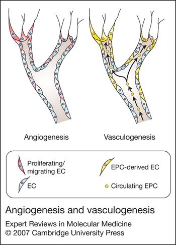
Normal blood vessels typically arise from one of two processes: angiogenesis or vasculogenesis (Fig. 3). Angiogenesis is defined as the growth of new blood vessels via extension and remodelling from existing vasculature (Ref. Reference Carmeliet25), while vasculogenesis involves the development of vessels that differentiate from precursor cells in situ. While numerous studies have attributed haemangioma pathogenesis to angiogenesis, more recent investigations suggest that vasculogenesis may have an important role in haemangioma development.
Angiogenesis and vasculogenesis. Normal blood vessels grow by one or a combination of these mechanisms. During angiogenesis, existing vessels branch and are extended through the action of local endothelial cells that proliferate and migrate in response to angiogenic factors. Vasculogenesis, in contrast, relies on progenitors, either circulating or in situ, as a source of new endothelium. EC, endothelial cell; EPC, endothelial progenitor cell.

The first endothelial structures to form in the embryo are the endocardium and great vessels (Ref. Reference Drake and Fleming26); both are examples of vasculogenesis. The cells of these structures originate from mesoderm-derived haemangioblasts, which are believed to function as progenitors to both haematopoietic and endothelial cells (Ref. Reference Choi27). This is relevant to understanding haemangioma pathophysiology since endothelial cells from haemangiomas coexpress haematopoietic and endothelial cell markers (see section ‘Progenitor cells in haemangioma’). Angiogenesis is active later in development and occurs in the normal adult under certain conditions such as in the endometrium during the female reproductive cycle and during wound healing. Angiogenesis plays an important role in many pathological conditions, including solid-tumour growth, arthritis and diseases of the eye (Ref. Reference Ferrara and Kerbel28). While some aspects of angiogenesis and/or vasculogenesis are likely operational in the growth of haemangiomas, the growth of these tumours differs in several important ways from typical angiogenesis or vasculogenesis.
Haemangioma pathogenesis: assembling the clues
Intrinsic defect hypothesis
Although no universal consensus exists, a number of hypotheses regarding haemangioma aetiology have emerged and some have gained significant support (Refs Reference Friedlander, Ritter and Friedlander29, Reference Bauland30). One hypothesis predicts that the basic defect in haemangiomas is a genetic mutation of the endothelial cell, or its progenitor. Linkage studies have identified six families that appear to have a predisposing genetic mutation and high incidence of haemangioma and/or vascular anomalies (Ref. Reference Blei31). In these families, haemangiomas segregated as an autosomal dominant trait and a subsequent study established a linkage to chromosome 5q31-33 (Ref. Reference Walter32), which harbours the genes for FGF receptor 4 (FGFR-4), PDGF receptor beta (PDGF-Rβ), and FMS-related tyrosine kinase 4 (FLT-4). These genes have known roles in regulating angiogenesis, but it remains to be determined if abnormalities in these genes can contribute to haemangioma pathogenesis. These genetic studies, while important, do not address the common sporadic form, leaving the causative defect in the majority of haemangiomas a mystery.
While previous investigations have presumed that haemangioma growth is the result of uncontrolled, disorganised angiogenesis, other studies support the hypothesis that haemangiomas result from a somatic mutation in a single endothelial (or progenitor) cell (Ref. Reference Bischoff33). This hypothesis predicts that haemangiomas should be clonal in nature and suggests that mechanisms other that angiogenesis may be involved. These studies used the X-linked human androgen receptor gene assay (HUMARA) (Ref. Reference Allen34), which exploits the fact that, in females, cells generally inactivate one copy of the X chromosome (to maintain appropriate gene dosing) in a random fashion. When a tissue or sample of isolated cells shows a bias towards inactivation of one allele, this is an indication that these cells may have been derived from a single parent cell. The first study used haemangioma endothelial cells isolated from seven heterozygous female patients. Cells grown in culture from all seven patients showed a skewing toward a single allele (Ref. Reference Boye35). A subsequent study analysed intact tissue with no selection of cells and similarly found some degree of allelic loss in 12 of 14 samples (Ref. Reference Walter36). Thus, two independent investigations suggest that haemangiomas may arise as a consequence of clonal expansion from a single cell. It remains possible, however, that sampling constraints biased these results. Since it is usually only possible and/or practical to sample a portion of the lesion, it is conceivable that clonality accounts for only portions of the lesion. When considering segmental haemangiomas, it would seem unlikely that this variety could arise from a single cell since the plaque-like nature of these lesions would require large numbers of cells spread over the region to expand simultaneously. Despite these limitations, the evidence of clonality in haemangiomas significantly impacts how we think of these lesions and may help focus future investigative efforts. These and other studies clearly suggest that haemangioma growth does not come about by typical angiogenesis.
Also predicted from the intrinsic defect hypothesis is that cells harbouring a mutation affecting their growth properties should behave abnormally when isolated from the tumour milieu. In one report, it was shown that haemangioma cells had higher expression of the angiopoietin 1 receptor TIE2 and increased response to angiopoietin 1, while angiopoietin 2 mRNA was downregulated in response to serum in endothelial cells from haemangiomas but not in normal endothelial cells (Ref. Reference Yu24). In another report it was demonstrated that haemangioma endothelial cells differ from normal endothelial cells in their proliferation rates and migration in vitro (Ref. Reference Boye35). Haemangioma cells proliferated two to three times faster and manifested enhanced migration in response to VEGF compared with normal dermal microvascular endothelial cells. In addition, treatment with the angiostatic collagen XVIII fragment endostatin increased migration of haemangioma endothelial cells rather than inhibiting migration as seen with normal cells.
A subsequent study compared endothelial progenitor cells (EPCs) isolated from haemangiomas to similar cells purified from normal cord blood (Ref. Reference Khan37). (See section ‘Progenitor cells in haemangioma’ for more on EPCs.) Haemangioma EPCs and cord blood EPCs, like haemangioma endothelial cells, displayed increased adhesion, migration and proliferation in response to endostatin, a protein that typically inhibits these processes in normal adult endothelial cells. Furthermore, it was shown that these two types of cells shared a common profile when 113 cell adhesion genes were analysed. Interestingly, a previous report of differential effects of endostatin suggested that differentiation status may determine the response: migration of mature umbilical-vein-derived endothelial cells was inhibited by endostatin, whereas migration of endothelial cells derived from embryonic stem cells was unaffected (Ref. Reference Schmidt, Addicks and Bloch38). The fact that haemangioma endothelial cells have altered responses to this factor might reflect the immature nature of these cells rather than the presence of a mutation or intrinsic defect. Could other differences between haemangioma endothelial cells and normal endothelial cells, such as unusual surface marker expression profiles (Ref. Reference Li39), be attributed to the fact that they are not fully differentiated or mature cells? Further comparison of haemangioma endothelial cells to immature progenitor-derived endothelial cells should clarify this issue.
The placenta connection
Multiple lines of evidence point to an involvement of the placenta in haemangioma pathogenesis. An increase in haemangioma incidence has been associated with transcervical chorionic villus sampling (CVS), but not with amniocentesis (Refs Reference Kaplan40, Reference Burton41), although a more recent study found no association with CVS (Ref. Reference Haggstrom4). It has been reasoned that the trauma of CVS could increase the number of placental cells released into the circulation and thus increase the likelihood that these cells might embolise to fetal vascular sites. Similarly, placental abnormalities, such as placenta praevia and preeclampsia, might also lead to higher risk of shed cells and haemangioma formation. Infants born prematurely have a higher risk of developing haemangiomas, and the incidence increases with lower birth weight (Refs Reference Amir3, Reference Powell42). Prematurity can be associated with placental complications as well, strengthening the link to the placenta.
Additional support for the idea of placental origin was provided in a report demonstrating that placental vasculature and haemangioma endothelium share expression of several surface markers (Refs Reference North43, Reference North, Waner and Brodsky44). It was shown that both tissues expressed merosin, Lewis Y, FcγRII (CD32) and glucose transporter 1 (GLUT-1, also expressed on endothelium of blood–brain barrier and red blood cells). None of these markers was found in other vascular anomalies or on nonhaemangioma tumour vasculature. Another study assessed whether haemangiomas were derived from trophoblasts, the invasive progenitor cells that mediate implantation into the uterine wall. Twelve haemangioma specimens were uniformly negative for a panel of trophoblast markers including human placental lactogen, placental alkaline phosphatase and cytokeratins 7, 8 and 17 (Ref. Reference Bree45). While these data might suggest that there is no relationship between placental and haemangioma tissue, the markers used in this study label undifferentiated trophoblasts, leaving open the possibility that haemangiomas may, in fact, be derived from these cells, but have differentiated and down-regulated expression of the markers studied.
More recent support for a placental connection was provided by the results of a study using large-scale genomic analysis (Ref. Reference Barnes46). This experimental approach uses DNA microarrays to measure mRNA levels of nearly the entire human genome, representing a powerful tool for highly detailed comparisons between different tissue types. In this study, intact tissues from placenta, haemangioma, normal skin, lung and brain, pulmonary tumours and a sclerodermal specimen were compared, accounting for genes expressed in endothelial cells as well as numerous nonendothelial cells within these samples. Each of the tissues used for comparison had at least one feature in common with haemangioma and/or placenta. For example, the lung tumours used have high levels of cell proliferation and are highly vascular. Clustering analysis showed that placenta and haemangioma were as similar as normal lung and lung tumours, raising the interesting possibility of viewing the placenta as ‘tumour’ and haemangioma as ‘metastasis’. When comparisons were restricted to genes with known expression in endothelial cells, the similarities were even higher; haemangiomas were more similar to placental samples than placental samples were to each other. These observations regarding similarities between haemangiomas and placenta are provocative and have generated new ideas about the origin of haemangiomas.
Nonrandom distribution of haemangiomas on the face
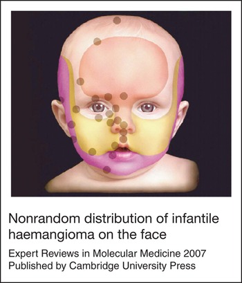
Clinical observations have contributed to our understanding of haemangiomas, as exemplified in a study that provided data supporting the long-held belief that sites of haemangioma occurrence on the face are not random (Ref. Reference Waner8). The authors first described two distinct types of haemangiomas: the more common focal, tumour-like type; and the less common segmental, or plaque-like, type. They found that focal haemangiomas had a nonrandom distribution, and identified 22 sites of occurrence on the face. Interestingly, these sites can be linked to boundaries between the different developmental units of the face, suggesting a rationale for haemangioma distribution. Haemangiomas were found near lines of fusion between mesenchymal growth centres (‘placodes’), or between placodes and facial ectoderm (Fig. 4). Perhaps these boundary areas are more likely to sustain haemangioma growth due to the presence of higher secreted levels of particular growth factors or a more favourable extracellular matrix composition. A similar study of segmental haemangiomas demonstrated that while lesions on the lower face conformed to locations previously described as developmental units, those found on the upper face did not (Ref. Reference Haggstrom9). Thus, when formulating a hypothesis regarding the origin of haemangioma, one must take into account that these lesions are often not randomly distributed, but in fact may be tied to early developmental events that determine where, and perhaps how, they are formed.
Nonrandom distribution of infantile haemangioma on the face. Focal sites of occurrence superimposed on the embryological map of facial placodes demonstrating the predilection for haemangioma formation at boundaries between developmental units. Reproduced, with permission, from Ref. Reference Waner8 [Waner, M. et al. (2003) The nonrandom distribution of facial hemangiomas. Arch Dermatol 139, 869-875] (Copyright © 2003 American Medical Association. All Rights reserved.).

Progenitor cells in haemangioma
Recent evidence suggests that several different types of progenitor cells may have roles in haemangioma progression. Normal bone marrow and peripheral blood contain progenitor cells that can differentiate into endothelial cells (Refs Reference Asahara47, Reference Yoder48). New vessel growth in the adult was long thought to depend on sprouting from existing vessels (i.e. angiogenesis), but more recent findings support a role for circulating bone-marrow-derived EPCs in this process. This concept was applied to haemangiomas where an increase of circulating EPCs was identified in patients with these lesions (Ref. Reference Kleinman49). The precise identity of EPCs has been controversial. Most agree that EPCs cannot be identified by expression of a single marker, but studies have shown that cells expressing both CD34 and AC133 (CD133) can differentiate into endothelial cells in vitro (Ref. Reference Peichev50). Analysis of peripheral blood samples from five haemangioma patients averaging just over 2 years of age revealed a 15-fold average increase in circulating CD34+CD133+ cells, versus five control subjects with an average age of 38 months.
A second study used samples from four haemangioma specimens to show that proliferating lesions contain mRNA for CD133, while involuting and involuted haemangiomas, haemangioma-derived endothelial cells, normal skin and normal endothelial cells did not contain CD133 mRNA (Ref. Reference Yu51). Combining antibodies against CD133 and CD34 to identify EPCs using flow cytometry, it was shown that 11 of 12 proliferating haemangiomas contained cells positive for both markers. Positive samples contained 0.1–2.9% CD133+CD34+ cells with one lesion showing a remarkable 21.4% double-positive cells. Although no EPCs were detected in control samples from venous or lymphatic malformations, the lack of cells coexpressing these markers in one of the haemangioma specimens studied suggests that EPCs may not be important in all haemangiomas. If EPCs could be shown to contribute directly to formation of haemangioma tissue, this would have significance for our understanding of the aetiology of these tumours.
Following haemangioma involution, the tissue at the site of the lesion is often composed of fibroadipose tissue, and a new study indicates that progenitor cells may have a role in the generation of this tissue. Mesenchymal stem cells were observed in haemangiomas more frequently than in normal skin, and their numbers were highest during the proliferative phase (Ref. Reference Yu52). The cells recovered from haemangiomas were found to have many of the characteristics of mesenchymal stem cells from other tissues, including surface marker expression and multilineage differentiation potential, and it was suggested that these cells may contribute to the generation of adipocytes during haemangioma involution. This study is among the first to shed light on the nature of the residuum left behind after haemangiomas regress.
The involvement of a haematopoietically derived endothelial progenitor would seem consistent with the finding that haemangioma endothelial cells express haematopoietic markers (Ref. Reference Ritter53). Specifically, haemangioma cells were found to express a subset of myeloid-specific markers, which leads to the speculation that perhaps this cell type could serve as a haemangioma progenitor. Supporting this idea, several reports have demonstrated that peripheral myeloid cells (monocytes) are capable of differentiating into cells with endothelial characteristics (Refs Reference Fernandez Pujol54, Reference Havemann, Pujol and Adamkiewicz55, Reference Bailey56). Alternatively, coexpression of haematopoietic markers on haemangioma cells could be an indication of a common progenitor between haematopoietic cells and endothelium, such as the haemangioblast. This concept has been discussed above, and may be an indication of the immature nature of the endothelial cells in haemangiomas, as suggested in a recent report showing that haemangioma endothelial cells express lymphatic endothelial hyaluronan receptor 1 (LYVE-1), a marker usually associated with the endothelium of lymphatic vessels (Ref. Reference Dadras57). This, combined with the finding that haemangioma cells also express endothelial markers CD31 and CD34, but not another lymphatic-specific protein, Prox-1, suggest that endothelial cells in haemangiomas are arrested in an early state of differentiation, and display features of both blood vascular and lymphatic endothelium. A follow-up study, however, reported that haemangioma endothelial cells were negative for LYVE-1, showing that LYVE-1 expression is restricted to perivascular cells expressing human leukocyte antigen DR (HLA-DR) with characteristics of dendritic cells (Ref. Reference Nguyen58). Such conflicting reports that rely solely on immunohistochemistry leave this issue open and raise the need for further investigation.
If not serving as precursor cells, the myeloid component of haemangiomas may still play a role in either stimulating initial proliferation or subsequent involution of haemangiomas. As discussed below, hypoxia has been hypothesised to have a role in haemangioma pathophysiology (Refs Reference Ritter53, Reference Hamlat59, Reference Kleinman, Blei and Gurtner60), and it is well established that myeloid cells such as macrophages can profoundly promote the growth of new vessels under low oxygen conditions.
A role for hypoxia in haemangioma development?
While the vast majority of infantile haemangiomas remain insignificant clinically, a fraction of these lesions demonstrate rapid growth in the first months of life. This sometimes alarming rate of growth could represent an exuberant response to one of the mechanisms driving cellular proliferation. Several pieces of evidence, at the molecular, cellular and clinical levels, suggest a possible role for hypoxia in haemangioma pathogenesis.
Hypoxia, or low levels of oxygen, is sensed by cells, which then respond by modifying gene expression. These modifications can enhance the ability of the cell to survive low oxygen conditions as well as induce expression of factors that promote the growth of new blood vessels. These new blood vessels then ultimately increase perfusion and oxygenation of the tissue. Hypoxia is a component of many disease states, but also has an important role in normal development and physiology (Ref. Reference Semenza61).
Hypoxia-inducible factor 1α (HIF-1α) is a transcription factor that serves as a major regulator of the cellular response to hypoxia. HIF-1α is involved in several cellular processes, including regulation of differentiation and myeloid cell function (Ref. Reference Cejudo-Martin and Johnson62), regulation of genes involved in angiogenesis, erythropoiesis, energy metabolism, inflammation, and cell survival decisions (Ref. Reference Semenza61). With respect to its role in angiogenesis, HIF-1α has been shown to regulate the expression of numerous factors including VEGF-A, angiopoietin 2, placental growth factor and PDGFB (Ref. Reference Manalo63). It is interesting to note that GLUT-1 and insulin-like growth factor 2 (IGF-2), two proteins shown to be present in haemangioma at relatively high levels, are both under the control of HIF-1α and induced by hypoxia (Refs Reference Feldser64, Reference Loike65), but VEGF is not found at high levels. The mechanisms regulating the high expression of IGF-2 were investigated recently in a study addressing the idea that a disruption in the epigenetic regulation of IGF-2 might be responsible for its high expression (Ref. Reference Yu66). IGF-2 expression is under the control of imprinting, a mechanism of silencing the maternal copy of a gene under normal conditions to maintain appropriate expression levels (Ref. Reference Burns, Jackson and Hassan67). A loss of imprinting can result in overexpression and contribute to pathogenesis, as has been shown in several tumours including Wilms tumour where IGF-2 is overexpressed (Ref. Reference Ogawa68). In haemangiomas, a normal pattern of imprinting was found in all 15 informative cases, indicating that a loss of imprinting is not involved and leaving open the possibility that an abnormal response to hypoxia could explain the high levels of IGF-2 in these lesions. Alternatively, high IGF-2 levels suggest another parallel to the placenta, where IGF-2 is also expressed (Ref. Reference Constancia69).
Some clinical features of haemangiomas suggest that reduced blood flow could be an important trigger in the growth of these lesions (Ref. Reference Hamlat59). During delivery, the infant is likely to experience some degree of hypoxia as a consequence of the birthing process. Systemic hypoxia could alter the levels of circulating factors and trigger or promote growth of cells overly sensitive to high levels of these factors. It is also possible that microenvironmental changes could promote the observed cellular proliferation, alone or in combination with systemic factors. Prior to the rapid growth that characterises haemangiomas, a pale or blanched area of skin may be observed that is subsequently found to define the borders of the lesion that will develop. Although the nature of this phenomenon is not known, it suggests that a local interruption or reduction of blood flow has occurred to the area. This event could represent the growth stimulus for normal or mutated cells residing in the area or for ectopic cells that have embolised to this site from other tissues such as the placenta. Alternatively, this hypoxia could signal circulating progenitor cells to deposit in the area and begin to proliferate, as has been shown in other systems (Ref. Reference Ceradini70). The cause of this putative interruption in blood flow would then become important to identify. Perhaps microanatomy exists on the boundaries between developmental units where vascular coverage is incomplete. Such a site could serve as a relatively hypoxic environment where circulating progenitor cells localise and proliferate.
Hypoxia has also been associated with tumours during pregnancy. Chorangioma, a proliferative lesion of the placenta, has some features in common with haemangioma. Both are primarily made up of endothelial cells (Ref. Reference Ogino and Redline71), and hypoxia has been reported to have an important role in the pathogenesis of chorangioma (Ref. Reference Reshetnikova72). Another association between these two lesions is the fact that pregnancies complicated by the presence of chorangiomas have a higher incidence of infants that develop haemangiomatosis (Ref. Reference Bakaris73), a condition characterised by multiple congenital haemangiomas, most often limited to the skin (Ref. Reference Metry and Hebert74). Could hypoxia, or a systemic defect in oxygen sensing or response, be at the root of both haemangiomas and chorangiomas?
What is causing involution?
Despite an increase in information relating to the biology of haemangiomas, and new evidence-based hypotheses on their origin, the precise cellular and molecular mechanisms underlying haemangioma pathogenesis remain unclear. Perhaps even more enigmatic than the aetiology of the tumours are the events that stimulate and govern the regression of these lesions. Apoptosis appears to be a major mechanism of cell death in the regressing tumour (Refs Reference Iwata75, Reference Mancini and Smoller76, Reference Razon77), but the events that trigger the apoptotic programme remain obscure. Two general routes to haemangioma involution have been proposed. The first might be termed ‘passive involution’ where inherent properties of the haemangioma endothelial cells place limits on their proliferative lifespan. It is also possible that the microenvironment supporting haemangioma growth/maintenance exists for a limited duration, perhaps being tied to neonatal development. The placenta has a clearly defined life cycle, and is designed to function solely for the period of gestation. Perhaps haemangioma cells also share this programmed lifespan (Refs Reference Halperin78, Reference Smith79). Another form of passive involution could involve the loss of a critical factor that supports proliferation and/or survival. IGF-2 is highly expressed in proliferating haemangiomas, but not in other vascular anomalies and could represent such a factor (Refs Reference Ritter20, Reference Yu66). mRNA levels of this factor decrease dramatically as haemangiomas begin to involute, suggesting a role for IGF-2 in haemangioma growth. However, IGF-2 levels appear to decrease as endothelial content decreases, raising the possibility that the observed decrease is related to the loss of the cellular source and may not, in fact, be critical for driving growth (Ref. Reference Yu66).
The second way of thinking about haemangioma involution involves the action of other cells, and may be thought of as ‘active involution’. The immune system would be a likely candidate to elicit a reaction leading to involution. Haemangioma cells, possessing abnormal surface marker expression, could be targeted by the immune system, which can be exquisitely sensitive to abnormal cells. Large numbers of CD8+ T cells have been found within haemangioma tissue (Ref. Reference Ritter80). This subtype of T cell is cytotoxic and capable of inducing apoptosis in target cells. There was no strong correlation, however, with the phase of the haemangioma lesion and the number of CD8+ T cells, which might be observed if these cells were, in fact, responsible for mediating involution. This study also described a possible role for indoleamine 2,3-dioxygenase (IDO) in haemangioma and showed, using large-scale expression analysis, that IDO was highly expressed in proliferating haemangiomas (Ref. Reference Ritter80), while a second study similarly described a possible role for IDO (Ref. Reference North81). This enzyme has an important role in preventing maternal rejection of the placenta through inhibition of T-cell function (Ref. Reference Mellor82). IDO catalyses the degradation of the amino acid tryptophan (Ref. Reference Takikawa83), an essential compound for optimal T-cell activity (Ref. Reference Frumento84). Is the tumour-killing function of the CD8+ T cells in haemangioma suppressed by the action of IDO? Perhaps this would explain the slow regression of these lesions, where it is not until IDO levels fall sufficiently that T cells can function effectively enough to induce apoptosis of haemangioma cells.
T cells are the major effectors of the adaptive immune response, but cells of the innate immune system may also have roles in mediating haemangioma regression. Mast cells are present in haemangioma and may play a role in haemangioma evolution (Refs Reference Glowacki and Mulliken85, Reference Tan86). The increased number of these cells during involution suggests a functional role in regression. Furthermore, haemangioma mast cells stain positively for clusterin/apolipoprotein J (Ref. Reference Hasan87), a secreted protein that may induce apoptosis in other cells. It is known from other work, however, that mast cells act directly and indirectly to promote angiogenesis (Ref. Reference Meininger and Zetter88), so understanding the precise role for these cells in haemangioma evolution will require further investigation.
Additional support for a role of the immune system in haemangioma regression comes from a series of studies examining the effect of the immune-response modifier imiquimod (Refs Reference Martinez15, Reference Welsh16, Reference Hazen17). When combining the results of three reports, topical administration appeared to accelerate involution in 12 of 13 patients treated. Although these are small, uncontrolled studies, it supports the idea that the immune system may be involved in haemangioma involution.
A better understanding of the mechanisms regulating haemangioma involution will provide opportunities to design treatments to accelerate this process. It is also possible that these findings might be applicable to other areas of vascular biology where regression of proliferating blood vessels is desirable.
The future: new models
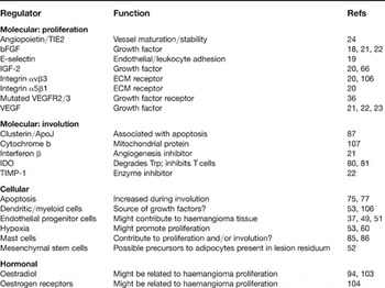
Potential regulators of haemangioma progression discussed in this article are summarised in Table 2. New information gathered from basic research will support efforts to develop better treatments for haemangiomas. A major impediment to the advancement of new therapies, however, has been the absence of useful models. The availability of an animal model that reproduces the major features of infantile haemangioma would provide a means by which potential therapeutic interventions could be tested, and permit an opportunity to better understand how existing therapies work. In vitro culture models will facilitate the development of high-throughput assays to screen candidate compounds.
Potential regulators of haemangioma progression

Abbreviations: ApoJ, apolipoprotein J; ECM, extracellular matrix; bFGF, basic fibroblast growth factor; IDO, indoleamine 2,3-dioxygenase; IGF2, insulin-like growth factor 2; TIMP-1, tissue inhibitor of metalloproteinase 1; Trp, tryptophan; VEGF(R), vascular endothelial growth factor (receptor).
There are numerous reports in the literature describing models for ‘haemangioma’, but most have limited utility. Models of endothelial tumours differing in important ways from infantile haemangioma include a spontaneously arising, immortalised haemangioendothelioma cell line (EOMA) (Ref. Reference Obeso, Weber and Auerbach89) and cells carrying polyoma virus middle T antigen (Ref. Reference Bautch90). Tumours generated using these cell lines have more malignant characteristics, are not obligately perinatal and do not spontaneously regress. While potentially useful as tools for investigating some endothelial tumours, caution should be used when extrapolating findings generated in these models to infantile haemangioma.
An ex vivo culture system has been described that has the advantage of using intact human haemangioma tissue and permits biochemical manipulation (Ref. Reference Tan91). The drawbacks of this model include loss of cellular features of haemangioma endothelial cells, such as GLUT-1 expression (Ref. Reference North92), and a mechanism of growth involving sprouting. Purified cultures of haemangioma-derived cells have been used to demonstrate their unusual behaviour in terms of growth and migration (Refs Reference Yu24, Reference Boye35), but this system, lacking the typical environmental milieu, removes potentially important soluble and insoluble molecules, such as growth factors and various extracellular matrix components, as well as any direct influence from surrounding cells.
It is possible that the complexity and multifactorial nature of haemangiomas may preclude the development of a faithful animal model. Numerous investigators have reported repeated failure at attempts to create models using a wide variety of approaches including the implantation of cells and tissues from haemangioma and placenta (Ref. Reference Frieden93). A recent report combined the hypothesis of an involvement of hormones in haemangioma with an attempt to create a mouse model (Ref. Reference Peng94). Using nude mice, which have severely compromised adaptive immunity and allow grafting of foreign tissues, fresh haemangioma tissue from a single patient was implanted subcutaneously into 16 mice. Weekly injection of 0.1 ng oestradiol was administered to eight mice and the growth of the implanted tumours was measured over the course of 90 days. Modest growth, or at least maintenance, of the tissue was reported in the animals receiving oestradiol, but none of the control animals. This study is of interest both as a possible indication of a requirement for oestradiol for haemangioma growth, and as a possible long-sought animal model. Follow-up confirmation of this work is needed, but if verified, it is an example of the insight that may be needed to generate a useful model of infantile haemangioma.
Clinical implications/applications
Several therapeutic approaches for haemangiomas have been suggested over the past decade. Interferon α has been used successfully in the treatment of complicated haemangiomas (Refs Reference Ezekowitz, Mulliken and Folkman95, Reference Chang96), but its use is associated with the development of spastic diplegia (Ref. Reference Barlow97) and is generally limited to life-threatening cases without other viable options. Initial reports indicated that the neurological effects were irreversible, but a subsequent study showed that these effects can resolve over time (Ref. Reference Greinwald98). Perhaps new therapies would employ angiostatic compounds already in use in other areas of medicine. A concern with this class of drugs is their use in the growing child where new vessel growth may be ongoing, highlighting the need for more targeted options.
Currently, the first-line therapy for troublesome infantile haemangiomas is corticosteroids. These drugs, while efficacious in many patients, carry undesired side effects such as temporary growth retardation, increased risk of infection and psychological changes (Ref. Reference Boon, MacDonald and Mulliken99). Corticosteroids have known anti-inflammatory activity, but their action in haemangioma is poorly understood. The finding that proliferating haemangiomas harbour large numbers of haematopoietic cells of the myeloid lineage (Ref. Reference Ritter53) suggests a possible mechanism whereby corticosteroids inhibit the speculated growth-promoting function of these cells. The idea of boosting the adaptive immune response against the tumour is worth exploring and has some limited experimental and clinical support. The discovery of IDO expression in haemangioma (Refs Reference Ritter80, Reference North81) suggests that inhibiting this enzyme and/or supplementing local tryptophan could lead to an increased T-cell response against the tumour. Further exploration of imiquimod or other compounds that modify the immune response is warranted (Ref. Reference Martinez15).
The impact of large haemangiomas has been shown to extend beyond simple mass-related effects such as encroachment of vital structures and disfigurement. Severe hypothyroidism in children with large haemangiomas was found to result from high type 3 iodothyronine deiodinase activity in the haemangioma tissue (Ref. Reference Huang100). The finding that large haemangiomas can have far-reaching effects highlights the need to find effective means to limit their growth.
Research in progress and outstanding research questions
The collective efforts of haemangioma researchers have raised numerous questions regarding the mechanisms behind the growth and regression of these tumours. The similarities and differences between haemangioma-derived cells, normal cells and immature normal cells should be further studied while exploring the intrinsic defect hypothesis. The evidence suggesting a connection to the placenta heightens interest in exploring the impact of placental complications such as placenta praevia, preeclampsia and chorangioma on haemangioma incidence and biology. One interesting hypothesis was recently removed from consideration by two studies investigating maternal–fetal chimaerism that showed haemangiomas were exclusively derived from the child, with no evidence of maternal origin (Refs Reference Pittman101, Reference Regnier102). Possible clues into haemangioma pathogenesis include the increased incidence in the head and neck regions and the predilection for the skin. Dermal haemangiomas come in two general forms: the plaque-like segmental form and the tumour-like focal form. This curious dichotomy begs the question of how these two types of lesions differ at the cellular and molecular levels. Speculation regarding the differences in segmental and focal haemangiomas suggests that segmental lesions may be derived from implantation of circulating placental/trophoblastic cells relatively early in development, whereas the focal variety arise from the same cells arriving late in development via the amniotic fluid (Ref. Reference Bree45). Only rarely are the segmental lesions candidates for surgical excision, leading to a paucity of this tissue for study. In fact, the limited supply of any haemangioma tissue is an ongoing concern. A centralised tissue bank for haemangiomas and other vascular anomalies would be of great value to the research community, making available these and other rare specimens that have been collected and stored using standardised protocols (Ref. Reference Frieden93). Based on evidence from varying sources, the idea of hypoxia as a trigger and driving force behind haemangioma growth deserves further study. Animal models incorporating hypoxia are available (Ref. Reference Ceradini70), and instrumentation is available to measure tissue oxygen levels in the clinic; thus, addressing this question would seem feasible. Another clue before us is the higher rate of haemangioma occurrence in females. Elevated levels of 17β-oestradiol (Ref. Reference Sasaki, Pang and Wittliff103) and oestrogen receptor (Ref. Reference Lui104) have been reported in children with haemangiomas. It was also speculated that corticosteroid treatment may negatively affect oestradiol action (Ref. Reference Sasaki, Pang and Wittliff103). Higher levels of angiogenic factors have been demonstrated in females, pointing to another possible explanation for the higher incidence in females (Ref. Reference Malamitsi-Puchner105). These findings support further investigations into possible hormonal regulation of haemangioma growth and the differences in factor expression between genders.
Despite the perception of some that infantile haemangiomas are merely birthmarks that will quietly regress without major complication, a significant number of children suffer physically and emotionally as a result of living with a haemangioma. For some, these lesions pose life-threatening complications that severely impact on their early years, while for others a permanent disfiguring scar leaves a constant reminder of these usually benign lesions. Further investigations will not only hopefully lead to better treatments, but also teach us more about endothelial biology and the basic processes of blood vessel growth and regression.
Acknowledgments and funding
We thank the reviewers of this article for their thoughtful critiques and suggestions, as well as those that have contributed to our haemangioma work, including Josh Ney, David Friedlander, Dana Copeland, Jeff Rubens, Mike Dorrell, Stacey Moreno and John Reinisch. Research in our laboratory is supported by a grant from the National Eye Institute (R01 EY11254, to M.F.) as well as by the MacTel Foundation and the V. Kann Rasmussen Foundation. M.R.R. was supported for a portion of this work by a Kirschstein National Research Service Award from the National Eye Institute. S.F.F. was supported by the Skaggs Scholar in Clinical Science Program.