INTRODUCTION
The term apraxia denotes “higher-order” disturbances of motor control. This definition embraces a wide variety of heterogeneous disturbances, but traditionally diagnosis and research on apraxia concentrate on three domains of human motor actions: imitation of gestures, performance of communicative gestures on command, and skillful use of tools and objects.
Compared to clinical assessment of other neuropsychological syndromes such as, for example, amnesia or dysexecutive syndrome, clinical diagnosis of apraxia is swift and easy. Several standardized screening tests evidence that a combination of some simple tests like, for example imitating hand postures or pantomiming the use of common tools, permit a reliable distinction between presence and absence of apraxia (Rothi, Raymer, & Heilman, Reference Rothi, Raymer and Heilman1997; Vanbellingen et al., Reference Vanbellingen, Kersten, Van de Winckel, Bellion, Baronti, Müri and Bohlhalter2011; Weiss, Kalbe, Kessler, & Fink, Reference Weiss, Kalbe, Kessler and Fink2013). However, the conclusion that apraxia is a single and unitary functional deficit that is either present or absent would be misleading. Detailed examinations of different manifestations of apraxia and of the lesions underlying them demonstrate double dissociations of behavioral deficits and of causal lesions between patients who had received the same global diagnosis of apraxia. Rather than constituting an obstacle to unequivocal diagnosis such dissociations offer insight into the cognitive and motor mechanisms leading to apraxia. They also have practical value for management of patients, because they can pinpoint their specific problems in daily living and thus guide the selection of therapeutic approaches. For example, a patient whose apraxia concerns predominantly use of tools and objects is likely to have more problems with household chores than another one in whom apraxia affects mainly the imitation of gestures.
Pantomiming tool use is a very widely, probably even the most widely, used test of apraxia. It is included in most protocols for systematic assessment of apraxia (Barde, Buxbaum, & Moll, Reference Barde, Buxbaum and Moll2007; Goldenberg & Randerath, Reference Goldenberg and Randerath2015; Goldstein, Reference Goldstein1928; Goodglass & Kaplan, Reference Goodglass and Kaplan1963; Rothi et al., Reference Rothi, Raymer and Heilman1997; Roy, Square-Storer, Hogg, & Adams, Reference Roy, Square-Storer, Hogg and Adams1991; Vanbellingen et al., Reference Vanbellingen, Kersten, Van de Winckel, Bellion, Baronti, Müri and Bohlhalter2011; Weiss et al., Reference Weiss, Kalbe, Kessler and Fink2016). For testing pantomime of tool use, the examiner names a tool and asks the patient to demonstrate the manual action of using this tool without holding it. As is generally the rule in apraxia testing, patients are asked to use the ipsi-lesional hand to rule out a possible influence of contra-lesional hemiparesis on dexterity. For aphasic patients, comprehension of the name of the tool can be facilitated by showing a picture of it. In right handed patients, defective pantomime is a symptom of left brain damage and associated with aphasia, but there are patients with aphasia without apraxia (Kertesz, Ferro, & Shewan, Reference Kertesz, Ferro and Shewan1984). By contrast, defective pantomime without accompanying aphasia is very rare and confined to patients with atypical lateralization of brain function (Alexander & Annett, Reference Alexander and Annett1996; Goldenberg, Reference Goldenberg2013).
THEORIES OF PANTOMIME
Scientific interest in defective pantomime is not limited to the reliable diagnosis of apraxia but aims at insights into principles of cognition and brain organization. Under this perspective pantomime turns out to be more intricate and controversial as it might appear in clinical diagnosis. Different theoretical approaches emphasize different properties of pantomiming as starting point for the investigation of underlying cognitive and neural mechanisms. They lead to different predictions concerning associations and dissociations between disturbed pantomime and other manifestations of apraxia, both on the behavioral and the anatomical level.
In the current literature, three properties have been proposed as starting point for speculations about the cognitive and neuronal mechanisms of pantomime: Absence of mechanical interaction between hand and objects, replication of motor programs of actual use; and gestural communication. We will review for each of these facets of pantomime whether it can explain the empirical data on associations and dissociations between defective pantomime and other manifestations of apraxia.
Absence of Mechanical Interaction between Hand and Objects
Pantomiming tool use is usually done without holding the tool and without contacting the recipient of the tool action (Goldenberg, Hentze, & Hermsdörfer, Reference Goldenberg, Hentze and Hermsdörfer2004; Randerath, Goldenberg, Spijkers, Li, & Hermsdörfer, Reference Randerath, Goldenberg, Spijkers, Li and Hermsdörfer2011). The absence of interaction with external objects gains theoretical significance under the assumption that control of deliberate motor actions is accomplished in two consecutive phases (Barbieri & De Renzi, Reference Barbieri and De Renzi1988; Buxbaum, Shapiro, & Coslett, Reference Buxbaum, Shapiro and Coslett2014; Heilman, Rothie, & Valenstein, Reference Heilman, Rothie and Valenstein1982; Liepmann, Reference Liepmann1908; Martin et al., Reference Martin, Beume, Kümmerer, Schmidt, Bormann, Dressing and Weiller2016; Roy & Hall, Reference Roy and Hall1992). In a first phase, a mental image of the intended action is created and in the second phase it is converted into motor commands.
Liepmann (Reference Liepmann1908), who introduced this dichotomy named the first phase “ideational” and the second “ideo-kinetic.” In the modern literature, the first phase has been named “conceptual” and the second “production” or “executive” phase. Probing pantomime has been acknowledged as a particularly demanding test for the integrity of the second phase, because the manual action of using the tool must be produced without support from mechanical interactions between hand, tool, and recipient of action. In actual use, these constraints reduce the degrees of freedom of the manual movements and hence the demands on planning and guiding them. For example, a key cannot be inserted into the lock elsewhere than in the keyhole; it cannot be guided into the hole accurately other than by a precision grip, and it cannot move inside the keyhole other than in rotation. Testing pantomime abolishes direct contact between hand and tool and thus invalidates these external constraints. The need to execute the motor actions without external support reveals insufficiencies of the conversion from mental images into motor programs.
Pantomime of tool use is not the only clinical test requiring production of gestures without support from mechanical interactions with external objects. Other tests with this constellation are the demonstration of emblematic gestures like “okay” or “silence” and the imitation of gestures. We are not aware of studies that compare the localizations of responsible lesions between emblematic gesture and pantomime. By contrast the localization of defective imitation has been investigated in a substantial number of patients and studies (De Renzi, Motti, & Nichelli, Reference De Renzi, Motti and Nichelli1980; Goldenberg, Reference Goldenberg1996; Goldenberg & Randerath, 2015; Kimura & Archibald, Reference Kimura and Archibald1974). They used predominantly meaningless gestures to make sure that imitation aimed at replicating the shape of the gesture and was not accomplished by comprehension and subsequent reproduction of their meaning. Like in pantomime, production of the imitated gesture is accomplished without support from mechanical constraints.
Furthermore, it has been argued that, since the shape of the intended action of the hand is provided by the demonstration, the only possible source of errors resides in the conversion of this image into motor commands (Barbieri et al., 1988; De Renzi & De Renzi, Reference De Renzi, Motti and Nichelli1980). The failure of transforming the idea of an action into its motor execution has been characterized by the traditional designation as “ideo-motor” apraxia. According to this tradition pantomime and imitation are affected by deficiencies in a common component of gesture production. This need not exclude the possibility that other components differ. The existence of such differences is proven by double dissociations between impairments of pantomime and of imitation (Barbieri et al., 1988; Goldenberg & Hagmann, Reference Goldenberg and Hagmann1997).
The assumption that pantomime and imitation test the same aspect of cognitive motor control constitutes a tacit prerequisite for screening tests of apraxia that compute sum scores of pantomime and imitation for diagnosing “ideomotor” apraxia (Rothi et al., Reference Rothi, Raymer and Heilman1997; Vanbellingen et al., Reference Vanbellingen, Kersten, Van de Winckel, Bellion, Baronti, Müri and Bohlhalter2011). Beyond this practical consequence, their presumed functional equivalence leads to the prediction that the typical localization of lesions disturbing pantomime should be congruent with those disturbing imitation.
Replication of Motor Programs of Actual Use
Another theoretical approach to pantomime of tool emphasizes the similarity between the manual actions of pantomime and of real use and maintains that both are directed by the same motor programs. More specifically it has been postulated that frequent execution of routine tool use, (e.g., hammering), leaves a permanent trace in form of a “visuo-kinetic” engram which can support replication of the routine actions without the tool that is normally involved [e.g., a hammering movement with the empty hand (Buxbaum et al., Reference Buxbaum, Shapiro and Coslett2014; Frey, Reference Frey2007; Martin et al., Reference Martin, Beume, Kümmerer, Schmidt, Bormann, Dressing and Weiller2016)]. In this view, pantomime is equivalent to running the routine motor program of tool use without holding the tool.
The postulated intimate relationship between motor programs for use and for pantomime leads to the expectation that their neural substrates are also closely coupled. The presumed similarity of motor programs for pantomime and real use has been exploited for exploration of neural substrates of object use by means of functional imaging. Bringing real three-dimensional tools and their recipients into the scanner and scanning subjects during their unrestricted use is technically difficult (Brandi, Wohlschläger, Sorg, & Hermsdörfer, Reference Brandi, Wohlschläger, Sorg and Hermsdörfer2014). Pantomime offers itself as a proxy eliciting the same activation pattern with less technical difficulties (Frey, Reference Frey2008; Hermsdörfer, Terlinden, Mühlau, Goldenberg, & Wohlschläger, Reference Hermsdörfer, Terlinden, Mühlau, Goldenberg and Wohlschläger2007; Króliczak & Frey, Reference Króliczak and Frey2009; Lewis, Reference Lewis2006; Vingerhoets et al., Reference Vingerhoets, Alderweireldt, Vandemaele, Cai, Van der Haegen, Brysbaert and Achten2013). However, the legitimacy of this substitution depends on confirmation of a substantial overlap between the neural substrates of pantomime and real use.
Communicative Gesture
Pantomimes are communicative gestures in the basic sense that they do not alter the material state of external objects but carry a meaning that can be understood by other persons (Novack & Goldin-Meadow, Reference Novack and Goldin-Meadow2016). They frequently accompany speech but are also a common component of nonverbal communication in daily life. They can be used for indicating objects and events in situations with restrictions on verbal communication like, for example, in noisy surroundings, and in communication with people who do not speak the same language. The range of gestures used for such functions is not restricted to pantomimes. They can be seamlessly combined with other types of communicative gestures like pointing to the locations of objects (“deictic” gestures) or drawing the outline of the indicated object with the finger on the table or in the air (“iconographic” gesture).
The recognizability of pantomimes can also be enhanced by the “body part as object” strategy where the absent object of the action is replaced by a part of the own body. For example cutting with scissors can be demonstrated more clearly by opening and closing the angle between the index and middle finger than by opening and closing the hand which is, however, more similar to the motor action of real scissoring.
The ability to pantomime the use of a tool is not contingent upon the competency of using it. Both daily life observations and experimental studies reveal substantial differences between individual abilities for actual tool use and the corresponding pantomime. For example, most people can produce comprehensible pantomimes of playing musical instruments such as a piano, a violin, or a trumpet, but only a minority masters the motor actions of their real use. A more subtle dissociation between pantomime and real use concerns the selection of features that are included in the pantomime. Inclusion or exclusion of distinct features of the motor action of use does not necessarily depend on their functional importance. When normal subjects pantomime grasping a glass they usually fail to demonstrate that in synchrony with the transport toward the glass the hand opens and then closes around the glass. By contrast they may exaggerate the amplitude of characteristic movements, like the oscillatory movement of sawing wood, or the inclination of the scoop for ladling (Goodale, Jakobson, & Keillor, Reference Goodale, Jakobson and Keillor1994; Hermsdörfer, Li, Randerath, Goldenberg, & Johannsen, Reference Hermsdörfer, Li, Randerath, Goldenberg and Johannsen2012; Laimgruber, Goldenberg, & Hermsdörfer, Reference Laimgruber, Goldenberg and Hermsdörfer2005) to increase the recognizability of the pantomime.
A likely explanation for the variability of pantomime and for its relative independence from motor acts of pantomime could be that pantomime is a communicative gesture whose aim is to transmit information about properties of objects and their use. The choice of which features are shown or neglected depends on how much they contribute to the comprehensibility of the gesture. In the clinical examination, patients are advised to show only motor acts that form part of the real use, but when pantomime is used for communication outside the clinical setting, subjects may prefer motor actions that do not participate in real use but demonstrate it less unequivocally, as, for example, by showing the body part as object.
Demonstration of the nature and properties of tools and their use requires retrieval, selection and combination of knowledge about them. Such knowledge is part of semantic memory. Arguably the inability to retrieve and combine it is the main cause for failure of pantomime. Pantomime of tool use can thus be considered as a test of semantic memory.
The conclusion that pantomimes are communicative gestures based on semantic memory does not rule out replications of motor routines. We conceive of semantic memory as an integrated multi-modal network providing unimpeded exchange and conversion between features from different modalities (Jefferies, Reference Jefferies2013; Meteyard, Cuadrado, Bahrami, & Vigliocco, Reference Meteyard, Cuadrado, Bahrami and Vigliocco2012; Vannuscorps, Andres, & Pillon, Reference Vannuscorps, Andres and Pillon2014). In such a network, motor routines can be implemented as one type of features and can be combined with features taken from other modalities. The accuracy and fidelity of replications of motor features of real tool use may be subject to the communicative context and strategic choices (Tessari, Canessa, Ukmar, & Rumiati, Reference Tessari, Canessa, Ukmar and Rumiati2007). For example, pantomime artists display rarely noticed features of motor routines like the opening and closing of the hand in synchrony with the transport toward a glass for creating the illusory impression of an external object withstanding the hand.
Lesion Analyses of Pantomime
In the previous section, we have derived predictions for commonalities of lesions sites depending on which property of pantomime is considered most important. We speculated that the absence of interactions with external objects is common to pantomime and imitation; that the use of routine motor programs is common to pantomime and real use, and that retrieval and combination of semantic knowledge is shared with other tests of semantic memory. According to these functional commonalities, we may expect anatomical commonalities between critical lesions for pantomime and imitation, pantomime and real use, and pantomime and semantic memory.
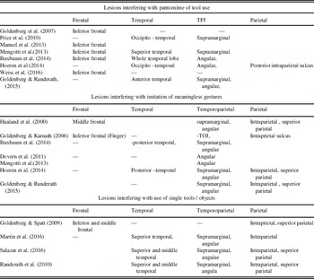
Table 1 lists recent results of voxel based lesion symptom mapping of manifestations of apraxia. The designations of lesion location are at the level of macro-anatomic structures. The territory at the temporo-parietal junction is subdivided into angular gyrus, supramarginal gyrus, and intraparietal gyrus.
Lesion analyses

Virtually all of the studies show an influence of lesions in supramarginal or angular gyrus on all manifestations of apraxia. The apparently indiscriminate influence of these regions on all manifestations of apraxia is only one aspect of their universal impact on disturbances of spatial, semantic, and linguistic processing symptoms. A possible anatomical basis for the manifoldness of functional consequences of angular and supramarginal lesions may be the richness of their fiber connections to adjoining and distant regions in temporal, parietal, and occipital regions (Seghier, Reference Seghier2013). Because of the ubiquity of the effects of damage to these regions, they cannot explain specific associations and dissociation between the different manifestations of apraxia, and we will not further discuss their roles.
The comparisons of lesions affecting pantomime with those underlying imitation does not support the hypothesis that their neuronal substrates are determined by the same basic problem of performing motor actions without external guidance. Lesions associated with defective pantomime are mainly located in temporal and inferior frontal regions. In only one of eight studies, the angular gyrus lesion extends into the posterior part of the intra-parietal sulcus. By contrast, defective imitation is associated with intra-parietal and superior parietal lesions in four out of seven studies whereas the temporal lobe is spared in five of them.
Evaluation of the hypothesis that neural substrates of pantomime and actual tool use reflect the commonality of their motor programs is less straightforward. In contrast to the neural substrate of pantomime the lesions associated with defective use of single tools always affect parietal regions. However, three of the four studies of tool use show also a participation of temporal lesions. This frequency is not conspicuously different from the studies exploring pantomime.
In the previous section, we hypothesized that employment of the same routine motor programs would lead to congruency between lesions underlying defective pantomime and defective tool use. The influence of temporal lesions on both of them accords with this prediction.
There are no lesion studies comparing pantomime to non-verbal tests of semantic memory, but there is one study that looked for anatomical overlap between lesions affecting pantomime and different aspects of language (Goldenberg et al., 2015). Linguistic competence was assessed by the Aachen Aphasia Test (AAT) that consists of subtests measuring naming, comprehension, repetition, written language, and the Token Test (Huber, Poeck, & Willmes, Reference Huber, Poeck and Willmes1984). The comparison revealed a zone at the anterior pole of the temporal lobe where lesions were associated with poor scores on pantomime and on all subtests of the AAT. By contrast, lesions at this site had no influence on imitation of meaningless gestures. The anterior temporal lobe has been identified as a central “hub” that stores amodal semantic information and distributes it to modality specific further processing (Jefferies, Reference Jefferies2013; Lambon Ralph, Reference Lambon Ralph2014). We reasoned that the need to access semantic memory is common to pantomime and to language. Although not all subtests of the AAT explicitly ask for semantic decisions, they use meaningful verbal stimuli. It is conceivable that access to the meaning of words facilitates also tasks that do not directly ask for meaning like repetition or writing to dictation.
CONCLUSIONS
We found little support for the dual hypothesis that the crucial property of pantomime is the need to perform skilled movements without external constraints, and that this need constitutes commonality between pantomime and imitation. VLSM supported the clinical observation of double dissociations between pantomime and imitation by revealing distinctly different and hardly overlapping lesions causing each of them. Whereas pantomime is mainly affected by temporal and inferior frontal lesions, defective imitation is bound to parietal lesions. The clear dissociation between pantomime and imitation casts doubts on the validity of sum scores adding up their results and on their traditional subsumption under the common entity of “ideomotor” apraxia.
Associations and dissociations between pantomime and real tool use are less straightforward. Similarities between the motor actions produced in both conditions are obvious but less stringent than would be expected if they were based on identity of motor programs. They are better compatible with the hypothesis that pantomime is a communicative gesture that uses manual actions for conveying information about objects and their use. The manual actions are constructed by selection and combination of distinctive features of tools and actions. They usually include replications of characteristic motor actions of real use, but the main criterion for selection and modification of features is the comprehensibility of the gestures rather than the accurate replication of the motor actions of real use.
The overlap of temporal lesions that interfere with pantomime and with real use does not necessarily indicate that they are based on the same motor programs. Alternatively, their common localization may derive from their common dependency on retrieval of functional knowledge from semantic memory. Functional knowledge specifies attributes of tools and objects like their purpose, their recipient, their typical action, or the social context of their application. Like semantic memory in general, functional knowledge is bound to temporal lobe integrity (Binder & Desai, Reference Binder and Desai2011; Bozeat, Patterson, & Hodges, Reference Bozeat, Patterson and Hodges2002; Corbett, Jefferies, & Lambon Ralph, Reference Corbett, Jefferies and Lambon Ralph2013; Goldenberg & Spatt, Reference Goldenberg and Spatt2009; Kubiak & Króliczak, Reference Kubiak and Króliczak2016; Silveri & Ciccarelli, Reference Silveri and Ciccarelli2009; Osiurak, Reference Osiurak2014). Functional knowledge provides the features that are selected and combined to comprehensible pantomimes. For real tool use functional knowledge is complemented by mechanical problem solving mediated by parietal regions (Goldenberg & Spatt, 2009; Spatt, Bak, Bozeat, Patterson, & Hodges, Reference Spatt, Bak, Bozeat, Patterson and Hodges2002), but the commonality between defective pantomime and defective tool use is due to their common dependency on retrieval of knowledge from semantic memory in temporal regions.
The conclusion that the crucial deficit in apraxia for pantomime concerns the retrieval of significant features from semantic memory and their combination to comprehensible gestures has consequences for clinical practice. Defective pantomime indicates problems of semantic retrieval and expression rather than of motor skill. It disrobes patients of a mean of nonverbal communication. The importance of this deprivation is enhanced by the regular co-occurrence of defective pantomime with aphasia (Hogrefe, Ziegler, Weidinger, & Goldenberg, Reference Hogrefe, Ziegler, Weidinger and Goldenberg2012).
Acknowledgment
There are no conflicts of interest