INTRODUCTION
Nematodes infect a high portion of the human and animal population. Current treatment is based on the use of anthelmintic drugs but the best alternative to reduce infections in the future is vaccination (Maizels et al. 1999). A major problem in the development of a vaccine is the identification of protective antigens. RNA interference (RNAi) could be a useful tool in this process. This technology has been used widely in Caenorhabditis elegans to define gene function (e.g. Kamath et al. 2003) and could help in the functional analysis of parasitic nematode genes and identify genes of vital importance (Kuwabara and Coulson, 2000). The technique has already been used, with some success, in Nippostrongylus brasiliensis (Hussein et al. 2002), Brugia malayi (Aboobaker and Blaxter, 2003), Onchocerca volvulus (Lustigman et al. 2004; Ford et al. 2005), Ascaris suum (Islam et al. 2005), Trichostrongylus colubriformis (Issa et al. 2005), Haemonchus contortus (Kotze and Bagnall, 2006; Geldhof et al. 2006) and Litomosoides sigmodontis (Pfarr et al. 2006). The RNAi methodology used predominantly was soaking the parasites in dsRNA. This was done both with larval (Issa et al. 2005; Geldhof et al. 2006) and adult life-stages (Hussein et al. 2002; Aboobaker and Blaxter, 2003; Kotze and Bagnall, 2006; Pfarr et al. 2006). Besides soaking, feeding L1/L2 larvae on dsRNA-producing bacteria and electroporation of L1 larvae have also been reported as effective methods to deliver dsRNA (Issa et al. 2005; Geldhof et al. 2006).
Successful RNAi has been performed on a wide variety of parasite genes, such as acetylcholinesterase in N. brasiliensis (Hussein et al. 2002), a RNA polymerase II gene, β-tubulin gene and sheath protein 1 gene in B. malayi (Aboobaker and Blaxter, 2003), cathepsin L and Z and serine protease inhibitor 1 and 2 in O. volvulus (Lustigman et al. 2004; Ford et al. 2005), pyrophosphatase in A. suum (Islam et al. 2005), ubiquitin and tropomyosin in T. colubriformis (Issa et al. 2005), β-tubulin, superoxide dismutase and sec-23 in H. contortus (Kotze and Bagnall, 2006; Geldhof et al. 2006) and actin in L. sigmodontis (Pfarr et al. 2006).
Despite these successes, concerns remain about the efficiency of this technology in parasitic nematodes. Some studies only used phenotypic examination or immunolocalization to evaluate the success of the RNAi treatment, so it is unclear to what extent gene expression levels were actually knocked down. In addition, the extensive studies in H. contortus (Kotze and Bagnall, 2006; Geldhof et al. 2006) have clearly indicated that although some genes can be readily silenced by RNAi, a number of other genes could not be efficiently knocked down. Similar effects have been observed in T. colubriformis (Issa et al. 2005). There are also concerns around the specificity of the different RNAi methods. The studies on B. malayi (Aboobaker and Blaxter, 2003) and O. volvulus (Lustigman et al. 2004) indicated non-specific effects on these parasites after soaking in non-sense dsRNA.
In order to use RNAi as a screening method for vaccine candidates in Ostertagia ostertagi, an important gastrointestinal nematode of cattle, we wanted to check the efficacy and specificity of RNAi in this parasite species. Eight different genes were targeted in L1 and exsheathed L3 larvae by electroporation and soaking, among which were genes successfully knocked down by RNAi in other parasites (β-tubulin (Oo-btub), tropomyosin (Oo-tropo), superoxide dismutase (Oo-sodc) and ubiquitin (Oo-ubi)). The specificity of observed RNAi effects was checked by semi-quantitative reverse transcriptase (RT-)PCR to determine the transcription level of a large set of genes not specifically targeted by the RNAi treatment.
MATERIALS AND METHODS
Parasite stages
To collect first-stage larvae (L1), eggs were isolated from the faeces of experimentally infected calves by a centrifugation flotation method, incubated at 28 °C for 20–22 h and hatched L1 larvae collected by filtration, and concentrated at 100 larvae per 10 μl water. Third-stage larvae (L3) were collected as described by Geldhof et al. (2000).
Cloning of Ostertagia ostertagi gene fragments for dsRNA production
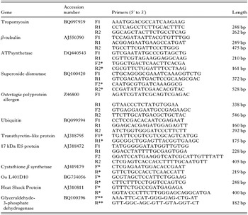
The Accession numbers of the targeted O. ostertagi genes are listed in Table 1. Fragments of 160 to 338 nucleotides were amplified by PCR on O. ostertagi L3 cDNA using gene-specific primers (Table 1). The homologue of the Haemonchus ATPsynthetase subunit was amplified using primers described previously for H. contortus (Geldhof et al. 2006). PCR products were cloned into the pGEM-t Easy vector (Promega) and DNA sequencing confirmed their identity. PCRs for all targeted genes were also performed on L1 cDNA to confirm transcription of the genes in this stage.

Production of dsRNA
The gene fragments were cloned into the double T7 promoter vector L4440 (designed by A. Fire) after which the vector was linearized by using unique restriction enzyme sites in the cloning site of the vector, except for Oo-sodc, where restriction sites were included in the PCR primers. T7 RiboMAX™ Express RNAi System (Promega) was used according to the manufacturer's instructions to produce single-stranded RNA. Complementary strands were pooled and heated to 70 °C for 10 min and left to cool at room temperature to allow annealing. Quantitation was done by measuring absorbance at 260 nm. For each targeted gene at least 3 different preparations of dsRNA were made, directed to the same fragment.
RNAi treatment
Soaking L3 larvae in dsRNA
One thousand exsheathed L3 larvae were incubated in a mixture of 30 μl of PBS, 5 μl of Lipofectin reagent (Invitrogen) and 5 μl of dsRNA (at a final concentration of 1 mg/ml) for 24, 48 or 72 h at 37 °C. For each gene 3 different preparations of dsRNA were tested in duplicate for each incubation time, except for a polyprotein allergen (Oo-opa) and a 17 kDa excretion-secretion (ES) protein (Oo-17 kDa).
Electroporation of L1 with dsRNA
One hundred larvae were suspended in 100 μl of trehalose electroporation buffer (272 mM trehalose, 7 mM KH2PO4 (pH 6), 1 mM MgSO4) containing 1 mg/ml dsRNA as described by Issa et al. (2005). Electroporation was done in 0·2 cm cuvettes at 100 V with a single pulse of 25 ms (Bio-Rad Gene Pulser II). Again, the 3 different preparations of dsRNA were used. The larvae were recovered in an Eppendorf tube, washed with distilled water and incubated in 80 μl of distilled water plus 20 μl of nutritive medium (10X Earle's saline solution fortified with a yeast extract as described by Hubert and Kerboeuf, 1984) at 23 °C for 24 h.
The negative control for both approaches consisted of larvae treated with different preparations of control dsRNA from the C. elegans yolk receptor (Ce-rme-2). The L4440 plasmid containing Ce-rme-2 was the kind gift of Dr Collette Britton, Glasgow University Veterinary School, UK (Britton and Murray, 2004).
Reverse Transcriptase PCR (RT-PCR)
The decrease in mRNA level of the targeted genes was measured using semi-quantitative RT-PCR. Double-stranded RNA-treated larvae were collected at determined time-points and extensively washed with PBS. Larvae were resuspended in 200 μl of lysis buffer (0·5% SDS, 5% 2-mercaptoethanol, 10 mM EDTA, 10 mM Tris-HCl, pH 7 and 0·5 g/ml proteinase K), frozen at −70 °C for at least 10 min and incubated at 55 °C for 1 h (Hashmi et al. 2002). Total RNA was extracted using Total RNA Isolation Reagent (ABgene). To perform the gene specific one-step RT-PCR reactions (SuperScript One-Step RT-PCR (Invitrogen)), at least 1 gene-specific primer targeting a region outside the region of dsRNA was used, except for a transthyretin-like protein (Oo-ttr) (Table 1). Twenty-eight cycles of amplification were done in the RT-PCRs for all genes except for Oo-ttr where 30 cycles were necessary to amplify the fragment. PCR on genomic DNA (gDNA) using the same primer sets confirmed the presence of introns that made the differentiation between cDNA and genomic contamination possible for all genes tested. The quality of the RNA was checked by RT-PCR (28 cycles of amplification) for the house-keeping gene glyceraldehyde-3-phosphate dehydrogenase (Oo-gapdh).
Monitoring the specificity of the observed RNAi effects
L3 larvae were soaked in Oo-tropo dsRNA or Ce-rme-2 dsRNA (control worms). Two dsRNA preparations for each gene were made and each preparation was tested in duplicate. After 24 h of incubation, larvae were collected and RNA was prepared as described above. Semi-quantitative RT-PCRs (28 to 30 cycles of amplification) were run for tropomyosin and 7 unrelated genes with different functions. The non-target genes tested were: ATPsynthetase (Oo-atps), cystathionine β-synthase (Oo-cystβs), heat shock protein (Oo-hsp), OoL401D10 gene (Oo-L401D10), Oo-sodc, Oo-ttr and Oo-ubi. Primers used are noted in Table 1.
Additionally, L3 larvae were cultured in the same way without addition of dsRNA. These larvae were collected after 24 h of incubation and the expression level of Oo-hsp and Oo-sodc was determined by semi-quantitative RT-PCR.
RESULTS
RNAi by soaking exsheathed L3 larvae
After 24, 48 and 72 h of incubation the larvae were examined microscopically to detect clear differences in phenotype, such as lethality or increased or decreased activity. Although the viability of the larvae was decreased after 72 h of incubation, there were no substantial differences between control and treated larvae (results not shown).
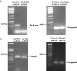
Decreases in mRNA levels were detected for 5 out of the 8 genes tested, varying from complete gene silencing for tropomyosin (Fig. 1A) and β-tubulin to smaller effects for ATP-synthase, superoxide dismutase and a polyprotein allergen. Expression levels of ubiquitin (Fig. 1B), a transthyretin-like protein and a 17 kDa ES protein were never affected by the RNAi treatments.

Fig.1. Transcript level of RNAi targeted genes (A: Oo-tropo, B: Oo-ubi) following soaking of Ostertagia ostertagi L3 larvae for 24 h. Control larvae were soaked in Ce-rme2 dsRNA. The right hand panel shows the transcription of the control gene Oo-gapdh in the same samples. λPst1 DNA marker or 100 bp ladder is shown.
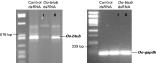
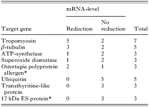
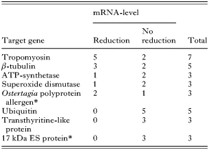
The reproducibility of the soaking method was analysed by repeating the RNAi experiments for each gene with at least 3 different dsRNA preparations, using each preparation in duplicate (except for Oo-opa and Oo-17 kDa). The results are shown in Table 2. While some dsRNA batches reduced target mRNA levels consistently and reproducibly, no reduction could be obtained with others. Results for beta-tubulin are shown in Fig. 2. Exheathed L3 larvae were soaked in different Oo-btub dsRNA samples for 48 h. A complete loss of Oo-btub transcript was observed for dsRNA batch I, while a much smaller effect was observed for dsRNA batch II compared to the control worms.

Fig. 2. Oo-btub mRNA level following soaking of Ostertagia ostertagi L3 larvae for 48 h in 2 different preparations of Oo-btub dsRNA (I and II) or in Ce-rme2 dsRNA as control. The level of Oo-gapdh in the same samples is shown for comparison. λPst1 DNA marker is shown.
Table 2. The number of experiments (each with a different dsRNA preparation) where a reduction or no reduction in mRNA level was obtained after soaking L3 larvae in Ostertagia ostertagi dsRNA, is shown for each gene (Each preparation was tested in duplicate (except for genes indicated with *) giving similar results.)
Soaking larvae for 48 or 72 h did not result in a further decrease in target transcript levels compared to 24 h of incubation, except for Oo-opa. A decrease in mRNA levels for this gene was only detected after 48 and 72 h incubation in 2 out of 3 experiments (not shown).
RNAi by electroporation of L1 larvae
Using the mentioned electroporation and culture conditions, L1 larvae survived the 24 h incubation period well. Longer incubation, however, resulted in a variable percentage of larvae that died or developed further to the (late) L2 stage (results not shown).
Targeting the same 8 genes with 3 different dsRNA preparations, a reduction of transcript of the target gene was noticed in 2 experiments, once for Oo-tropo and once for Oo-btub, but these results were not reproducible. All other experiments did not lead to reduction of gene product (results not shown).
Specificity of observed RNAi effects
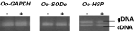
The influence of dsRNA on the larvae was controlled by comparing the expression level of the heat shock protein and the cytoplasmic superoxide dismutase in larvae cultured without dsRNA and larvae cultured with addition of non-sense dsRNA. The mRNA levels of these genes were comparable in all samples (Fig. 3).

Fig. 3. mRNA level of Oo-sodc and Oo-hsp in larvae cultured in the absence (−) or presence (+) of non-sense dsRNA. The mRNA level of Oo-gapdh is shown to verify equal RNA levels in all samples.
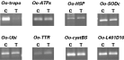
The specificity of the RNAi response was checked in exsheathed L3 larvae treated with tropomyosin dsRNA. This was done by comparing mRNA levels of 7 non-target genes between the RNAi-treated worms and control worms (incubated in Ce-rme-2 dsRNA). A semi-quantitative RT-PCR clearly showed an almost complete knock down of tropomyosin expression (Fig. 4). The mRNA levels of the 7 non-target genes were similar in both tropomyosin-treated worms and control worms (Fig. 4).

Fig.4. RT-PCR results for 8 different Ostertagia genes, using RNA of larvae that were soaked for 24 h in Oo-tropo dsRNA (T) or in Ce-rme2 dsRNA (control) (C).
DISCUSSION
In order to evaluate the potential of RNA interference as a screening method in O. ostertagi, 8 genes were targeted in L1 and L3 larvae by electroporation and soaking respectively. In L3 larvae, variable decreases in mRNA levels were detected for 5 out of the 8 target genes after 24 or 48 h of soaking. Almost complete gene silencing was obtained in some experiments for tropomyosin and β-tubulin, smaller effects were observed for ATP-synthase, superoxide dismutase and a polyprotein allergen. The expression levels of ubiquitin, a transthyretin-like protein and a 17 kDa ES protein were not affected by the RNAi treatments. Except for the polyprotein allergen, longer incubation times did not result in a further decrease of transcript levels. Although it was shown in T. colubriformis (Issa et al. 2005) and H. contortus (Geldhof et al. 2006) that electroporation of L1 larvae was more successful compared to soaking of L3 larvae, this was not the case for O. ostertagi. Reductions in target transcript levels were noticed in only 2 electroporation experiments, once for tropomyosin and once for β-tubulin, and were not reproducible. This indicates that although a technique is successful in one parasitic nematode, it is not guaranteed to be efficient in other parasite species.
The results on L3 larvae show that it is feasible to interfere with the expression of genes by RNAi. However, for unknown reasons, some genes are more sensitive to RNAi than others. For example, the expression of tropomyosin in L3 larvae was almost completely knocked out in 5 out of 7 experiments. Transcript levels of ubiquitin on the other hand were never down-regulated. Similar observations were made by Geldhof et al. (2006) in H. contortus. It could be a reflection of differences in location, stability and/or levels of target transcript. In addition, it has been suggested that the accessibility of a target transcript to the RNAi machinery can depend on the secondary structure of the mRNA and the sequestration in the cell (Timmons et al. 2001). Little is known about these factors in parasitic nematodes. It will be necessary to investigate this if we want to increase the efficiency of the RNAi technology in parasitic nematodes such as O. ostertagi and H. contortus.
Next to the low efficiency, there was also a problem with the reproducibility of RNAi effects in O. ostertagi. Although tropomyosin and β-tubulin expression was significantly reduced in some experiments, no reduction was seen in others. Geldhof et al. (2006) reported similar problems with H. contortus and suggested that differences between dsRNA batches or culture conditions could be responsible for these inconsistencies. Our data now indicate that the dsRNA preparation is an important factor. It was clear that the efficiency of down-regulation of gene expression varied considerably between dsRNA preparations. Some samples induced complete silencing whereas others only induced minor down-regulation, even when tested on the same batch of worms. The reason for this is unclear. The production of dsRNA is a straightforward lab procedure in which every step is quality controlled. The problem to reproduce successful RNAi experiments has now been observed in 2 different parasites by different groups (Geldhof et al. 2006), which suggests that it is not due to a simple technical error in the dsRNA preparation method. This is supported by the finding that dsRNA to a H. contortus cathepsin L showed the capability to completely silence expression of the parasite gene in transgenic C. elegans, although it did not have any effect in H. contortus itself (Geldhof et al. 2006).
Pfarr et al. (2006) recently showed that high concentrations of non-sense dsRNA were stressful to L. sigmodontis worms, as measured by the upregulation of a heat shock protein. The stress response in O. ostertagi larvae seems to be minimal or absent after 24 h of incubation in dsRNA. The expression levels of 2 stress-inducible genes, heat shock protein and superoxide dismutase, were similar in worms cultured with or without dsRNA. However, we have only tested 2 genes. It is possible that other stress related genes are upregulated.
The specificity of the RNAi soaking method was analysed by measuring changes in expression levels of a panel of non-target genes in worms silenced for tropomyosin. Some of these genes, such as the heat shock protein, superoxide dismutase and ATPsynthase, are assumed to have important roles in the basic metabolic pathways of stress response and energy production. No differences in mRNA levels for these genes were detected between the RNAi-treated worms and worms soaked in control dsRNA, indicating a specific down-regulation of the target gene. The results also suggest that down-regulation of tropomyosin did not cause a stress response in L3 larvae. However, the worms were collected only 24 h after the RNAi treatment. It is possible that a prolonged down-regulation of tropomyosin would cause additional responses in the worm and that a phenotypic effect would become visible. This has been reported for β-tubulin down-regulation in H. contortus (Kotze and Bagnall, 2006).
In conclusion, the aim of the present study was to verify if RNAi methods such as electroporation and soaking could be used as a screening method to identify essential genes and vaccine targets in O. ostertagi. These methods were previously reported as successful and useful in parasitic nematodes. However, a putative screening tool should be applicable on most genes and, more important, should give consistent results. Based on our data, we have to conclude that at this moment, the RNAi techniques do not meet these requirements for O. ostertagi. There were problems with the efficiency and reproducibility of both the electroporation and soaking methods. Unless we can further optimize the in vitro culture conditions, dsRNA preparation and delivery route, the application of RNAi in O. ostertagi is limited.
The authors would like to thank Stijn Casaert for excellent technical assistance. This work was supported by a grant from Research Foundation – Flanders (FWO). P.G. is the recipient of a Marie Curie Intra-European Fellowship (no. 500859) and a Marie Curie reintegration grant (no. 028870).