Introduction
Auditory verbal hallucinations (AVH) are a cardinal symptom of schizophrenia, but can also occur in several other neuropsychiatric disorders, as well as in the healthy population (Daalman et al., Reference Daalman, Diederen, Derks, van Lutterveld, Kahn and Sommer2012). Despite transdiagnostic similarities (Daalman et al., Reference Daalman, Boks, Diederen, de Weijer, Blom, Kahn and Sommer2011), pathological substrates of AVH remain largely elusive, with contradictory findings as to specific brain regions and pathophysiological mechanisms involved.
Magnetic resonance imaging (MRI) studies comparing schizophrenia patients with and without AVH implicate volume and gray matter thickness (GMT) changes in predominantly language-related gray matter areas, such as temporal cortical regions, Broca's area, and prefrontal regions in the pathology of AVH (Kühn and Gallinat, Reference Kühn and Gallinat2012; Palaniyappan et al., Reference Palaniyappan, Balain, Radua and Liddle2012). These findings cohere with a hypothesis that the generation of AVHs are caused by disrupted corollary discharges (Mathalon and Ford, Reference Mathalon and Ford2008). This hypothesis posits that the abnormal integration of activity in cortical areas responsible for speech production and auditory perception results in the inability to accurately perceive self-generated signals as arising from external/internal sources or intrinsic activity (Catani and Ffytche, Reference Catani and Ffytche2005; Stephan et al., Reference Stephan, Friston and Frith2009).
In addition to pathology affecting gray matter regions, functional disintegration may result from disrupted anatomical connectivity involving the brain's white matter architecture (Whitford et al., Reference Whitford, Ford, Mathalon, Kubicki and Shenton2010a, Reference Whitford, Mathalon, Shenton, Roach, Bammer, Adcock, Bouix, Kubicki, De Siebenthal, Rausch, Schneiderman and Ford2011). Evidence for white matter pathology derives from diffusion MRI studies, reporting changes in diffusion tensor imaging (DTI) indices such as the fractional anisotropy (FA). However, little consensus exists regarding the location of abnormalities, with some studies reporting changes in circuits connecting speech areas (Seok et al., Reference Seok, Park, Chun, Lee, Cho, Kwon and Kim2007; Catani et al., Reference Catani, Craig, Forkel, Kanaan, Picchioni, Toulopoulou, Shergill, Williams, Murphy and McGuire2011; Kubicki et al., Reference Kubicki, Alvarado, Westin, Tate, Markant, Terry, Whitford, De Siebenthal, Bouix, McCarley, Kikinis and Shenton2011; Ćurčić-Blake et al., Reference Ćurčić-Blake, Nanetti, van der Meer, Cerliani, Renken, Pijnenborg and Aleman2015; Oestreich et al., Reference Oestreich, McCarthy-Jones and Whitford2016; Psomiades et al., Reference Psomiades, Fonteneau, Mondino, Luck, Haesebaert, Suaud-Chagny and Brunelin2016), and others in commissural fibers and subcortical–cortical connections (Hubl et al., Reference Hubl, Koenig, Strik, Federspiel, Kreis, Boesch, Maier, Schroth, Lovblad and Dierks2004; Mulert et al., Reference Mulert, Kirsch, Whitford, Alvarado, Pelavin, McCarley, Kubicki, Salisbury and Shenton2012; Xi et al., Reference Xi, Guo, Li, Chang, Sun, Zhu, Liu, Cui, Chen and Wang2016; Zhang et al., Reference Zhang, Gao, Zhu, Wang, Fan, Ma, Ma and Yang2018), which are outside language-linked circuitry. In addition, some studies report increased (Hubl et al., Reference Hubl, Koenig, Strik, Federspiel, Kreis, Boesch, Maier, Schroth, Lovblad and Dierks2004; Mulert et al., Reference Mulert, Kirsch, Whitford, Alvarado, Pelavin, McCarley, Kubicki, Salisbury and Shenton2012), and others, decreased FA (Seok et al., Reference Seok, Park, Chun, Lee, Cho, Kwon and Kim2007; Ćurčić-Blake et al., Reference Ćurčić-Blake, Nanetti, van der Meer, Cerliani, Renken, Pijnenborg and Aleman2015; Oestreich et al., Reference Oestreich, McCarthy-Jones and Whitford2016) in schizophrenia patients with AVH. Thus, it seems that white matter pathology is associated with AVH in schizophrenia patients; however, the nature of microstructural deficits and specific tracts involved remains unclear.
We point to three primary reasons for heterogenous findings across prior DTI studies. First, schizophrenia is diagnostically characterized by complex clinical phenotypes, broadly categorized into positive, negative, and cognitive symptoms. Thus, previous observations of imaging marker abnormalities in groups of schizophrenia patients with AVH may not be specific to hallucinations, but rather relate to other symptom dimensions or illness-related factors (Ford et al., Reference Ford, Morris, Hoffman, Sommer, Waters, McCarthy-Jones, Thoma, Turner, Keedy and Badcock2014). Second, age-related changes have been shown to substantially impact white and gray matter abnormalities in schizophrenia (Jones et al., Reference Jones, Catani, Pierpaoli, Reeves, Shergill, O'sullivan, Golesworthy, McGuire, Horsfield and Simmons2006; Cropley et al., Reference Cropley, Klauser, Lenroot, Bruggemann, Sundram, Bousman, Pereira, Di Biase, Weickert, Weickert, Pantelis and Zalesky2016; Schnack et al., Reference Schnack, van Haren, Nieuwenhuis, Hulshoff Pol, Cahn and Kahn2016; Di Biase et al., Reference Di Biase, Cropley, Baune, Olver, Amminger, Phassouliotis, Bousman, McGorry, Everall, Pantelis and Zalesky2017, Reference Di Biase, Cropley, Cocchi, Fornito, Calamante, Ganella, Pantelis and Zalesky2018). As such, mixed findings in previous AVH studies may reflect heterogeneity in age range and illness duration (which are intricately linked), where findings in older schizophrenia patients are confounded by more evolved pathologies associated with prolonged illness and cumulative medication effects (Ford et al., Reference Ford, Morris, Hoffman, Sommer, Waters, McCarthy-Jones, Thoma, Turner, Keedy and Badcock2014). Analyzing younger cohorts with shorter illness durations could potentially mitigate these confounding effects to elucidate AVH-specific phenomena. Third, methodological limitations may contribute to the lack of consensus, such that traditional DTI indices may not be able to distinguish specific microstructural features and as such, caution is warranted when interpreting FA changes, which may reflect various, non-specific underlying biological mechanisms across the studies (O'Donnell and Pasternak, Reference O'Donnell and Pasternak2015). For example, recent studies suggest that reduced FA is mostly ascribed to excess extracellular free-water (FW) fractions in patients with a first-episode of psychosis (Pasternak et al., Reference Pasternak, Westin, Bouix, Seidman, Goldstein, Woo, Petryshen, Mesholam-Gately, McCarley, Kikinis, Shenton and Kubicki2012; Lyall et al., Reference Lyall, Pasternak, Robinson, Newell, Trampush, Gallego, Fava, Malhotra, Karlsgodt and Kubicki2017), whereas in chronic schizophrenia patients, reduced FA reflects cellular-specific white matter deficits (Pasternak et al., Reference Pasternak, Westin, Dahlben, Bouix and Kubicki2015; Oestreich et al., Reference Oestreich, Lyall, Pasternak, Kikinis, Newell, Savadjiev, Bouix, Shenton, Kubicki, Whitford and McCarthy-Jones2017). It follows that FA changes associated with AVH could either originate from extracellular or cellular mechanisms, which may reflect distinct pathological processes (Pasternak et al., Reference Pasternak, Kubicki and Shenton2016).
One way to address the first limitation, and isolate the contribution of AVH from the broader collection of schizophrenia sequelae, is to identify shared neurobiological deficits across voice-hearers, including schizophrenia patients (SCZ-AVH) and healthy individuals experiencing AVH who are otherwise healthy (i.e. no clinical diagnosis) and are not treatment-seeking (H-AVH; Larøi, Reference Larøi2012; Baumeister et al., Reference Baumeister, Sedgwick, Howes and Peters2017). In an earlier study focused on the arcuate fasciculus (AF), we found altered white matter microstructure in SCZ-AVH, but not in H-AVH, possibly suggesting that FA alterations of the AF reflect disease-specific processes (de Weijer et al., Reference de Weijer, Neggers, Diederen, Mandl, Kahn, Pol, Hilleke and Sommer2013). Here, we extend our examination to include both white and gray matter spanning the entire brain. To comprehensively characterize the structural neuropathology underlying AVH, multimodal imaging was used to probe overlapping abnormalities across both GMT and white matter microstructure (FA) between H-AVH and SCZ-AVH. As FA abnormalities may either reflect changes in extracellular FW pools or cellular-specific changes within the white matter tissue, we applied FW imaging to separately examine these distinct biological compartments and to provide insight into the microstructural underpinnings of any shared FA abnormalities. Moreover, to reduce patient heterogeneity associated with age-related effects and pinpoint AVH-specific pathology, all analyses were repeated on younger subjects with shorter illness durations and cumulative medication exposure.
Methods
Participants
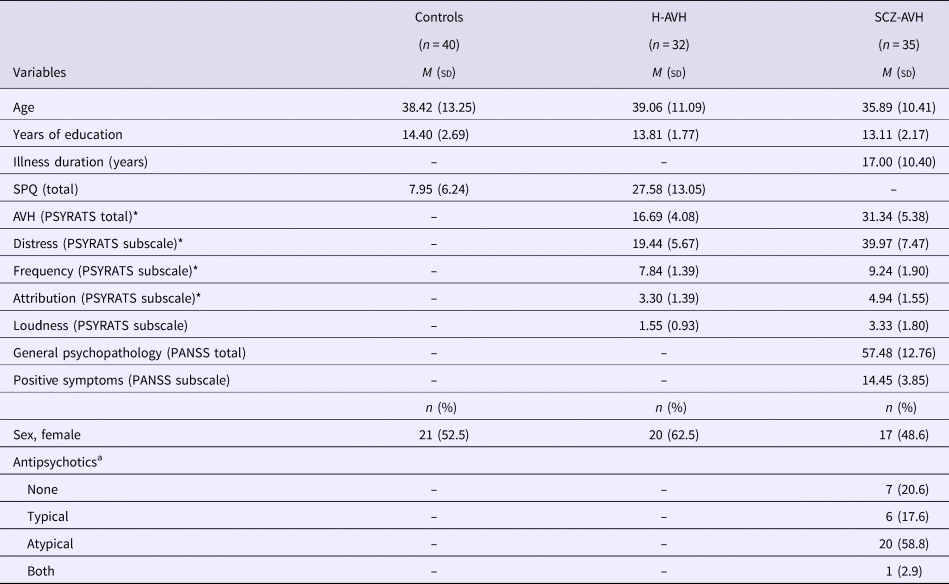
This study was approved by the Human Ethics Committee of the University Medical Center, Utrecht. All participants provided written informed consent, consistent with the Helsinki Declaration of 1975, as revised in 2008. A total of 107 participants were included in this study: 35 patients with a schizophrenia-spectrum disorder (SCZ-AVH; mean age = 35.89 ± 10.41), 32 healthy voice-hearers (H-AVH; mean age = 39.06 ± 11.09) and 40 healthy controls (HC), without a history of AVH (mean age = 38.42 ± 13.25). Most subjects overlapped with subjects comprising the de Weijer et al. (Reference de Weijer, Neggers, Diederen, Mandl, Kahn, Pol, Hilleke and Sommer2013) investigation (30/35 SCZ-AVH; 32/35 H-AVH; 35/40 controls). Key demographic and clinical characteristics are presented in Table 1.
Sample population and clinical characteristics: overall study population

* Significant difference between H-AVH and SCZ-AVH at the level of p < 0.05.
a Missing data for one patient.
Detailed recruitment and sample characteristics are described elsewhere (Daalman et al., Reference Daalman, Boks, Diederen, de Weijer, Blom, Kahn and Sommer2011). In brief, eligible SCZ-AVH patients were diagnosed with a schizophrenia-spectrum disorder according to the Diagnostic and Statistical Manual of Mental Disorders, Fourth Edition (DSM-IV; First and Gibbon, Reference First, Gibbon, Hersen, Haynes, Goldstein, Heiby, Hilsenroth, Beers, Segal and Thomas2004) and presented with AVHs. Diagnoses were confirmed with the Comprehensive Assessment of Symptoms and History (CASH; Andreasen et al., Reference Andreasen, Flaum and Arndt1992) by an independent psychiatrist. The Positive and Negative Syndromes Scale (PANSS; Kay et al., Reference Kay, Flszbein and Opfer1987) was used to assess general psychopathology (PANSS total) and positive symptoms (PANSS positive subscale) in the patient group.
The H-AVH subjects were recruited via an online self-report questionnaire (www.verkenuwgeest.nl), which quantifies hallucination tendencies in healthy individuals (Larøi et al., Reference Larøi, Marczewski and Van der Linden2004). Respondents endorsing voice-hearing items were selected for telephone screening by trained psychologists and invited to participate if they met the following criteria: (i) voices were distinct from thoughts; (ii) voices were experienced at least once a month, for over 1 year; (iii) no lifetime history of diagnosis or treatment for psychiatric disorders; (iv) no alcohol or drug abuse for at least 3 months; and (v) no lifetime history of a chronic somatic disorder (Daalman et al., Reference Daalman, Boks, Diederen, de Weijer, Blom, Kahn and Sommer2011). Individuals fulfilling these criteria were invited to undergo a psychiatric interview. For both healthy groups (H-AVH and HC), the absence of current or history of diagnosable psychopathology, including substance abuse, was confirmed with CASH (Andreasen et al., Reference Andreasen, Flaum and Arndt1992) and with the Structured Clinical Interview for Personality Disorders (First and Gibbon, Reference First, Gibbon, Hersen, Haynes, Goldstein, Heiby, Hilsenroth, Beers, Segal and Thomas2004) by an independent psychiatrist.
For both AVH groups, detailed characteristics of hallucinations were assessed using the Psychotic Symptom Rating Scale (PSYRATS; Haddock et al., Reference Haddock, McCarron, Tarrier and Faragher1999). Four hallucination dimensions were measured with PSYRATS (Woodward et al., Reference Woodward, Jung, Hwang, Yin, Taylor, Menon, Peters, Kuipers, Waters and Lecomte2014): Distress (Distress/Negative Content/Control), Frequency (Frequency/Duration/Disruption), Attribution (Attribution of voices: Location and Origin), and Loudness (Loudness). All clinical ratings were performed on the scan day by trained interviewers.
MRI acquisition and pre-processing
All diffusion MRI and T1-weighted scans were acquired between 2007 and 2010 at the University Medical Center, Utrecht, on a 1.5 T Philips Achieva MR scanner using an eight-channel SENSE head-coil. No scanner upgrades were performed during the study lifetime. For diffusion MRI, 30 volumes of diffusion-weighted images (DWI) with non-collinear diffusion gradient directions, and a baseline (b0) volume were obtained with single-shot EPI-DTI imaging sequence. Acquisition parameters were as follows: b-value = 1000 s/mm2, isotropic 2 mm × 2 mm × 2 mm voxels, image matrix = 128 × 128, repetition time/echo time = 7035/68 ms, FOV = 240 mm. A second set of diffusion volumes were acquired with the same parameters as the first set, but with a reversed k-space readout (anterior direction), to facilitate EPI distortion correction in the pre-processing pipeline. The parameters for T1-weighted images were as follows: slice thickness = 1 mm (without gap), field of view = 244 mm × 160 mm × 168 mm, repetition time/echo time = 9.87/4.6 ms, flip angle = 8°, data matrix size = 256 × 256.
Tract-based spatial statistics pipeline
Diffusion MRI data were pre-processed with FMRIB Software Library (FSL; Jenkinson et al., Reference Jenkinson, Beckmann, Behrens, Woolrich and Smith2012). This involved visual inspection for artifacts, during which eight scans were removed from the analysis (two controls, two H-AVH and four SCZ-AVH) due to poor image quality (movement artifacts, ghosting and signal drops). The remaining scans (sample population described in Table 1) underwent correction for EPI distortions by combining the phase flipped images; motion and gradient-induced eddy currents using affine registration to the first b0 image; brain masking performed with FSL's Brain Extraction Tool (BET; Jenkinson et al., Reference Jenkinson, Beckmann, Behrens, Woolrich and Smith2012) and subsequent manual editing in Slicer (https://www.slicer.org); computation of a FA volume by fitting a single tensor to the DWI data [as previously done in de Weijer et al. (Reference de Weijer, Neggers, Diederen, Mandl, Kahn, Pol, Hilleke and Sommer2013)], as well as computation of the FW fraction and tissue-specific FA (FAT) volumes by applying FW imaging (Pasternak et al., Reference Pasternak, Sochen, Gur, Intrator and Assaf2009). FW imaging applies a regularization framework to fit a two-compartment (bi-tensor) model that separates the contribution of FW from that of tissue-specific diffusivity (Pasternak et al., Reference Pasternak, Sochen, Gur, Intrator and Assaf2009). FW constitutes unrestricted extracellular water molecules found in cerebrospinal fluid, blood, or the interstitial space. Reduced FA may reflect increased partial volume of FW pools (e.g. in fibers adjacent to ventricles), rather than changes in the white matter microstructure. As such, disambiguating these distinct biological compartments (i.e. into FW and FAT) provides insight into the extracellular and microstructural underpinnings of FA deficits.
We used FSL's tract-based spatial statistics (TBSS; Smith et al., Reference Smith, Jenkinson, Johansen-Berg, Rueckert, Nichols, Mackay, Watkins, Ciccarelli, Cader, Matthews and Behrens2006) and ENIGMA-DTI protocols (http://enigma.ini.usc.edu/protocols/dti-protocols/) as a data-driven approach, which preclude the need for a priori hypotheses regarding tract/region-specific changes, in light of mixed findings across previous AVH studies. ENIGMA-DTI protocols were used to extract average diffusion (FA, FAT, FW) estimates from white matter regions of interest (ROIs; Jahanshad et al., Reference Jahanshad, Kochunov, Sprooten, Mandl, Nichols, Almasy, Blangero, Brouwer, Curran, de Zubicaray, Duggirala, Fox, Hong, Landman, Martin, McMahon, Medland, Mitchell, Olvera, Peterson, Starr, Sussmann, Toga, Wardlaw, Wright, Hulshoff Pol, Bastin, McIntosh, Deary, Thompson and Glahn2013). In brief, FA maps were registered to the common ENIGMA-DTI target, derived from 400 adult brains (Jahanshad et al., Reference Jahanshad, Kochunov, Sprooten, Mandl, Nichols, Almasy, Blangero, Brouwer, Curran, de Zubicaray, Duggirala, Fox, Hong, Landman, Martin, McMahon, Medland, Mitchell, Olvera, Peterson, Starr, Sussmann, Toga, Wardlaw, Wright, Hulshoff Pol, Bastin, McIntosh, Deary, Thompson and Glahn2013). Registration followed a two-step procedure performed with FSL (Jenkinson et al., Reference Jenkinson, Beckmann, Behrens, Woolrich and Smith2012): first, an affine transformation was computed to match the FA volume to the template (Andersson et al., Reference Andersson, Jenkinson and Smith2007); next, non-linear normalization applied the deformation field that warps the original FA volume to the template, setting the previous affine transformation as the starting estimate (Andersson et al., Reference Andersson, Jenkinson and Smith2010). The ENIGMA-DTI target was skeletonized with TBSS (Smith et al., Reference Smith, Jenkinson, Johansen-Berg, Rueckert, Nichols, Mackay, Watkins, Ciccarelli, Cader, Matthews and Behrens2006) and FA voxels were projected onto the skeleton mask. The FA-derived transformations and projection parameters were used to project FAT and FW maps onto the skeleton mask. Resulting skeletonized maps (FA, FAT, FW) were used to extract values from a subset of white matter regions comprising the ICBM-DTI white matter labels atlas (38 ROIs in total: 17 left and right lateralized respectively, as well as four midline structures; online Supplementary Table S1; Mori et al., Reference Mori, Wakana, Van Zijl and Nagae-Poetscher2005). To define ROIs, diffusion metrics were averaged over skeleton voxels traversing a given atlas region. Diffusion metrics were obtained from deep white matter structures (i.e. the skeleton) to minimize partial volume effects and from ROIs (rather than voxels), to reduce multiple comparisons and bolster statistical power, given the modest sample size.
GMT estimation
T1-weighted images were processed using the FreeSurfer automated neuroanatomical segmentation software (version 5.3; Fischl and Dale, Reference Fischl and Dale2000) to generate a three-dimensional model of the cortical surface for each participant. Surface-based cortical reconstruction was performed to derive the white matter and pial surfaces, from which GMT estimates were derived (Dale et al., Reference Dale, Fischl and Sereno1999; Fischl et al., Reference Fischl, Sereno and Dale1999, Reference Fischl, Salat, Busa, Albert, Dieterich, Haselgrove, Van Der Kouwe, Killiany, Kennedy and Klaveness2002, Reference Fischl, van der Kouwe, Destrieux, Halgren, Ségonne, Salat, Busa, Seidman, Goldstein and Kennedy2004; Fischl and Dale, Reference Fischl and Dale2000). All images were visually inspected to verify the accuracy of reconstructions and identify errors, including skull-strip errors, gross segmentation errors and white/gray matter and pial surface inaccuracies. Mean GMT was extracted from each region comprising the Desikan–Killiany atlas (68 cortical regions in total: 34 left and right lateralized, respectively; Desikan et al., Reference Desikan, Ségonne, Fischl, Quinn, Dickerson, Blacker, Buckner, Dale, Maguire and Hyman2006) using standard procedures for ROI extraction, provided by FreeSurfer. Cortical thickness was examined in ROIs (rather than voxels), to reduce multiple comparisons and bolster statistical power, given the modest sample size.
Statistical analyses
Between-group differences and overlap
All statistical analyses were performed in MATLAB (version 2017b). Diffusion and GMT metrics were normally distributed, as assessed with one-sample Kolmogorov–Smirnov test and homogeneity of variances was checked by Levene's test. The FA and GMT measures were initially compared between SCZ-AVH and HC groups at each white matter ROI by general linear models (GLMs) to regress out variance associated with sex and age. The false discovery rate (FDR; Benjamini and Heller, Reference Benjamini and Heller2007) was used to provide multiple comparisons error control across the regions (38 FA regions; 68 GMT regions). Regions showing a significant group effect after FDR correction were then compared by GLMs between H-AVH and HC groups. Regions surviving FDR correction were taken to reflect shared pathology between H-AVH and SCZ-AVH cohorts. To determine the nature of any overlapping FA deficits, subsidiary analyses examined FAT and FW diffusion metrics in regions with overlapping FA abnormalities between H-AVH and SCZ-AVH. Support for a two-step GLM procedure was based on our aim to identify pathology affecting both AVH groups relative to HCs, irrespective of differences occurring between AVH groups (e.g. significantly lower FA in SCZ-AVH compared with H-AVH).
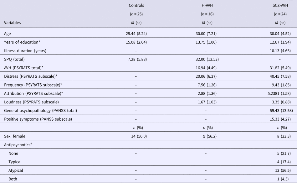
All analyses were repeated on a subsample of younger subjects (<40 years of age) to minimize the effects of aging, cumulative medication exposure, illness chronicity, or repeated AVH experiences. A threshold of 40 years of age was used to coincide with the median split – a robust statistical means to dichotomize data (Iacobucci et al., Reference Iacobucci, Posavac, Kardes, Schneider and Popovich2015), as well as the timing of peak white matter maturation, as shown through diffusion imaging lifespan studies (Westlye et al., Reference Westlye, Walhovd, Dale, Bjørnerud, Due-Tønnessen, Engvig, Grydeland, Tamnes, Østby and Fjell2009; Cropley et al., Reference Cropley, Klauser, Lenroot, Bruggemann, Sundram, Bousman, Pereira, Di Biase, Weickert, Weickert, Pantelis and Zalesky2016). Refer to Table 2 for demographic and clinical characteristics of the younger subsample.
Sample population and clinical characteristics: younger subset

* Significant difference between H-AVH and SCZ-AVH at the level of p < 0.01
a Missing data for one patient.
Demographic and clinical characteristics
Analysis of variance was used to test differences in age and number of years educated between the three study groups. Differences in clinical variables (PYSRATS Total and four subscales) between H-AVH and SCZ-AVH groups were analyzed with independent t tests. Partial correlations were used to test relationships between clinical variables (PYSRATS Total and four subscales) and diffusion metrics across the pooled H-AVH and SCZ-AVH groups, after controlling for group membership as a binary variable. To investigate potential substrates of hallucinatory phenomena, correlations were confined to regions with overlapping pathology between H-AVH and SCZ-AVH groups. The Bonferroni correction provided error control across the correlation analyses (5 symptom scores) and thus, an adjusted p-value of 0.01 was considered statistically significant.
Results
Demographic and clinical characteristics
There were no significant differences in age or sex between SCZ-AVH, H-AVH, and HC groups (p > 0.05). Compared with controls, H-AVH and SCZ-AVH were educated for fewer years (p < 0.05; see Table 1). Hallucination severity differed between SCZ-AVH and H-AVH groups, with SCZ-AVH showing significantly higher total PSYRATS scores, as well as Distress, Frequency, and Attribution subscale scores compared with the H-AVH group (p < 0.05; see Table 1). The SCZ-AVH and H-AVH groups did not differ with respect to Loudness (PSYRATS subscale) scores (p > 0.05). There was no relationship between hallucination severity (total PSYRATS scores, as well as Distress, Frequency, and Attribution subscale scores) and age (p > 0.05). Furthermore, hallucination severity did not differ between younger and older subgroups within SCZ-AVH and H-AVH cohorts, respectively (online Supplementary Table S2).
Microstructural changes in SCZ-AVH and H-AVH
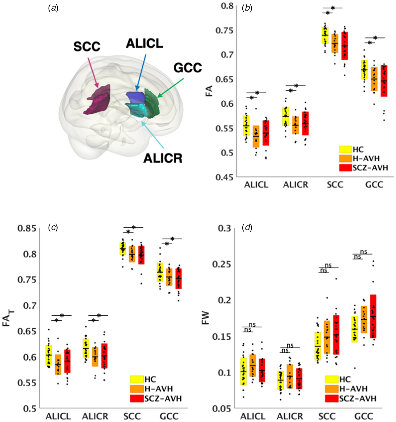
Compared with HCs, schizophrenia patients showed reduced FA in 34/38 ROIs (FDRp < 0.05; online Supplementary Table S3). Post-hoc analyses constrained to these regions did not detect any differences between H-AVH and HCs (FDRp > 0.05). In the younger subgroups, fewer regions (24/38) showed reduced FA in SCZ-AVH patients relative to HCs (FDRp < 0.05; online Supplementary Table S4). Post-hoc comparisons constrained to these regions revealed lower FA in younger H-AVH compared with HCs in four regions (Fig. 1a, b): (i) left (t = 3.62, FDRp = 0.001); and (ii) right anterior limbs of the internal capsule (ALIC; t = 3.22, FDRp = 0.003); (iii) splenium of the corpus callosum (SCC; t = 2.87, FDRp = 0.007); and (iv) genu of the corpus callosum (GCC; t = 2.85, FDRp = 0.007). Supplemental unconstrained comparisons (i.e. across all 38 white matter regions) did not reveal differences localized elsewhere in H-AVH subjects, suggesting that alterations were specifically confined to regions associated with schizophrenia pathology.
Overlapping deficits between younger healthy voice-hearers and schizophrenia patients. (a) Surface rendering displays regions with overlapping FA and FAT pathology. (b) Boxplots represent regions with overlapping FA pathology for healthy controls (HC), healthy voice-hearers (H-AVH), and schizophrenia patient (SC-AVH) groups across. (c) Regions with overlapping FA reductions were investigated for (c) cellular-specific (FAT) and (d) free-water volume (FW) changes. Boxes represent standard deviations and dots reflect data points for each subject across the groups, after multiple comparison correction with the false discovery rate. Stars denote significant differences and ns, not significant. Overlapping pathology reflects regions where the H-AVH and SCZ-AVH patients displayed common significant differences from controls.

To investigate the nature of shared FA abnormalities, FAT and FW diffusion metrics were examined within these four white matter regions. Significantly lower FAT was found in SCZ-AVH and H-AVH relative to controls across these four regions (FDRp < 0.05; Fig. 1c), indicating shared cellular-specific white matter pathology between SCZ-AVH and H-AVH groups. No FW differences emerged between SCZ-AVH and H-AVH relative to HC (FDRp > 0.05; Fig. 1d). The impact of sex was not significant across any region or diffusion metric (FDRp > 0.05).
GMT changes in SCZ-AVH and H-AVH
Compared with HCs, schizophrenia patients displayed reduced GMT spanning 55/68 ROIs (FDRp < 0.05; online Supplementary Table S5). Across these regions associated with schizophrenia pathology, no significant differences were found between H-AVH and controls. However, two regions displayed reduced GMT in H-AVH subjects before correcting for multiple comparisons, including the left bank of the superior temporal gyrus (t = 2.00, p = 0.049, uncorrected) and the right fusiform gyrus (t = 2.15, p = 0.035, uncorrected). In the younger study groups, 37/68 ROIs showed lower GMT in SCZ-AVH compared with HCs (FDRp < 0.05; online Supplementary Table S6). Within these regions, no significant GMT reductions were observed for younger H-AVH subjects compared with HCs (FDRp > 0.05). Similar to the overall H-AVH group, younger H-AVH subjects displayed reduced GMT in three temporal regions before correcting for multiple comparisons: (i) the left bank of the superior temporal gyrus (t = 2.48, p = 0.018, uncorrected); (ii) the right fusiform gyrus (t = 2.26, p = 0.029, uncorrected); and (iii) the left middle-temporal gyrus (t = 2.08, p = 0.044, uncorrected). The impact of sex was not significant across any region (FDRp > 0.05).
Clinical correlations with overlapping pathology in white matter microstructure
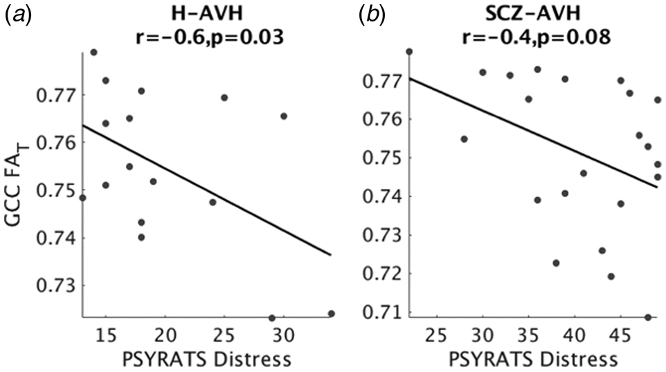
In the younger (<40 years) H-AVH and SCZ-AVH groups who displayed overlapping white matter pathology, there was a significant negative correlation between FAT in the GCC and the degree of distress associated with AVHs (r = −0.6, p = 0.007, Bonferroni corrected). This relationship was significant in H-AVH subjects (r = −0.6, p = 0.03; Fig. 2a) and trended toward significance in SCZ-AVH patients (r = −0.4, p = 0.08; Fig. 2b). There was no relationship between FA and hallucination symptom severity (PSYRATS total or subscale scores). No overlapping deficits emerged across FW or GMT metrics, and were hence, omitted from symptom correlations.
Associations between FAT and hallucination distress. Decreased FAT in the genu of the corpus callosum (GCC) was associated with increased hallucination distress in (a) healthy voice-hearers (H-AVH) and (b) schizophrenia patients (SCZ-AVH). Correlations controlled for age.

Discussion
We tested for overlapping abnormalities in regional white matter microstructure and GMT between schizophrenia patients and healthy voice-hearers, relative to age- and sex-matched HC. While significant differences were not identified in healthy voice-hearers overall, younger AVH groups displayed overlapping microstructural deficits across several white matter regions, regardless of a clinical diagnostic status. These abnormalities were driven by cellular-specific white matter deficits, rather than changes in the extracellular FW compartment and were correlated with the degree of distress associated with hallucinatory experiences. Schizophrenia patients also displayed globally reduced GMT, as well as more extensive white matter deficits than healthy voice-hearers, which could reflect more evolved AVH symptomatology or the compounding of other biological/psychological disease processes in schizophrenia.
Our findings indicate a crucial role for white matter deficits in AVH. Consistent with several previous studies (Hubl et al., Reference Hubl, Koenig, Strik, Federspiel, Kreis, Boesch, Maier, Schroth, Lovblad and Dierks2004; Xi et al., Reference Xi, Guo, Li, Chang, Sun, Zhu, Liu, Cui, Chen and Wang2016; Zhang et al., Reference Zhang, Gao, Zhu, Wang, Fan, Ma, Ma and Yang2018), white matter deficits affected the corpus collosum and internal capsule, confirming involvement of widely distributed commissural, frontal, reciprocal thalamo-cortical and auditory-linked circuitry in the generation of AVH. However, different white matter tracts have been implicated across prior reports, including the superior longitudinal fasciculus (Seok et al., Reference Seok, Park, Chun, Lee, Cho, Kwon and Kim2007; Ćurčić-Blake et al., Reference Ćurčić-Blake, Nanetti, van der Meer, Cerliani, Renken, Pijnenborg and Aleman2015), inferior longitudinal fasciculus (Seok et al., Reference Seok, Park, Chun, Lee, Cho, Kwon and Kim2007), inferior fronto-occipital fasciculus (Ćurčić-Blake et al., Reference Ćurčić-Blake, Nanetti, van der Meer, Cerliani, Renken, Pijnenborg and Aleman2015; Oestreich et al., Reference Oestreich, McCarthy-Jones and Whitford2016), anterior thalamic radiation (Ćurčić-Blake et al., Reference Ćurčić-Blake, Nanetti, van der Meer, Cerliani, Renken, Pijnenborg and Aleman2015) and AF (the AF was not examined in the current study; Hubl et al., Reference Hubl, Koenig, Strik, Federspiel, Kreis, Boesch, Maier, Schroth, Lovblad and Dierks2004; Psomiades et al., Reference Psomiades, Fonteneau, Mondino, Luck, Haesebaert, Suaud-Chagny and Brunelin2016). As nearly all previous studies examined AVH in the context of schizophrenia, abnormalities likely reflected a combination of disease processes and AVH-related phenomena. Indeed, we previously found decreased FA within the AF of schizophrenia patients, but not healthy voice-hearers (de Weijer et al., Reference de Weijer, Mandl, Diederen, Neggers, Kahn, Pol and Sommer2011). It is further notable that several previous studies specifically focused on various language-linked white matter fibers of interest (de Weijer et al., Reference de Weijer, Mandl, Diederen, Neggers, Kahn, Pol and Sommer2011; Ćurčić-Blake et al., Reference Ćurčić-Blake, Nanetti, van der Meer, Cerliani, Renken, Pijnenborg and Aleman2015; Oestreich et al., Reference Oestreich, McCarthy-Jones and Whitford2016) and hence, may have missed abnormalities localized elsewhere.
Our findings, when considered together with previous work, suggest that white matter pathology underlying AVH involves pathways beyond language and auditory-linked circuits. The specific connections disrupted in younger AVH subjects could implicate auditory perception failures in AVH manifestation. Specifically, reduced interhemispheric (GCC) and thalamocortical (ALIC) information transfer to frontal regions could underlie attention and impulse control deficits, resulting in self-monitoring failures (Hugdahl et al., Reference Hugdahl, Løberg, Specht, Steen, van Wageningen and Jørgensen2008). In addition, the SCC carries fibers to temporal, parietal and occipital lobes and thus, deficits in this structure could interrupt information flow between regions which are crucial for delivering and coordinating speech percepts (Friederici et al., Reference Friederici, von Cramon and Kotz2007). These notions accord with a more sophisticated view of AVH as emerging from comprised structural connections between multiple extra-sensory, association and perception areas, as opposed to pathways specific to the sensory modality in question (Penfield and Perot, Reference Penfield and Perot1963; Stephan et al., Reference Stephan, Friston and Frith2009).
After determining the spatial extent of AVH-specific pathology, we next sought to examine the possible mechanisms underlying connectional deficits. To this end, we applied FW imaging, allowing for greater interrogation of distinct biological compartments – not previously possible with conventional DTI metrics. Using this method, we could ascribe FA abnormalities to cellular-specific FAT changes, i.e. decreased anisotropy of water diffusion in the tissue, as opposed to an increase in extracellular volume. Although FAT cannot establish the exact tissue-specific pathology, taken together with repeated neuropathologic findings of altered oligodendrocyte morphology in schizophrenia patients (Uranova et al., Reference Uranova, Vikhreva, Rachmanova and Orlovskaya2011, Reference Uranova, Vikhreva, Rachmanova and Orlovskaya2014), it is possible microstructural changes in AVH result from a myelin deficit. Indeed, myelin pathology has been proposed as the primary mechanism accounting for hallucinatory phenomena, by way of disrupting temporal coordination between spatially disparate neuronal populations, for example, through slowing of the conduction velocity and inducing synaptic changes (Whitford et al., Reference Whitford, Ford, Mathalon, Kubicki and Shenton2010a). With regard to the corollary discharge system, conduction delays could slow ‘efference copies’ of neural transmission of motor commands, which are consequently received too late by the auditory cortex to accurately predict and suppress the sensations of an impending action (Whitford et al., Reference Whitford, Ford, Mathalon, Kubicki and Shenton2010a). Of relevance to our findings, thalamic pathway involvement in corollary discharge failures can occur by way of impairing the updating of sensory information, resulting in less coherent percepts (Bellebaum et al., Reference Bellebaum, Daum, Koch, Schwarz and Hoffmann2005). Furthermore, the corollary discharge mechanism has been shown to rely on cross-hemispheric transfer for relaying corollary discharge signals (Colby et al., Reference Colby, Berman, Heiser and Saunders2005) and integrating auditory percepts (Westerhausen and Hugdahl, Reference Westerhausen and Hugdahl2008). Therefore, our findings may accord with accumulating evidence that AVH involves dysfunctional corollary discharge mechanisms, and in particular, suggest that tissue-specific white matter changes involving key corollary discharge circuits contribute to the pathophysiology underlying AVH.
Decreased FAT in the GCC was correlated to AVH-related distress (sum of PSYRATS items: negative content, distress, and control), which was significant in healthy voice-hearers and showed a tendency toward significance in patients. This association was not detected with the more general FA metric, suggesting that cellular-specific white matter changes are more sensitive to AVH symptom dimensions. White matter of the GCC interconnects interhemispheric prefrontal regions involved in a wide range of somatosensory, attention, and language processes (Hinkley et al., Reference Hinkley, Marco, Findlay, Honma, Jeremy, Strominger, Bukshpun, Wakahiro, Brown, Paul, Barkovich, Mukherjee, Nagarajan and Sherr2012; Edwards et al., Reference Edwards, Sherr, Barkovich and Richards2014) and disruptions to this region were previously associated with stress (Jackowski et al., Reference Jackowski, Douglas-Palumberi, Jackowski, Win, Schultz, Staib, Krystal and Kaufman2008), early psychosis (Di Biase et al., Reference Di Biase, Cropley, Baune, Olver, Amminger, Phassouliotis, Bousman, McGorry, Everall, Pantelis and Zalesky2017), and positive symptoms in schizophrenia (Whitford et al., Reference Whitford, Kubicki, Schneiderman, O'Donnell, King, Alvarado, Khan, Markant, Nestor, Niznikiewicz, McCarley, Westin and Shenton2010b). Extending this work, our findings indicate that anterior callosal fibers may be vulnerable to AVH-induced distress. Notably, greater self-reported distress accompanying psychosis symptoms may increase risk for developing full-blown psychotic symptoms and transition to other mental health difficulties (Yung et al., Reference Yung, Buckby, Cotton, Cosgrave, Killackey, Stanford, Godfrey and McGorry2005; Baumeister et al., Reference Baumeister, Sedgwick, Howes and Peters2017). Consistent with this finding, we observed significantly higher distress scores in patients compared with healthy voice-hearers. Interestingly, this was accompanied by comparable FAT reductions within the GCC across younger clinical and non-clinical AVH groups compared with controls, indicating that greater distress likely involves disruption to additional brain regions, as seen in patient groups. Given that self-reported distress increases risk for developing psychosis (Yung et al., Reference Yung, Buckby, Cotton, Cosgrave, Killackey, Stanford, Godfrey and McGorry2005; Baumeister et al., Reference Baumeister, Sedgwick, Howes and Peters2017) and that GCC deficits transcend diagnostic status, white matter deficits involving the GCC in particular may signify poor outcomes for younger individuals with AVH symptoms.
Overlapping pathology and symptom correlations in this study were notably constrained to younger AVH groups, as no differences emerged between healthy voice-hearers and controls in the overall study population. We reasoned that pathological variance could increase with age, stress of repeated AVH experiences, and general medication exposure. These confounds could result in pathology that is differentially localized in spatial extent or severity, precluding group-level findings. Indeed, we observed greater variance in average FA across the overall sample (sd = 0.022) compared with the younger subset (sd = 0.019), in line with our expectation. Such inter-subject heterogeneity, combined with the subtle alterations that characterize these otherwise healthy individuals, may explain our null result in the overall healthy voice-hearing population. In contrast, we observed overlap across younger AVH subgroups, where homogenous pathology may relate more purely to AVH symptom constructs, rather than extraneous age, stress, or other chronicity effects. However, it is possible that similarities across younger subgroups might not relate to AVH as an isolated construct, but rather as part of a general vulnerability to psychosis or schizophrenia-spectrum disorders (Sommer et al., Reference Sommer, Daalman, Rietkerk, Diederen, Bakker, Wijkstra and Boks2010). For example, it is conceivable that symptoms experienced by younger healthy voice-hearers worsen overtime or that other psychosis symptoms transpire, bringing about clinical risk for schizophrenia-spectrum disorders (Yung et al., Reference Yung, Buckby, Cotton, Cosgrave, Killackey, Stanford, Godfrey and McGorry2005). However, given that most younger healthy voice-hearers missed the peak window of risk for psychosis onset (i.e. late adolescence to early adulthood; McGrath et al., Reference McGrath, Saha, Al-Hamzawi, Alonso, Andrade, Borges, Bromet, Oakley Browne, Bruffaerts, Caldas de Almeida, Fayyad, Florescu, de Girolamo, Gureje, Hu, de Jonge, Kovess-Masfety, Lepine, Lim, Navarro-Mateu, Piazza, Sampson, Posada-Villa, Kendler and Kessler2016), and did not display other dimensions of psychopathology at the time of this study; it is reasonable to ascribe our observed white matter deficits specifically to dimensions of AVH, rather than psychosis risk more generally.
Insofar as we did not detect significant gray matter abnormalities in the overall or younger healthy voice-hearers, our findings do not support a central role for GMT alterations in AVH. However, it is noteworthy that the largest differences in healthy voice-hearers relative to controls, involved thinning of key brain regions for language processing and recognition, including the superior temporal gyrus, right fusiform gyrus, and left middle-temporal gyrus. Reduced thickness and volume in extra-sensory auditory regions are consistently reported in association with AVH within a schizophrenia context (Allen et al., Reference Allen, Larøi, McGuire and Aleman2008; Palaniyappan et al., Reference Palaniyappan, Balain, Radua and Liddle2012) and are proposed to reflect neuropil loss, which could underlie dysfunctional cortical activations (van Swam et al., Reference van Swam, Federspiel, Hubl, Wiest, Boesch, Vermathen, Kreis, Strik and Dierks2012). It may be speculated that our results did not survive multiple comparison correction because cortical thinning is mild in healthy voice-hearers. Notably, in addition to cortical thinning, the years of education and clinical presentation (i.e. AVH severity) of the healthy voice-hearers in this study exhibited intermediate properties of the patient and HC groups, in line with a quasi-dimensional view (Claridge and Beech, Reference Claridge, Beech, Raine, Lencz and Mednick1995) of AVH and other psychotic symptoms. However, it is alternatively possible that reduced thickness across these regions was due to chance and hence, non-essential to the pathophysiology underlying AVH. If gray matter abnormalities are indeed involved in the pathophysiology of AVH, it is our contention that they are spatially constrained to auditory association areas, relative to more widely distributed connectional deficits.
Limitations
Our findings of cellular-specific white matter changes were limited to younger study groups, comprising relatively small samples. Furthermore, a portion of these healthy voice-hearers may develop clinical risk or frank psychosis, and hence, we cannot discount the possibility that the observed changes relate to psychosis risk or other illness dimensions. Key risk factors that mediate vulnerability to psychosis and AVH manifestation, including environmental (e.g. trauma exposure; Fusar-Poli et al., Reference Fusar-Poli, Tantardini, De Simone, Ramella-Cravaro, Oliver, Kingdon, Kotlicka-Antczak, Valmaggia, Lee, Millan, Galderisi, Balottin, Ricca and McGuire2017) and genetic (e.g. family history of psychiatric illness; Agerbo et al., Reference Agerbo, Sullivan, Vilhjalmsson, Pedersen, Mors, Børglum, Hougaard, Hollegaard, Meier and Mattheisen2015) factors, were unavailable for examination by the current study. Therefore, assessment of white matter pathology in younger individuals with AVH in larger samples utilizing risk assessments and longitudinal designs is warranted, in order to map symptom-linked white matter trajectories. Finally, while we observed cellular-specific deficits in AVH, we cannot disentangle the specific mechanisms accounting for changes within the tissue compartment (e.g. demyelination and axonal degeneration), or the relationship to functional or corollary discharge mechanisms. Future multimodal studies that combine advanced white matter mapping with functional parameters will help to resolve the precise pathophysiological mechanisms, the relationship to corollary discharge failures, as well as the temporal course of functional v. anatomical connectivity alterations invoked by AVH experiences.
Conclusions
In summary, we found evidence for overlapping white matter abnormalities across younger schizophrenia patients and healthy voice-hearers. These commonalities were characterized by cellular-specific deficits in white matter microstructure, involving callosal, frontal, and thalamo-cortical circuitry. However, the structural basis of AVH could not be straightforwardly ascribed to all AVH subjects, implying that advancing age and other illness-related contributions to brain structure possibly dilute associations with AVH. Future studies could address this complexity by longitudinally mapping the progression of white and gray matter abnormalities in individuals experiencing AVH.
Supplementary material
The supplementary material for this article can be found at https://doi.org/10.1017/S0033291719000205
Author ORCIDs
Maria Angelique Di Biase, 0000-0002-7100-651X.
Acknowledgements
We wish to gratefully acknowledge all participants for making this study possible.
Financial Support
This work was supported by the following National Institutes of Health (NIH) grants: R01MH108574, R01MH102377, R01MH074794, P41EB015902.
Conflict of interest
None.
Ethical standards
The authors assert that all procedures contributing to this work comply with the ethical standards of the relevant national and institutional committee on human experimentation with the Helsinki Declaration of 1975, as revised in 2008. The study protocol was approved by the ethics committee of each participating institution and all participants provided written informed consent, consistent with the Helsinki Declaration of 1975, as revised in 2008.