Introduction
Neurocysticercosis (NCC) is a common infection of the central nervous system (CNS) with a larval stage of the cestode Taenia solium in humans and pigs. It is considered to be a frequent but neglected parasitic disease worldwide (Garcia et al., Reference Garcia, Gilman, Gonzalez, Verastegui, Rodriguez, Gavidia, Tsang, Falcon, Lescano, Moulton, Bernal and Tovar2003a; Fleury et al., Reference Fleury, Escobar, Fragoso, Sciutto and Larralde2010; Del Brutto, Reference Del Brutto2012). The life cycle of the parasite involves humans as the definitive host for the adult tapeworm, which is known as taeniosis, whereas pigs are the main intermediate hosts for the larval tapeworm, commonly referred to as a cyst (Carpio, Reference Carpio2002). Humans acquire taeniosis through the consumption of raw or undercooked infected pork (Pawlowski, Reference Pawlowski, Singh and Prabhakar2002). On the other hand, pigs become infected with cysticercosis following ingestion of viable T. solium eggs that develop into cysts in different parts of the body, including the CNS (De Aluja & Vargas, Reference De Aluja and Vargas1988; Fleury et al., Reference Fleury, Morales, Bobes, Dumas, Yánez, Piña, Carrillo-Mezo, Martínez, Fragoso, Dessein, Larralde and Sciutto2006). There is an increasing number of reported cases of human cysticercosis following accidental ingestion of eggs from contaminated food or water (Del Brutto, Reference Del Brutto2012; Mwanjali et al., Reference Mwanjali, Kihamia, Kakoko, Lekule, Ngowi, Johansen, Thamsborg and Willingham2013). In developed countries, NCC prevalence is rising due to an increase in human mobility (Garcia et al., Reference Garcia, Gilman, Gonzalez, Verastegui, Rodriguez, Gavidia, Tsang, Falcon, Lescano, Moulton, Bernal and Tovar2003a).
The condition of NCC in humans might be asymptomatic or symptomatic with variable clinical presentations (Chavarria et al., Reference Chavarria, Fleury, Garcia, Marquez, Fragoso and Sciutto2005). The heterogeneity in clinical presentations might be related to the location, developmental stage of the parasite and the degree of host immune reaction, among other factors (White, Reference White2000). Patients with cysts in the parenchyma of the brain may show symptoms such as epileptic seizures and headaches (Kerstein & Massey, Reference Kerstein and Massey2010). Patients with extra-parenchymal cysts may experience a block in the outflow of cerebrospinal fluid, leading to increased intracranial pressure and hydrocephalus (Garcia et al., Reference Garcia, Gilman, Gonzalez, Verastegui, Rodriguez, Gavidia, Tsang, Falcon, Lescano, Moulton, Bernal and Tovar2003a; Suri et al., Reference Suri, Goel, Ahmad, Vellimana, Sharma and Mahapatra2008). Finally, patients having NCC with cysts in the spinal cord often suffer back pain and paralysis of the lower body parts (Rossi et al., Reference Rossi, Sestari and Cerioni2006; Gupta et al., Reference Gupta, Singh, Gupta, Singh and Azam2009).
Clinical signs of NCC in pigs are not common, although occasional heavy infections do occur. This could be explained by pigs being slaughtered before the death of tapeworm cysts, suggesting that inflammation of the host tissues does not occur, which is unlike the case in humans (Garcia et al., Reference Garcia, Evans, Nash, Takanayagui, White, Botero, Rajshekhar, Tsang, Schantz, Allan, Flisser, Correa, Sarti, Friedland, Martinez, Gonzalez, Gilman and Del Brutto2002). Another explanation could be linked to differences in the location of cysts in humans and pigs, with different inflammatory responses being invoked (Saenz et al., Reference Saenz, Ramirez, Aluja, Escobar, Fragoso, Morales, Perez-Tamayo, Rosetti, Larralde, Sciutto and Fleury2008). The difference might also be attributed to a lack of solid clinical studies focusing on describing the pathogenesis and clinical signs in T. solium cyst-infected pigs. Only a single study has reported clinical signs of NCC in pigs, as being excessive salivation and blinking as well as lacrimation (Prasad et al., Reference Prasad, Chawla, Prasad, Tripathi, Husain and Gunta2006).
The link between cyst localization, inflammation and clinical manifestations in human NCC has been reported, supporting the hypothesis of possible compartmentalization of the inflammatory response within the CNS (Chavarria et al., Reference Chavarria, Fleury, Garcia, Marquez, Fragoso and Sciutto2005). This suggests that a specific inflammatory response is elicited locally to the invading cysts, leading to heterogeneity in clinical manifestations (Pittella, Reference Pittella1997; Rahalkar et al., Reference Rahalkar, Shetty, Kelkar, Kelkar, Kinare and Ambardekar2000; White, Reference White2000). Cases of human NCC with multiple cysts in different developmental stages have been observed (Fleury et al., Reference Fleury, Dessein, Preux, Dumas, Tapia, Larralde and Sciutto2004), suggesting that local immune activity could be responsible (Fleury et al., Reference Fleury, Dessein, Preux, Dumas, Tapia, Larralde and Sciutto2004; Chavarria et al., Reference Chavarria, Fleury, Garcia, Marquez, Fragoso and Sciutto2005). However, histopathological lesions corresponding to cyst localization have not been described in porcine NCC. In addition, geographical genetic variation of T. solium cysts has been reported, with possible differences in pathology and clinical symptoms (Ito et al., Reference Ito, Yamasaki, Nakao, Sako, Okamoto, Sato, Nakaya, Margono, Ikejima, Kassuku, Sonia, Afonso, Ortiz, Plancarte, Zoli, Geerts and Craig2003; Vega et al., Reference Vega, Pinero, Ramanankandrasana, Dumas, Bouteille, Fleury, Sciutto, Larralde and Fragoso2003).
Studies on inflammatory responses in the human brain have been limited by the chronic progression of the disease, limited specimen availability (Cardona et al., Reference Cardona, Restrepo, Jaramillo and Teale1999; Grewal et al., Reference Grewal, Kaur, Bhatti, Sawhney, Ganguly, Mahajan and Malla2000; Restrepo et al., Reference Restrepo, Alvarez, Castano, Arias, Restrepo, Trujillo, Colegial and Teale2001) and ethical considerations. In view of the fact that it is difficult to conduct an experimental infection in pigs (Verastegui et al., Reference Verastegui, Gonzales, Gilman, Gavidia, Falcon, Bernal and Garcia2000), the use of naturally infected pigs seems to be the best option for understanding the pathogenesis of this parasitic disease. This study was conducted to further describe the distribution and localization of T. solium cysts and the corresponding host inflammatory cells in different brain compartments of naturally infected pigs.
Materials and methods
Collection and examination of cysts
Pigs employed in this study were purchased from rural smallholder pig farmers in Kongwa district, Dodoma, Tanzania, where the gross evaluation of cysts was carried out. The histopathology work was conducted at the Faculty of Veterinary Medicine, Sokoine University of Agriculture (SUA), Morogoro, Tanzania.
The study involved 17 pigs naturally infected with T. solium, eight males and nine females. All pigs were crosses between local pigs and large white pigs. The inclusion criteria were age of 12–24 months, and having five or more T. solium tongue cysts (Phiri et al., Reference Phiri, Dorny, Gabriel, Willingham, Sikasunge, Siziya and Vercruysse2006). Pregnant or lactating sows were excluded. The selected pigs were observed for the presence of clinical signs related to NCC and monitored for at least 24 h before slaughter. Additional health information of the studied pigs was sought from their owners. Pigs were slaughtered by a local butcher in Mlali village and, after the completion of bleeding, they were decapitated at the level of the atlanto-occipital joint. A procedure described by Agerholm (Reference Agerholm and Jensen2011) was adopted to remove the brain from the cranium and the spinal cord from the vertebral canal.
The cerebral part of the brain was divided into its left and right hemispheres to evaluate the distribution of cysts. Then, each of the hemispheres was divided into the frontal, temporal, parietal and occipital lobes using the large sulci as landmarks. The cerebellum was also longitudinally sectioned into right and left parts. Serial transverse sections at intervals of about 0.5–1.0 cm were made on the brain parts to examine and enumerate the cysts. The cysts were evaluated to determine whether they were located in the extra-parenchymal part, i.e. dorsal subarachnoid, subarachnoid base or intraventricular (in other words, those parts which were not completely surrounded by the brain tissue), or in the parenchymal part of the frontal lobe, temporal lobe, parietal lobe, occipital lobe and cerebellum. In addition, the status of cysts was evaluated to establish whether they were viable or dead. A viable cyst contained an intact vesicular membrane filled with transparent fluid with an invaginated scolex that appeared as a small white spot.
Histopathology of cysts
A total of seven pig brains were randomly selected from the 17 examined brains for histopathology. About 5 cm3 of brain tissues with a cyst from the subarachnoid or parenchymal locations were collected and preserved in 10% formalin for 24 h before they were transferred to 70% ethanol, in which they remained until paraffin embedded. Paraffin-embedded blocks were cut into sections of thickness 5 μm, mounted on examination slides and stained with haematoxylin and eosin for histopathology. The slides were examined under a light microscope. The types of cells and the extent of inflammation around the cysts were evaluated and the lesions were classified into stages I, III and V as proposed by Pérez-Torres et al. (Reference Pérez-Torres, Ustarroz, Constantino, Villalobos and De2002). In stage I, the viable parasite is surrounded with minimal inflammatory cells and a thin layer of type I collagen. In stage III the lesion shows increased inflammation leading to the destruction of the parasite and, in stage V, the disintegrating parasite is surrounded by an extensive granulomatous infiltrate. Stage I is referred to as an early lesion and stages III and V as late lesions (Sikasunge et al., Reference Sikasunge, Phiri, Johansen, Willingham and Leifsson2008).
Data analysis
Data were analysed using Microsoft Excel 2010 (Microsoft Corp., Redmond, Washington, USA) and Epi Info version 7 (Centers for Disease Control and Prevention; http://wwwn.cdc.gov/epiinfo/7/). Descriptive statistics were used to compute the means and proportions of cyst counts in different parts of the brain. For comparison of the means of the total cysts per distribution/localization, the non-parametric Mann–Whitney or Wilcoxon test was used. The relationship between clinical signs and sex was determined using the χ2 test. All values of P< 0.05 were considered statistically significant.
Results
The 17 selected pigs did not show signs of diseases other than cysticercosis. However, dullness and sluggishness were observed in four of the 17 pigs. In addition, the pig owners reported some further clinical signs, such as somnolence, apathy and occasional loss of consciousness for about 5–10 min among the pigs that had sluggishness. Additionally, hypersensitivity to sound and touch was reported in one of the studied pigs. The observed clinical signs were not associated with the sex of the pigs (P= 0.87), total cyst count (P= 0.26) or the anatomical locations of the cysts, such as dorsal subarachnoid (P= 0.15), subarachnoid base (P= 0.16), parenchymal (P= 0.20) and ventricular (P= 0.23).
All pigs that were selected on the basis of the presence of five or more tongue cysts had NCC at necropsy, and all the cysts that were grossly evaluated were viable. A total of 1030 cysts were evaluated and enumerated in 17 pig brains. The parasitic load varied from 5 to 160 cysts per brain, with a mean of 61 (SD 48). The 1030 cysts were distributed in five regions of the brain, namely, 39.73% in the frontal lobe, 19.67% in the temporal lobe, 20.25% in the parietal lobe, 19.96% in the occipital lobe and 0.39% in the cerebellum. However, there were no significant differences in the proportions of cyst distribution when different locations were compared, except for the cerebellum. The variation in the total number of cysts located in the two cerebral hemispheres was not statistically significant (P= 0.72). No cysts were found in the spinal cord. In pigs with a high number of cysts, the cysts were more widely distributed. As for localization, the cysts were found more or less equally in the dorsal subarachnoid and parenchyma, 47.9% and 46.9%, respectively, with 4.4% in the subarachnoid base and 0.9% in the ventricles.
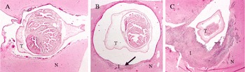
Moreover, a total of 96 cysts were evaluated histopathologically in seven of the studied pig brains. Histopathology results revealed cysts in a more or less similar inflammatory stage, regardless of the anatomical locations and the total number of cysts in the brains. All the cysts were in a vesicular stage with a slight variation in the degree of surrounding host tissue reaction, except for one pig, 2 of the 10 lesions of which showed an increased immune reaction (fig. 1). The inflammatory cells commonly involved were macrophages, eosinophils and lymphocytes. Of the 96 examined lesions, 94 were in stage I and only two were in stage III.
Histopathology of three regions of the same pig brain infected with cysts of Taenia solium, showing: (A) the dorsal region of the frontal subarachnoid without inflammatory cells; (B) temporal parenchyma with inflammatory cells (arrowed); and (C) occipital parenchyma with an intense inflammatory reaction around a disintegrating cyst. T, cyst of T. solium; N, nervous tissue; I, inflammatory cells.

Discussion
The present study revealed no significant difference in the type of cells and the extent of inflammation with respect to anatomical locations and the total number of cysts in the brain of naturally infected pigs. Cysts were distributed in all cerebral lobes, a few in the cerebellum and none in the spinal cord. The cysts were localized equally in the dorsal subarachnoid and in the parenchyma, and a few were in the subarachnoid base and in the ventricles.
In this study, dullness, sluggishness, somnolence, apathy and loss of consciousness were the reported clinical signs of the infected pigs. However, it was not possible to link the signs directly with NCC despite the heavy parasitic load in the brains of some of the examined pigs. In another study, lacrimation, excessive salivation and excessive blinking were reported to be important signs of porcine NCC (Prasad et al., Reference Prasad, Chawla, Prasad, Tripathi, Husain and Gunta2006). A study in Latin America reported an absence of clinical signs despite severe immune reactions around the parasite in some pigs with NCC (De Aluja et al., Reference De Aluja, Gonzalez, Carbajal and Flisser1989). This phenomenon probably led to speculation that humans and pigs differ in their intrinsic pathways for handling the invading cysts, and hence the clinical outcomes (Saenz et al., Reference Saenz, Ramirez, Aluja, Escobar, Fragoso, Morales, Perez-Tamayo, Rosetti, Larralde, Sciutto and Fleury2008). Since some of the clinical signs reported in this study were based on the observations of farmers, it is recommended that a more thorough clinical examination over a longer period of time should be conducted.
It is also important to note that cysts were found to be distributed in all cerebral lobes at varying proportions, with very few in the cerebellum. The cysts were mostly located in dorsal subarachnoid and parenchymal parts and a few in the subarachnoid base and the ventricles. Observations made in this study are comparable with those reported in Mexican pigs regarding the proportions of cyst distribution and the localization of cysts in the parenchyma and in the dorsal subarachnoid parts (Saenz et al., Reference Saenz, Ramirez, Aluja, Escobar, Fragoso, Morales, Perez-Tamayo, Rosetti, Larralde, Sciutto and Fleury2008). However, the two studies differ in the proportions of cysts located in the subarachnoid base. In the Mexican pigs no cysts were found in the subarachnoid base whereas in this study cysts were found, although in a low proportion. The determinant of the observed patterns of cyst distribution and localization is not clear and there has been speculation that blood flow dynamics might contribute partly to this phenomenon. It is therefore recommended that further studies should be performed to investigate this.
The anatomical location of the cysts strongly influences the immunopathology and the resulting clinical outcomes in human NCC (Pittella, Reference Pittella1997; Chavarria et al., Reference Chavarria, Fleury, Garcia, Marquez, Fragoso and Sciutto2005). In experimental infection of mice with a T. solium-related parasite, Mesocestoides corti, an early and increased inflammatory response was observed around extra-parenchymal cysts as compared with those located in parenchymal parts (Cardona et al., Reference Cardona, Restrepo, Jaramillo and Teale1999). In human NCC, cysts in the ventricles or basal cisternae may cause a severe inflammatory reaction, leading to ependymitis or arachnoiditis, which may, in turn, block the circulation of cerebrospinal fluid, leading to hydrocephalus in addition to other clinical complications (Bandres et al., Reference Bandres, White, Samo, Murphy and Harris1992; Garcia et al., Reference Garcia, Evans, Nash, Takanayagui, White, Botero, Rajshekhar, Tsang, Schantz, Allan, Flisser, Correa, Sarti, Friedland, Martinez, Gonzalez, Gilman and Del Brutto2002, Reference Garcia, Gonzalez, Evans and Gilman2003b; Ramirez-Zamora & Alarcon, Reference Ramirez-Zamora and Alarcon2010). Parenchymal cysts usually manifest themselves through seizures and epilepsy (Garcia et al., Reference Garcia, Evans, Nash, Takanayagui, White, Botero, Rajshekhar, Tsang, Schantz, Allan, Flisser, Correa, Sarti, Friedland, Martinez, Gonzalez, Gilman and Del Brutto2002). The absence of cysts in the spinal cord in this study may be due to the small number of pigs studied (Rossi et al., Reference Rossi, Sestari and Cerioni2006; Gupta et al., Reference Gupta, Singh, Gupta, Singh and Azam2009; Jae-Won et al., Reference Jae-Won, Jung-Kil, Jae-Hyun, Bo-Ra and Soo-Han2010). One explanation of this scenario could be that the arteries supplying the spinal cord are too small to allow the passage of oncospheres to the area during the infection.
The histopathological examination showed no differences in the type of cells and the severity of inflammatory response with respect to anatomical locations and the total number of cysts in all the pigs evaluated. It has been suggested that differences in cyst location are an important factor, responsible for the observed differences in clinical manifestations of NCC between humans and pigs (Saenz et al., Reference Saenz, Ramirez, Aluja, Escobar, Fragoso, Morales, Perez-Tamayo, Rosetti, Larralde, Sciutto and Fleury2008). The subarachnoid base and ventricular locations are commonly associated with a stronger immune reaction with clinical signs in human NCC (Saenz et al., Reference Saenz, Ramirez, Aluja, Escobar, Fragoso, Morales, Perez-Tamayo, Rosetti, Larralde, Sciutto and Fleury2008). With regard to observations made in the present study, the anatomical locations were not the only factor determining the differences in immune response and the resulting clinical signs between the two species. Lack of a severe immune reaction around cysts in this study might be due to immune evasion strategies employed by viable cysts to prevent a host immune attack (Molinari et al., Reference Molinari, Mejia, White, Garrido, Borgonio, Baig and Tato2000; Solano et al., Reference Solano, Cortés, Copitin, Tato and Molinari2006; Sikasunge et al., Reference Sikasunge, Phiri, Johansen, Willingham and Leifsson2008; Mkupasi et al., Reference Mkupasi, Sikasunge, Ngowi, Leifsson and Johansen2013). However, pigs might be a well-adapted intermediate host, hence mounting a less severe immune response to the invading cysts (Saenz et al., Reference Saenz, Ramirez, Aluja, Escobar, Fragoso, Morales, Perez-Tamayo, Rosetti, Larralde, Sciutto and Fleury2008). The clinical signs observed in this study could not be linked to the anatomical localization of the cysts as reported in humans (Fleury et al., Reference Fleury, Dessein, Preux, Dumas, Tapia, Larralde and Sciutto2004; Saenz et al., Reference Saenz, Ramirez, Aluja, Escobar, Fragoso, Morales, Perez-Tamayo, Rosetti, Larralde, Sciutto and Fleury2008). However, the presence of intracranial pressure resulting from subarachnoid cysts that leads to headaches cannot be denied, because it is difficult to detect in pigs. The clinical manifestation of NCC may be a result of complex interactions between the host and the parasite (Fleury et al., Reference Fleury, Escobar, Fragoso, Sciutto and Larralde2010).
Older pigs, aged 1–2 years, were deliberately selected in order to find degenerating cysts, especially because in endemic areas pigs are usually infected early in life (De Aluja et al., Reference De Aluja, Martinez and Villalobos1998). However, all the cysts were found to be viable with mild to moderate inflammation surrounding the lesions, except for one pig, which had increased inflammation, most probably a result of differences in exposure time. Similar observations were made by other scholars (e.g. Saenz et al., Reference Saenz, Ramirez, Aluja, Escobar, Fragoso, Morales, Perez-Tamayo, Rosetti, Larralde, Sciutto and Fleury2008; Sikasunge et al., Reference Sikasunge, Johansen, Phiri, Willingham and Leifsson2009; Fleury et al., Reference Fleury, Escobar, Fragoso, Sciutto and Larralde2010), who suggested the possible occurrence of multiple cysts at different developmental stages in the same individual in both humans and pigs. Although the present results lack evidence to associate cyst location with the nature and severity of host tissue reaction, the idea of compartmentalization of inflammatory response cannot be disregarded as the lesions evaluated were at an early stage (Gonzalez et al., Reference Gonzalez, Rodriguez-Carbajal, Aluja and Flisser1987). Possibly, pigs older than the ones used in this study could have revealed a different immune reaction, as noted by De Aluja & Vargas (Reference De Aluja and Vargas1988).
However, the longevity of cysts may vary greatly depending on many factors, such as the host species. Pigs might be more adapted, and hence mount less of an immune response compared with humans (Saenz et al., Reference Saenz, Ramirez, Aluja, Escobar, Fragoso, Morales, Perez-Tamayo, Rosetti, Larralde, Sciutto and Fleury2008). The age at the time of exposure is also important, as it has been noted that, in humans, older individuals are more likely to have vesicular cysts with less of an inflammatory reaction compared to young individuals (Fleury et al., Reference Fleury, Dessein, Preux, Dumas, Tapia, Larralde and Sciutto2004; Praet et al., Reference Praet, Speybroeck, Rodriguez-Hidalgo, Benitez-Ortiz, Berkvens, Brandt, Saegerman and Dorny2010). Repeated exposure may induce a stronger inflammatory response due to immunological memory, and the presence of other brain infections may induce some kind of immunity to the invading cysts (Fleury et al., Reference Fleury, Dessein, Preux, Dumas, Tapia, Larralde and Sciutto2004).
The limitation of this study was the use of naturally infected pigs in which the researchers had no control over age and dose of the infection. However, we do believe that the pig is a relevant model for human NCC due to many anatomical and functional similarities of the pig and human brain (Sauleau et al., Reference Sauleau, Lapouble, Val-Laillet and Malbert2009; Swindle et al., 2012).
Clinical signs observed in the present study cannot be used as a reliable indicator of porcine NCC as they were neither specific nor consistent. Clinical manifestations associated with porcine NCC remain to be elucidated. A difference in the type and severity of the host inflammatory response with respect to anatomical locations and the number of cysts was not demonstrated. The cysts were distributed in all cerebral lobes, but with a limited proportion in the cerebellum and none in the spinal cord. More studies should focus on explaining the determinants of cyst distribution in the CNS and describing the clinical signs of porcine NCC.
Acknowledgements
We are thankful to Z. Mbilu and M. Mlekwanyuma of the Department of Veterinary Pathology, Faculty of Veterinary Medicine, SUA, Morogoro, Tanzania, for their technical assistance during the performance of histopathology.
Financial support
Financial support by the SLIPP project (Securing rural Livelihoods through Improved Smallholder Pig Production in Mozambique and Tanzania), funded by the Danish International Development Agency (DANIDA; grant number 09-07-LIFE), is gratefully acknowledged.
Conflict of interest
None.
Ethical standards
Permission to conduct this study was granted by Sokoine University of Agriculture, reference no. SUA/ADM/RR.1/8.